



Abstract
Preoperative radiotherapy (RT) for non-metastatic rectal cancer reduces local recurrence rates but can cause pelvic insufficiency fractures. Despite the high morbidity from RT-induced skeletal injuries, predictive and preventive measures are lacking. How these injuries are reflected by bone biomarkers are largely unknown. The aim was to assess longitudinal changes in bone biomarkers and their relation to RT-related bone injuries in women with rectal cancer. This longitudinal cohort study includes 47 women with non-metastatic rectal cancer treated with surgery ± preoperative RT with or without chemotherapy. Sclerostin, bioactive sclerostin, C-terminal telopeptide cross-links of collagen type I (CTX), bone-specific alkaline phosphatase (BALP), and type I procollagen intact N-terminal propeptide (PINP) were measured at baseline, after RT, and 1 yr postoperatively. Pelvic magnetic resonance imaging was used for detection of skeletal injury. Sixteen of 36 (44%) irradiated women had radiation-induced bone injuries and were compared to 11 women (RT–) and 20 women (RT+) without bone injuries. Serum CTX, BALP, and PINP increased during the first year after RT in women with radiation-induced bone injuries. The difference in mean change of CTX (p=.037) and BALP (p=.042) was conferred by longitudinal regression analyses adjusted for serum estradiol. Serum sclerostin and bioactive sclerostin remained stable over time. Taken together, bone markers may be of interest for future research on fracture prediction or preventive measures in women susceptible to radiation-induced bone injury. Due to few measure points, the full pattern cannot be captured regarding the relation over time between bone biomarkers and skeletal injury from irradiation.
Keywords: bone injury, bone-specific alkaline phosphatase, bone turnover markers, radiotherapy, sclerostin
Introduction
The majority of patients with non-metastatic rectal cancer are treated with preoperative radiotherapy (RT),1,2 since the local recurrence rate is reduced by about 50% in comparison with surgery alone.3 Recent studies report a complete tumor response from RT combined with chemotherapy in selected individuals,4–6 in whom extensive surgery can be avoided.7,8 Accordingly, RT has gained an increasing interest with an expected growing number of irradiated rectal cancer survivors, emphasized by the steadily rising incidence of early onset colorectal cancer in high-income countries.9,10
Although effective on the tumor, RT comes with troublesome adverse toxic effects on the surrounding healthy tissue that is exposed to the radiation field. Pathophysiological injury from irradiation includes early red bone marrow differentiation into fat cells,11 late vascular injury,12 microarchitectural changes in corticoid and trabecular bone,13–15 with osteocyte loss16 and damage to osteoblasts with a subsequent decrease in collagen production resulting in impaired bone quality.12,17 A clinical research goal is to identify factors that predict and diagnose RT-induced skeletal lesions. Clinical risk factors have been linked to higher age, post-menopausal status, corticosteroid therapy, and diabetes mellitus, that is, risk factors similar to post-menopausal and age-related fractures.18,19 Bone biomarkers have been suggested as potential surrogate markers, in addition to radiographic imaging, to report on altered bone properties and the presence of insufficiency fractures associated with pelvic RT.18,20 However, further studies are needed to understand the response of bone biomarkers in relation to the involved bone cells and bone injuries after RT. Biochemical markers of bone turnover are useful for monitoring anabolic and antiresorptive treatments for osteoporosis, to assess compliance to therapy, and possibly to predict risk of fractures and identify secondary osteoporosis.21,22
Three different types of bone cells produce and maintain bone through the remodeling process that can be assessed by biochemical markers of bone turnover.22 Osteoclasts resorb mature bone tissue and can be assessed by the bone degradation product C-terminal telopeptide cross-links of collagen type I (CTX). Osteoblasts synthesize new osteoid matrix and are crucial for adequate mineralization, reflected as type I procollagen intact N-terminal propeptide (PINP) and bone-specific alkaline phosphatase (BALP). Osteocytes, the most abundant bone cells, orchestrate bone remodeling in response to both hormonal and mechanical cues.23 Mature osteocytes produce sclerostin, which is a pivotal regulator of bone formation through its inhibitory effect on the Wnt signaling pathway. Sclerostin is not considered a conventional marker of bone turnover since it is a regulatory molecule rather than a product from the resorption and formation processes. Several immunoassays have been developed for the measurement of circulating sclerostin, and second-generation assays for intact sclerostin (a.k.a. bioactive sclerostin) have recently become available that might improve the clinical significance of sclerostin as a bone biomarker.23
Although it is well established that RT has detrimental effects on bone tissue metabolism, leading to subsequent bone injuries, it is not understood how this is reflected by bone biomarkers.17,20 The primary aim of the current study was to assess baseline and longitudinal changes in bone biomarkers in a cohort of women with and without RT-related bone injuries detected by MRI. The secondary aim was to investigate a possible association between bone biomarkers and bone injury, which might be of interest for the evaluation of risk for insufficiency fractures after preoperative RT.
Materials and methods
Study participants and design
The original multi-center cohort study included 139 women treated with surgery for non-metastatic rectal cancer to compare adverse effects of preoperative RT on sexual function,24 ovarian androgen production,25 and bone biomarkers.26 In the latter study, RT was associated with an increase in PINP during the first year after cancer treatment. For many participants of the irradiated group, the radiologic follow-up included only CT scans, which are inferior to MRI for detection of radiation-induced insufficiency fractures.27–30 The current study is restricted to 50 women included at Ersta Hospital (Stockholm, Sweden) as this center performed pelvic MRI in addition to CT of the thorax, abdomen, and pelvis for follow-up of rectal cancer. Magnetic resonance imaging was used to compare changes in bone biomarkers between women with and without radiological signs of radiation-induced bone injuries.
Inclusion criteria were women with non-metastatic rectal cancer, planned for abdominal resection of the rectum ± preoperative RT with or without oral capecitabine. Recommendations regarding oncological regimens and surgical procedures were made at multidisciplinary team conferences dependent on patient and tumor characteristics. Metastatic disease, inability to give informed consent, and treatment with bisphosphonates were exclusion criteria.22
Preoperative RT was delivered with so-called box-technique, that is, 3 or 4 fields of photon irradiation (anterior–posterior, posterior–anterior, left–right, right–left), either as short-course (5 × 5.0 Gray (Gy)) or long-course (25–28 × 1.8–2.0 Gy) RT, comprising the regional lymph nodes at risk of subclinical disease. The surrounding pelvic tissue is to a large extent exposed to irradiation.31 Following RT, selected women underwent either “immediate” surgery within a week or “delayed” surgery after a waiting time of 6–8 wk. Surgical procedures were anterior resection of the rectum with or without a colorectal anastomosis, or abdominoperineal excision of the rectum. Selected women were treated with adjuvant oral capecitabine or combination chemotherapy with 5-fluorouracil and oxaliplatin for up to 6 mo postoperatively.
Fasting venous blood samples were collected at baseline before start of any rectal cancer treatment, on the day before surgery (only participants with preoperative RT), and 1 yr postoperatively. The change in bone biomarkers was compared between women with and without signs of radiation-induced bone injuries on MRI within 3 yr. Non-irradiated women were analyzed in the group without radiation-induced bone injury. Variables regarding clinical and pathological characteristics as well as data on treatment and outcome were longitudinally collected in a separate database and validated against medical records. This study was approved by the Regional Ethics Committee in Stockholm (2008/247-31/3, 2012/1730-32/3, 2020-02148).
Radiologic imaging
Magnetic resonance imaging of the lower abdomen and pelvis, and CT of the abdomen and chest were performed at baseline, 4–6 wk after RT before surgery in women with delayed surgery, and annually for 3 yr postoperatively.
Magnetic resonance imaging was performed by using a 3 Tesla MRI scanner (Verio, Siemens Healthcare, Erlangen, Germany). A standard protocol for rectal cancer was used, without contrast agent. Sequences include T1-weighted images in the transverse plane and T2-weighted images in all 3 dimensions. Coronal and transverse planes were angulated perpendicular to the tumor axis. The section thickness was 4–5 mm in the transverse plane and 3–5 mm in the sagittal and coronal plane, with a 0–0.5 mm interscan gap. The upper border was cranial to the intervertebral disc between the lumbar vertebra 5 and sacrum, and the lower border caudal to the soft tissue covering tuber ischii. The images were scrutinized for signs of local tumor recurrence and metastases as well as radiation-induced changes in bone. The latter were classified as insufficiency fractures, osteoradionecrosis in cortical bone, and signal changes in bone marrow. Insufficiency fractures were diagnosed in women with a fracture line displayed with low signal intensity in T1-weighted images. Fractures were combined with bone marrow edema, detected by low signal intensity in T1 and high in T2-weighted images. Signal changes in bone marrow have a typical appearance with an initially mottled heterogeneous pattern, usually detectable early, from 3 wk post-irradiation. This represents the gradual replacement of hematopoietic tissue by fat, which may proceed to a more advanced fatty replacement with a homogenous high signal.32–34 All radiological examinations were reviewed by 2 MRI-radiologists with more than 20 yr of experience.
Biochemical measurements
Fasting blood samples were drawn between 08.00 and 10.00 h, and serum aliquots were stored at –20°C until analysis. Serum intact PINP was assessed with the UniQ radioimmunoassay (Aidian Oy), with an assay performance of: analytical range 5 to 250 μg/L, intra-assay coefficient of variation (CV) of <5%, and interassay CV of <6% at 53 μg/L. Serum BALP was measured by the MicroVue BAP quantitative ELISA (Quidel Corp.), with an assay performance of: analytical range 0.7 to 140 U/L, intra-assay CV of <6%, and interassay CV of <8% at 12 U/L. Serum CTX was assessed by the serum CrossLaps® ELISA (Immunodiagnostic Systems Holdings PLC), with an assay performance of: analytical range 20 to 3380 ng/L, intra-assay CV of <6%, and interassay CV of <10% at 450 ng/L. Serum sclerostin and bioactive sclerostin were measured by quantitative ELISA (Biomedica). The sclerostin assay (BI-20492) had an assay performance of: analytical range 3.2 to 240 pmol/L, intra-assay CV of <7%, and interassay CV <10% at 32 pmol/L. The bioactive sclerostin assay (BI-20472) had an assay performance of: analytical range 1.9 to 320 pmol/L, intra-assay CV of ≤2%, and interassay CV of ≤5% at 19 pmol/L. Serum estradiol was determined by radioimmunoassay (Estradiol-Ultrasensitive-Coated tube RIA, Cisbio Bioassays), with an assay performance of: analytical range 5 to 2000 pmol/L and CVtot of 16% at 57 pmol/L. All manual assays were performed in full accordance with the manufacturers’ instructions, and all samples were run in duplicates. Follicle-stimulating hormone (FSH) was measured by immunofluorescence using the PerkinElmer AutoDELFIA® system (PerkinElmer). The CVtot for FSH was 3% at 7.6 IU/L, 4% at 22 IU/L and 38 IU/L. Serum albumin and creatinine were analyzed using the Cobas 8000 instrument (Roche Diagnostics Scandinavia AB) by routine clinical laboratory assays at the Department of Clinical Chemistry (Swedac accredited no. 1886), Karolinska University Hospital, Sweden, with all intra-assay and interassay CVs <7%.
Statistical analysis
Statistical analyses were performed with Stata/IC 14.2 (StataCorp LLC) with an alpha error set at 0.05. The change in bone markers within groups was tested by the Wilcoxon matched-pairs signed rank test. Longitudinal data were analyzed with general equation estimation (GEE)-based regression models including the interaction between time point of bone marker measurement and group assignment (no bone injury vs bone injury). Final models were displayed graphically by margin plots. Adjustment for serum estradiol was preferred over menopausal status and FSH as participants could be pre-menopausal or treated by hormonal replacement therapy. Adjustment for the elapsed time between the end of RT and surgery, as well as for chemotherapy, minimally changed the estimates and was omitted.
Results
Three of 50 women were excluded from the current study due to treatment with bisphosphonates (n = 1) or loss to follow-up (n = 2). Clinical and treatment-related characteristics of the 47 participants are presented in Table 1. Median age for the total study group was 62 yr (26–90 yr). Results are displayed separately for the group of 16 women with subsequent radiation-induced bone injuries and the group of 31 women without bone injuries. RT treatment was given to all women with subsequent injuries and to 20 (65%) of women without bone injury. There were no significant differences between the groups (ie, with or without radiation-induced bone injuries) regarding age, BMI, American Society of Anesthesiologists classification of physical status, smoking status, menopausal status, chemotherapy treatment, type of surgery or tumor stage (Table 1).
Table 1.
Patient, tumor, and treatment-related characteristics for the study cohort.
| Radiation-induced bone injury (n = 16) |
No bone injury (n = 31) |
p-value | |
|---|---|---|---|
| Age (yr) | 62 (46–77) | 61 (26–90) | .884a |
| BMI (kg/m2) | 23.3 (18.6–35.9) | 25.5 (19.3–36.1) | .426a |
| missing | 1 | 0 | |
| ASA classification | |||
| I | 6 | 7 | .256b |
| II | 9 | 16 | |
| III | 1 | 8 | |
| Smokingc | |||
| Yes | 3 | 1 | .196b |
| No | 8 | 16 | |
| Former | 5 | 14 | |
| Menopausal at baseline | 1.000b | ||
| Yes | 13 | 26 | |
| No | 3 | 5 | |
| Menopause 1 yr postop | .340b | ||
| Yes | 15 | 31 | |
| No | 1 | 0 | |
| Preoperative RT | NA | ||
| No RT | 0 | 11 | |
| Short-course RT (5 Gy × 5) | 11 | 15 | |
| Long-course RT (2.0 Gy × 25 or 1.8 Gy × 28) | 5 | 5 | |
| Preoperative chemotherapyd | .416b | ||
| Yes | 4 | 4 | |
| No | 12 | 27 | |
| Postoperative chemotherapye | 1.000b | ||
| Yes | 3 | 7 | |
| No | 13 | 24 | |
| Type of surgery | .696b | ||
| Anterior resection of rectum | 10 | 15 | |
| Abdominoperineal excision of rectumf | 6 | 15 | |
| Local excision of rectal cancer | 0 | 1 | |
| Surgical approach | .455b | ||
| Laparoscopic | 2 | 8 | |
| Open | 14 | 22 | |
| pTNM | |||
| 0–I | 7 | 9 | .636b |
| II | 4 | 6 | |
| III | 5 | 15 | |
| IVg | 0 | 1 | |
Abbreviations: ASA, American Society of Anesthesiologists classification of physical status; Gy, gray; pTNM, pathological tumor-node-metastasis staging; RT, radiotherapy.
NA = not applicable since radiation-induced bone injury only affects irradiated participants.
Values are shown as median (minimum–maximum) for continuous variables and numbers for categorical variables.
p-values calculated with Wilcoxon rank sum test.
p-values calculated with Fisher’s exact test.
Yes, present or former <1 yr before inclusion; No, never; Former, >1 yr before inclusion.
Oral capecitabine.
Oral capecitabine (6 pts) or combination chemotherapy with 5-fluorouracil and oxaliplatin (4 pts).
Including one Hartmann’s procedure.
Distant metastases shortly after inclusion.
Radiological follow-up with MRI or CT (n = 41) or CT only (n = 6) was available in all individuals at least 1 yr postoperatively. Radiation-induced pelvic bone injuries were present in 16 (44%) of 36 irradiated women (Table 2). Changes were classified as insufficiency fractures with fracture lines in cortical bone (n = 11), osteoradionecrosis (n = 1), and signal changes in bone marrow (n = 15). Bone-related changes were detected between 1 mo after RT and 2 yr postoperatively.
Table 2.
Characteristics of radiation-induced injuries to cortical bone and bone marrow.
| Patient no. |
Age
(yr) |
Type of RT | Time from radiation to diagnosis of first injury |
Cortical bone
injury |
Signal changes in trabecular bone and/or bone marrow | Localization and description a |
|---|---|---|---|---|---|---|
| 1 | 71 | 5 × 5 Gy | 1 yr | ✓ | ✓ | Bilateral, vertical sacral fractures. Unilateral pubic rami sup and inf fractures |
| 2 | 62 | 5 × 5 Gy | 1 yr | ✓ | ✓ | H-shaped sacral fracture (bilateral vertical and horizontal fracture lines). Bilateral pubic rami sup and inf fractures |
| 3 | 69 | 25 × 2 Gy | 1 yr | ✓ | ✓ | H-shaped sacral fracture (bilateral vertical and horizontal fracture lines) |
| 4 | 59 | 5 × 5 Gy | 1.5 yr | ✓ | ✓ | Bilateral, vertical sacral fractures |
| 5 | 76 | 5 × 5 Gy | 1 yr | ✓ | ✓ | Bilateral, vertical sacral fractures |
| 6 | 61 | 5 × 5 Gy | 1 yr | ✓ | ✓ | Bilateral, vertical sacral fractures |
| 7 | 51 | 5 × 5 Gy | 2 yr | ✓ | ✓ | Bilateral, vertical sacral, and iliac fractures |
| 8 | 69 | 5 × 5 Gy | 2 yr | ✓ | ✓ | Bilateral, vertical sacral, and iliac fractures |
| 9 | 66 | 5 × 5 Gy | 1 yr | ✓ | ✓ | Unilateral, vertical sacral fracture |
| 10 | 63 | 5 × 5 Gy | 1 yr | ✓ | ✓ | Unilateral, vertical sacral fracture |
| 11 | 48 | 25 × 2 Gy | 2 yr | ✓ | – | Horizontal, sacral fracture |
| 12 | 46 | 25 × 1.8 Gy | 1 yr | ✓ | ✓ | 7 mm osteoradionecrosis in corticalis of os ilium and unilateral signal changes in sacrum |
| 13 | 53 | 25 × 2 Gy | 1 mo | – | ✓ | Bilateral signal changes in sacrum and os ilium |
| 14 | 47 | 25 × 1.8 Gy | 1.5 yr | – | ✓ | Bilateral signal changes in sacrum and os ilium |
| 15 | 64 | 25 × 1.8 Gy | 1 mo | – | ✓ | Bilateral signal changes in sacrum and os ilium |
| 16 | 77 | 5 × 5 Gy | 1 yr | – | ✓ | Unilateral signal changes in acetabulum |
Abbreviations: Gy, gray; RT, radiotherapy.
Fractures in cortical bone and signal changes in sacrum and os ilium are parallel to sacroiliacal joints.
Medication with potential influence on bone metabolism
Treatments with drugs with a possible influence on bone metabolism are presented in Table 3. In total, 4 women had postoperative treatment with betamethasone for chemotherapy-induced nausea, including 2 women without and 2 with skeletal injury (woman number 2, injury detected 4 mo after betamethasone treatment; and woman number 13, injury detected before betamethasone treatment (Table 2)). One woman had only one dose of betamethasone 9 mo before and the remaining 3 stopped the treatment between 6 and 4 mo before the 1-yr follow-up sampling. No women had been treated with anti-osteoporosis medications.
Table 3.
Use of drugs in the total cohort with possible impact on bone metabolism.
|
Baseline
N |
1-yr postop
N |
|
|---|---|---|
| Betamethasone | 0 | 4a |
| Menopausal hormone therapy | 3 | 4 |
| Oral contraceptives | 1 | 0 |
| Prednisolone (2.5–5 mg/d) | 1 | 2 |
| Calcium carbonate | 4 | 5 |
| Cholecalciferol | 3 | 3 |
| Metformin | 2 | 2 |
| Insulin | 2 | 2 |
| Levothyroxine | 3 | 3 |
Abbreviation: N, number of patients.
Betamethasone treatment for chemotherapy-induced nausea, stopped 4–9 mo before the 1-yr follow-up.
Biochemical assessments
Results of the bone biomarkers, estradiol, FSH, creatinine, and albumin analyses are presented in Table 4. The baseline levels were comparable according to cross-sectional tests and longitudinal regression models between both groups (p>.30). Serum levels of CTX (p=.030), BALP (p=.016), and PINP (p =.019) increased during the first year after RT in women with radiation-induced bone injuries and remained stable in women without radiation-induced bone injuries. Serum levels of sclerostin and bioactive sclerostin were rather constant throughout the study period in both groups and within the reported reference interval with exception of one participant with decreased sclerostin and normal bioactive sclerostin. The difference in mean change of CTX (p=.037) and BALP (p=.042) between the 2 groups during the first year after cancer treatment was conferred by the longitudinal GEE regression analyses adjusted for serum estradiol and displayed in Table 5 and Figure 1. Adjustment of the final models of CTX and BALP with elapsed time between end of RT and blood sampling for the second study visit resulted in minimal changes of point estimates. The GEE models for PINP, sclerostin, and bioactive sclerostin were omitted due to non-significant global Wald tests, indicating that inclusion of further variables besides the baseline value did not improve fit of the model.
Table 4.
Comparison of bone biomarkers, estradiol, and FSH for women with and without radiation-induced bone injury.
| Radiation-induced bone injury | No bone injury | |||||
|---|---|---|---|---|---|---|
| N | Serum levels | Longitudinal comparisona p-valueb |
N | Serum levels | Longitudinal comparisona p-valueb |
|
| Bioactive sclerostin (pmol/L) | ||||||
| Baseline | 14 | 71 (43–100) | 26 | 82 (22–184) | ||
| After RT | 16 | 73 (40–155) | 18 | 71 (30–181) | ||
| 1 yr | 14 | 67 (41–111) | .422 | 29 | 77 (28–236) | .300 |
| Sclerostin (pmol/L) | ||||||
| Baseline | 14 | 35 (21–50) | 26 | 32 (3.2–84) | ||
| After RT | 16 | 34 (16–76) | 18 | 32 (16–75) | ||
| 1 yr | 14 | 33 (23–64) | .184 | 29 | 33 (3.2–92) | .518 |
| CTX (ng/L) | ||||||
| Baseline | 14 | 150 (60–350) | 26 | 165 (50–450) | ||
| After RT | 16 | 170 (100–330) | 18 | 140 (50–320) | ||
| 1 yr | 14 | 215 (110–570) | .030 | 29 | 170 (70–660) | .747 |
| BALP (U/L) | ||||||
| Baseline | 14 | 12.0 (5.2–17.2) | 26 | 10.4 (5.1–24.8) | ||
| After RT | 16 | 12.5 (7.9–25.6) | 17 | 10.2 (6.1–24.8) | ||
| 1 yr | 14 | 16.0 (10.0–30.8) | .016 | 29 | 12.1 (6.0–22.6) | .080 |
| PINP (μg/L) | ||||||
| Baseline | 14 | 48 (11–78) | 26 | 57 (20–106) | ||
| After RT | 16 | 50 (28–149) | 18 | 53 (16–157) | ||
| 1 yr | 14 | 62 (38–143) | .019 | 29 | 57 (25–173) | .443 |
| Estradiol (pmol/L) | ||||||
| Baseline | 16 | 68 (29–343) | 30 | 72 (33–2000) | ||
| After RT | 16 | 49 (10–149) | 20 | 68 (29–314) | ||
| 1 yr | 16 | 46 (23–88) | <.001 | 30 | 55 (10–180) | .007 |
| FSH (IU/L) | ||||||
| Baseline | 16 | 50 (1.8–82) | 30 | 52 (1.1–98) | ||
| After RT | 16 | 50 (4.6–104) | 20 | 54 (6.6–101) | ||
| 1 yr | 16 | 52 (4.2–125) | .044 | 30 | 56 (33–94) | .162 |
| Creatinine (μmol/L) | ||||||
| Baseline | 16 | 59 (50–114) | 31 | 67 (43–109) | ||
| After RT | 16 | 60 (48–106) | 23 | 63 (44–115) | ||
| 1 yr | 14 | 64 (38–113) | .046 | 26 | 68 (38–126) | .706 |
| Albumin (g/L) | ||||||
| Baseline | 16 | 44 (34–49) | 31 | 43 (36–51) | ||
| After RT | 16 | 38 (32–46) | 20 | 42 (30–48) | ||
| 1 yr | 16 | 41 (35–45) | .038 | 29 | 41 (32–47) | .005 |
Abbreviations: BALP, bone-specific alkaline phosphatase; CTX, C-terminal telopeptide cross-links of collagen type I; FSH, follicle-stimulating hormone; N, number of patients; PINP, type I procollagen intact N-terminal propeptide; RT, radiotherapy.
Values are shown as median (minimum–maximum).
Longitudinal measures are calculated as baseline vs 1 yr after surgery.
Wilcoxon signed rank test.
Table 5.
GEE regression analysis comparing change in bone biomarkers between participants with and without bone injury.
| Time period |
Difference in CTX
(ng/L) |
Difference in BALP
(U/L) |
|
|---|---|---|---|
| Unadjusted analysis | Short | 48 (–23 to 120) p =.182 |
1.7 (–2.1 to 5.6) p=.380 |
| Long | 67 (–1 to 135) p=.054 |
3.8 (0.0 to 7.5) p=.049 |
|
| Adjusted for serum estradiol | Short | 54 (–17 to 126) p=.136 |
1.9 (–2.0 to 5.8) p=.346 |
| Long | 73 (4 to 141) p=.037 |
3.9 (0.1 to 7.7) p=.042 |
Abbreviations: BALP, bone-specific alkaline phosphatase; CTX, C-terminal telopeptide cross-links of collagen type I; RT, radiotherapy.
Short time period: from baseline to the day before surgery, which implies 0–79 d after the last day of preoperative RT.
Long time period: from baseline to 1 yr after surgery.
The estimates represent the difference (95% CI) in mean change of bone biomarkers over time between participants with and without bone injury (reference group: no bone injury).
Figure 1.
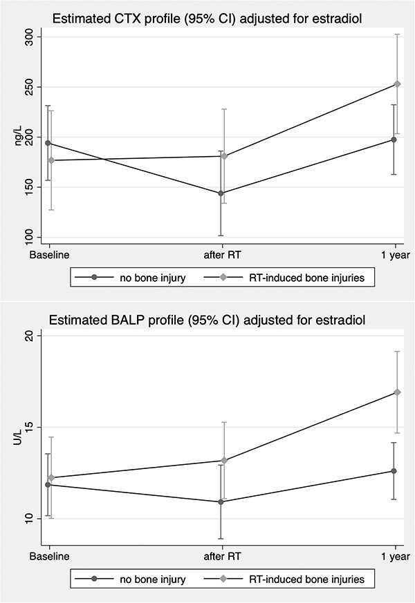
Estimated mean CTX and BALP with 95% CI derived from longitudinal regression models adjusted for serum estradiol illustrating the difference in mean change between women with vs without radiation-induced bone injuries. CTX, C-terminal telopeptide cross-links of collagen type I; RT, radiotherapy; BALP, bone-specific alkaline phosphatase.
Discussion
This longitudinal cohort study explored bone biomarkers in women with and without MRI-detected bone injury from preoperative RT for non-metastatic rectal cancer. Sixteen (44%) of the irradiated women had RT-induced bone injuries and increased bone biomarkers. In the regression analysis, adjusted for serum estradiol, changes in CTX and BALP were associated with radiation-induced bone injury. This is new knowledge that may be of interest for risk evaluation and preventive measures in women susceptible to radiation-induced bone injury.
RT, with curative intent or as neoadjuvant therapy before surgery, is an established treatment for rectal, anal, cervical, and prostate cancer although associated with negative side effects including damage to bone tissue and bone marrow.35 Weakened bone after RT is susceptible for insufficiency fractures caused by normal stress.12 Symptoms include pelvic and back pain, and immobility, which can affect quality of life substantially.35 Reported incidences vary largely depending on, inter alia, method for definition and detection of fractures. MRI, which is a more sensitive method in comparison with conventional CT,27–29 detected pelvic insufficiency fractures in 34% of irradiated rectal cancer patients 3 yr postoperatively in a nationwide Danish study.28 This finding coincides with the current study where 16 women (44%) had radiation-induced injuries among whom 12 (33%) had fractures in pelvic cortical bone. Today, it is not possible to predict the individual risk of insufficiency fractures, but there are some established risk factors including radiation dose13 and technique36 as well as patient characteristics, that is, high age, female sex, postmenopausal status, low BMI, osteoporosis,35,36 and glucocorticoid use.15,34 These risk factors were all evenly distributed between groups with and without radiation-induced bone injury in the present cohort. Bone biomarkers are of potential predictive use but the information in the literature is limited.37 According to a Cochrane analysis from 2018, there is a significant lack of evidence regarding preventive measures of radiation-induced pelvic bone injury.18 These authors identified a need for interventional trials including individuals planned for pelvic irradiation, applying bone turnover markers as surrogate markers of bone health through prospective, and repeated sampling during treatment and follow-up, in addition to radiology and BMD measurements.18
In our previous study, focusing on the impact of RT on bone biomarkers in a larger cohort including the current,26 RT was associated with an increase only in PINP during the first postoperative year, interpreted as a possible recovery process following bone injury. Encouraged by this new finding, further analyses were made and presented here, regarding the association between bone biomarkers and radiation-induced skeletal injury. In addition, bioactive sclerostin was added to the serum analyses. Serum levels of sclerostin and bioactive sclerostin remained, however, quite stable over time and between the 2 groups with and without skeletal lesions. This was somewhat surprising since it has been reported, in animal and in vitro models, that osteocytes are highly sensitive to radiation exposure since it induces osteocyte apoptosis.37,38 However, neither sclerostin nor expression of the SOST gene (encodes sclerostin), was investigated in these studies. Experimental murine models with PTH 1–34 treatment and neutralization antibodies against sclerostin have shown that both these anabolic therapies alleviates radiation-induced bone loss through the Wnt signaling pathway.39,40
Regarding the assessed bone turnover markers, significant differences were found between the groups. In the unadjusted analyses (Table 4), both resorption (CTX) and formation (PINP, BALP) markers increased over time in the group with bone injury but not in the group without; however, an increase in BALP was nearly significant in the latter. After adjustment in longitudinal GEE regression analyses (Table 5), increases in CTX and BALP were associated with radiation-induced bone injury. The findings regarding CTX and BALP may be of future interest in fracture prediction, or in selecting patents for prophylactic treatment of radiation damage to bone. This is not routinely performed today, but the concept of prophylactic treatment and the relation with bone turnover markers is under investigation in a randomized controlled trial from the United Kingdom, including individuals with cervical cancer. Intervention with oral bisphosphonates, in addition to calcium and vitamin D, is used in individuals considered at high-risk for radiation-induced bone injury.41 Bone turnover markers are measured as a secondary outcome. Results from the current study may be applicable for other pelvic cancers. Some clinical evidence suggests that intravenous zoledronic acid administered before irradiation of skeletal metastases may have a protective effect against radiation-induced bone toxicity.42,43
Strengths and limitations
The analyzed study population is younger and healthier than the general population of women with rectal cancer since the original cohort study focused on sexual function, and participants of the current study were included at a center not treating locally advanced rectal cancer. However, the reported findings are important for a younger population in the light of the worldwide increasing incidence of early onset rectal cancer and expanding use of RT in early cancer stages for organ preservation. The venous blood samples were collected prospectively and analyzed in batches to reduce variability and information bias. The reported intervals in time (baseline, after RT, and 1 yr) are long and future studies should include more frequent and longer follow-up to enhance biochemical traceable changes of radiation-induced bone injuries, repair, and remodeling. Confounding by age, menopausal status, and estrogen treatment was assessed in the longitudinal regression analysis, and adjustment for serum levels of estradiol seemed to account for these confounders. A limitation of this study is that we only have data on radiation doses to tumors and not data on the absorbed dose to bones at different locations.
Conclusions
In this prospective exploratory study, bone turnover markers increased during the first postoperative year in women undergoing curative treatment for non-metastatic rectal cancer with adverse radiation-related side effects on bone detected with MRI. In the adjusted analyses, an association was found between the increase in CTX and BALP and radiation-induced bone injury. The findings may be of interest for further studies evaluating fracture risk and selection for prophylactic treatment. Due to few measure points, it was not possible to describe the relation in time between laboratory data and radiological findings.
Acknowledgments
The authors thank colleagues at Ersta Hospital, professor Lennart Blomqvist at Karolinska University Hospital, research nurses Nina Blommé, Madelene Ahlberg, Berit Legestam, and biologist Tana Tomic from the Department of Clinical Chemistry, Linköping University Hospital. We thank Biomedica Medizinprodukte GmbH., Vienna, Austria, for providing the bioactive sclerostin ELISA kits.
Contributor Information
Per Magnusson, Department of Clinical Chemistry, and Department of Biomedical and Clinical Sciences, Linköping University, Linköping, SE-581 85, Sweden.
Maria Sääf, Department of Molecular Medicine and Surgery, Karolinska Institutet, Stockholm, SE-171 77, Sweden.
Anna Martling, Department of Molecular Medicine and Surgery, Karolinska Institutet, Stockholm, SE-171 77, Sweden; Department of Pelvic Cancer, Unit of Gastrointestinal Oncology and Colorectal Surgery, Karolinska University Hospital, Stockholm, SE-171 76, Sweden.
Annika Svanström Röjvall, Department of Molecular Medicine and Surgery, Karolinska Institutet, Stockholm, SE-171 77, Sweden; Department of Surgery and Oncology, Unit of Gastrointestinal Oncology, Capio S:t Göran’s Hospital, Stockholm, SE-112 81, Sweden.
Diana Atanasova, Department of Clinical Chemistry, and Department of Biomedical and Clinical Sciences, Linköping University, Linköping, SE-581 85, Sweden.
Franciszek Wilamowski, Department of Radiology, Ersta Hospital, Stockholm, SE-116 91, Sweden.
Angelique Flöter Rådestad, Department of Hereditary Cancer, Karolinska University Hospital, Stockholm, SE-171 76, Sweden; Department of Women’s and Children’s Health, Karolinska Institutet, Stockholm, SE-171 77, Sweden.
Christian Buchli, Department of Molecular Medicine and Surgery, Karolinska Institutet, Stockholm, SE-171 77, Sweden; Department of Pelvic Cancer, Unit of Gastrointestinal Oncology and Colorectal Surgery, Karolinska University Hospital, Stockholm, SE-171 76, Sweden.
Josefin Segelman, Department of Molecular Medicine and Surgery, Karolinska Institutet, Stockholm, SE-171 77, Sweden; Department of Surgery, Ersta Hospital, Stockholm, SE-116 91, Sweden.
Author contributions
Per Magnusson (Conceptualization, Data curation, Formal analysis, Funding acquisition, Investigation, Methodology, Project administration, Resources, Supervision, Validation, Writing—original draft, Writing—review and editing), Maria Sääf (Conceptualization, Investigation, Methodology, Writing—original draft, Writing—review and editing), Anna Martling (Conceptualization, Data curation, Funding acquisition, Investigation, Methodology, Resources, Supervision, Writing—review and editing), Annika Svanström Röjvall (Conceptualization, Data curation, Formal analysis, investigation, Methodology, Validation, Writing—original draft, Writing—review and editing), Diana Atanasova (Conceptualization, Data curation, Investigation, Validation, Writing—review and editing), Franciszek Wilamowski (Conceptualization, Data curation, Formal analysis, Investigation, Methodology, Validation, Writing—review and editing), Angelique Flöter Rådestad (Conceptualization, Funding acquisition, Investigation, Resources, Writing—review and editing), Christian Buchli (Conceptualization, Data curation, Formal analysis, Investigation, Methodology, Validation, Visualization, Writing—original draft, Writing—review and editing) and Josefin Segelman (Conceptualization, Data curation, Formal analysis, Funding acquisition, Investigation, Methodology, Project administration, Resources, Supervision, Validation, Writing—original draft, Writing—review and editing)
Funding
Financial support was provided through the Swedish Cancer Society, the Stockholm Cancer Society, the Bengt Ihre Research Fellowship, the Bengt Ihre Foundation, and ALF grants from the Stockholm County Council, Karolinska Institutet, and Region Östergötland.
Conflicts of interest
P.M., M.S., A.M., A.S.R., D.A., F.W., A.F.R., C.B., and J.S. report no conflicts of interest.
Data availability
The data underlying this article will be shared on reasonable request to the corresponding author.
References
- 1. Bahadoer RR, Bastiaannet E, Claassen YHM, et al. One-year excess mortality and treatment in surgically treated patients with colorectal cancer: a EURECCA European comparison. Eur J Surg Oncol. 2021;47(7):1651–1660. 10.1016/j.ejso.2021.01.011 [DOI] [PubMed] [Google Scholar]
- 2. Pellino G, Alós R, Biondo S, et al. Trends and outcome of neoadjuvant treatment for rectal cancer: a retrospective analysis and critical assessment of a 10-year prospective national registry on behalf of the Spanish Rectal Cancer Project. Eur J Surg Oncol. 2021;47(2):276–284. 10.1016/j.ejso.2020.04.056 [DOI] [PubMed] [Google Scholar]
- 3. Abraha I, Aristei C, Palumbo I, et al. Preoperative radiotherapy and curative surgery for the management of localised rectal carcinoma. Cochrane Database Syst Rev. 2018;2018(10):CD002102. 10.1002/14651858.CD002102.pub3 [DOI] [PMC free article] [PubMed] [Google Scholar]
- 4. Petrelli F, Trevisan F, Cabiddu M, et al. Total neoadjuvant therapy in rectal cancer: a systematic review and meta-analysis of treatment outcomes. Ann Surg. 2020;271(3):440–448. 10.1097/SLA.0000000000003471 [DOI] [PubMed] [Google Scholar]
- 5. Bahadoer RR, Dijkstra EA, van Etten B, et al. Short-course radiotherapy followed by chemotherapy before total mesorectal excision (TME) versus preoperative chemoradiotherapy, TME, and optional adjuvant chemotherapy in locally advanced rectal cancer (RAPIDO): a randomised, open-label, phase 3 trial. Lancet Oncol. 2021;22(1):29–42. 10.1016/S1470-2045(20)30555-6 [DOI] [PubMed] [Google Scholar]
- 6. Conroy T, Bosset J-F, Etienne P-L, et al. Neoadjuvant chemotherapy with FOLFIRINOX and preoperative chemoradiotherapy for patients with locally advanced rectal cancer (UNICANCER-PRODIGE 23): a multicentre, randomised, open-label, phase 3 trial. Lancet Oncol. 2021;22(5):702–715. 10.1016/S1470-2045(21)00079-6 [DOI] [PubMed] [Google Scholar]
- 7. Nilsson PJ, Ahlberg M, Kordnejad S, Holm T, Martling A. Organ preservation following short-course radiotherapy for rectal cancer. BJS Open. 2021;5(5):zrab093. 10.1093/bjsopen/zrab093 [DOI] [PMC free article] [PubMed] [Google Scholar]
- 8. van der Valk MJM, Hilling DE, Bastiaannet E, et al. Long-term outcomes of clinical complete responders after neoadjuvant treatment for rectal cancer in the International Watch & Wait Database (IWWD): an international multicentre registry study. Lancet. 2018;391(10139):2537–2545. 10.1016/S0140-6736(18)31078-X [DOI] [PubMed] [Google Scholar]
- 9. Mauri G, Sartore-Bianchi A, Russo A-G, Marsoni S, Bardelli A, Siena S. Early-onset colorectal cancer in young individuals. Mol Oncol. 2019;13(2):109–131. 10.1002/1878-0261.12417 [DOI] [PMC free article] [PubMed] [Google Scholar]
- 10. Petersson J, Bock D, Martling A, Smedby KE, Angenete E, Saraste D. Increasing incidence of colorectal cancer among the younger population in Sweden. BJS Open. 2020;4(4):645–658. 10.1002/bjs5.50279 [DOI] [PMC free article] [PubMed] [Google Scholar]
- 11. Wang C, Qin X, Gong G, Wang L, Su Y, Yin Y. Correlation between changes of pelvic bone marrow fat content and hematological toxicity in concurrent chemoradiotherapy for cervical cancer. Radiat Oncol. 2022;17(1):70. 10.1186/s13014-022-02029-y [DOI] [PMC free article] [PubMed] [Google Scholar]
- 12. Oh D, Huh SJ. Insufficiency fracture after radiation therapy. Radiat Oncol J. 2014;32(4):213–220. 10.3857/roj.2014.32.4.213 [DOI] [PMC free article] [PubMed] [Google Scholar]
- 13. Okoukoni C, Randolph DM, McTyre ER, et al. Early dose-dependent cortical thinning of the femoral neck in anal cancer patients treated with pelvic radiation therapy. Bone. 2017;94:84–89. 10.1016/j.bone.2016.10.021 [DOI] [PubMed] [Google Scholar]
- 14. van Wulfften PO, Jee K-W, Bramer JAM, Hornicek FJ, Chen Y-LE, Schwab JH. What is the effect of high-dose radiation on bone in patients with sacral chordoma? A CT study. Clin Orthop Relat Res. 2018;476(3):520–528. 10.1007/s11999.0000000000000063 [DOI] [PMC free article] [PubMed] [Google Scholar]
- 15. Lim Y, Baek KH, Kim H-J, Lee S, Lee JW, Kang M-I. Changes in trabecular bone score and bone mineral density following allogeneic hematopoietic stem cell transplantation. Bone. 2019;124:40–46. 10.1016/j.bone.2019.04.004 [DOI] [PubMed] [Google Scholar]
- 16. Akashi M, Hashikawa K, Wanifuchi S, et al. Heterogeneity of necrotic changes between cortical and cancellous bone in mandibular osteoradionecrosis: a histopathological analysis of resection margin after segmental mandibulectomy. Biomed Res Int. 2017;2017:3125842. 10.1155/2017/3125842 [DOI] [PMC free article] [PubMed] [Google Scholar]
- 17. Dekker H, Schulten EAJM, Lichters I, et al. Osteocyte apoptosis, bone marrow adiposity, and fibrosis in the irradiated human mandible. Adv Radiat Oncol. 2022;7(4):100951. 10.1016/j.adro.2022.100951 [DOI] [PMC free article] [PubMed] [Google Scholar]
- 18. van den Blink QU, Garcez K, Henson CC, Davidson SE, Higham CE, Cochrane Gynaecological, Neuro-oncology and Orphan Cancer Group . Pharmacological interventions for the prevention of insufficiency fractures and avascular necrosis associated with pelvic radiotherapy in adults. Cochrane Database Syst Rev. 2018;2018(4):CD010604. 10.1002/14651858.CD010604.pub2 [DOI] [PMC free article] [PubMed] [Google Scholar]
- 19. Sakaguchi M, Maebayashi T, Aizawa T, Ishibashi N. Risk factors for sacral insufficiency fractures in cervical cancer after whole pelvic radiation therapy. Anticancer Res. 2019;39(1):361–367. 10.21873/anticanres.13120 [DOI] [PubMed] [Google Scholar]
- 20. Donaubauer A-J, Deloch L, Becker I, Fietkau R, Frey B, Gaipl US. The influence of radiation on bone and bone cells-differential effects on osteoclasts and osteoblasts. Int J Mol Sci. 2020;21(17):6377. 10.3390/ijms21176377 [DOI] [PMC free article] [PubMed] [Google Scholar]
- 21. Eastell R, Pigott T, Gossiel F, Naylor KE, Walsh JS, Peel NFA. Diagnosis of endocrine disease. Bone turnover markers: are they clinically useful? Eur J Endocrinol. 2018;178(1):R19–R31. 10.1530/EJE-17-0585 [DOI] [PubMed] [Google Scholar]
- 22. Schini M, Vilaca T, Gossiel F, Salam S, Eastell R. Bone turnover markers: basic biology to clinical applications. Endocr Rev. 2023;44(3):417–473. 10.1210/endrev/bnac031 [DOI] [PMC free article] [PubMed] [Google Scholar]
- 23. Omran A, Atanasova D, Landgren F, Magnusson P. Sclerostin: from molecule to clinical biomarker. Int J Mol Sci. 2022;23(9):4751. 10.3390/ijms23094751 [DOI] [PMC free article] [PubMed] [Google Scholar]
- 24. Svanström Röjvall A, Buchli C, Bottai M, et al. Effect of radiotherapy for rectal cancer on female sexual function: a prospective cohort study. Br J Surg. 2020;107(5):525–536. 10.1002/bjs.11373 [DOI] [PubMed] [Google Scholar]
- 25. Segelman J, Buchli C, Svanström Röjvall A, et al. Effect of radiotherapy for rectal cancer on ovarian androgen production. Br J Surg. 2019;106(3):267–275. 10.1002/bjs.10980 [DOI] [PMC free article] [PubMed] [Google Scholar]
- 26. Svanström Röjvall A, Buchli C, Sääf M, et al. Impact of radiotherapy on bone health in women with rectal cancer - a prospective cohort study. Eur J Surg Oncol. 2022;48(12):2509–2517. 10.1016/j.ejso.2022.06.028 [DOI] [PubMed] [Google Scholar]
- 27. Cabarrus MC, Ambekar A, Lu Y, Link TM. MRI and CT of insufficiency fractures of the pelvis and the proximal femur. AJR Am J Roentgenol. 2008;191(4):995–1001. 10.2214/AJR.07.3714 [DOI] [PubMed] [Google Scholar]
- 28. Jørgensen JB, Bondeven P, Iversen LH, Laurberg S, Pedersen BG. Pelvic insufficiency fractures frequently occur following preoperative chemo-radiotherapy for rectal cancer - a nationwide MRI study. Color Dis. 2018;20(10):873–880. 10.1111/codi.14224 [DOI] [PubMed] [Google Scholar]
- 29. Palm HG, Lang P, Hackenbroch C, Sailer L, Friemert B. Dual-energy CT as an innovative method for diagnosing fragility fractures of the pelvic ring: a retrospective comparison with MRI as the gold standard. Arch Orthop Trauma Surg. 2020;140(4):473–480. 10.1007/s00402-019-03283-8 [DOI] [PubMed] [Google Scholar]
- 30. Zhong X, Zhang L, Dong T, et al. Clinical and MRI features of sacral insufficiency fractures after radiotherapy in patients with cervical cancer. BMC Womens Health. 2022;22(1):166. 10.1186/s12905-022-01758-2 [DOI] [PMC free article] [PubMed] [Google Scholar]
- 31. Syk E, Torkzad MR, Blomqvist L, Nilsson PJ, Glimelius B. Local recurrence in rectal cancer: anatomic localization and effect on radiation target. Int J Radiat Oncol Biol Phys. 2008;72(3):658–664. 10.1016/j.ijrobp.2008.01.063 [DOI] [PubMed] [Google Scholar]
- 32. Stevens SK, Moore SG, Kaplan ID. Early and late bone-marrow changes after irradiation: MR evaluation. Am J Roentgenol. 1990;154(4):745–750. 10.2214/ajr.154.4.2107669 [DOI] [PubMed] [Google Scholar]
- 33. Ugurluer G, Akbas T, Arpaci T, Ozcan N, Serin M. Bone complications after pelvic radiation therapy: evaluation with MRI. J Med Imaging Radiat Oncol. 2014;58(3):334–340. 10.1111/1754-9485.12176 [DOI] [PubMed] [Google Scholar]
- 34. Meixel AJ, Hauswald H, Delorme S, Jobke B. From radiation osteitis to osteoradionecrosis: incidence and MR morphology of radiation-induced sacral pathologies following pelvic radiotherapy. Eur Radiol. 2018;28(8):3550–3559. 10.1007/s00330-018-5325-2 [DOI] [PubMed] [Google Scholar]
- 35. Higham CE, Faithfull S. Bone health and pelvic radiotherapy. Clin Oncol (R Coll Radiol). 2015;27(11):668–678. 10.1016/j.clon.2015.07.006 [DOI] [PubMed] [Google Scholar]
- 36. Sapienza LG, Salcedo MP, Ning MS, et al. Pelvic insufficiency fractures after external beam radiation therapy for gynecologic cancers: a meta-analysis and meta-regression of 3929 patients. Int J Radiat Oncol Biol Phys. 2020;106(3):475–484. 10.1016/j.ijrobp.2019.09.012 [DOI] [PubMed] [Google Scholar]
- 37. Wright LE. Radiotherapy-induced osteoporosis. In: Bilezikian JP, ed. Primer on the Metabolic Bone Diseases and Disorders of Mineral Metabolism. 9th ed. Wiley-Blackwell; 2019:788–792 10.1002/9781119266594.ch102. [DOI] [Google Scholar]
- 38. Wright LE, Buijs JT, Kim H-S, et al. Single-limb irradiation induces local and systemic bone loss in a murine model. J Bone Miner Res. 2015;30(7):1268–1279. 10.1002/jbmr.2458 [DOI] [PMC free article] [PubMed] [Google Scholar]
- 39. Chandra A, Lin T, Zhu J, et al. PTH1-34 blocks radiation-induced osteoblast apoptosis by enhancing DNA repair through canonical Wnt pathway. J Biol Chem. 2015;290(1):157–167. 10.1074/jbc.M114.608158 [DOI] [PMC free article] [PubMed] [Google Scholar]
- 40. Chandra A, Lin T, Young T, et al. Suppression of sclerostin alleviates radiation-induced bone loss by protecting bone-forming cells and their progenitors through distinct mechanisms. J Bone Miner Res. 2017;32(2):360–372. 10.1002/jbmr.2996 [DOI] [PMC free article] [PubMed] [Google Scholar]
- 41. Chatzimavridou Grigoriadou V, Barraclough LH, Baricevic-Jones I, et al. RadBone: bone toxicity following pelvic radiotherapy - a prospective randomised controlled feasibility study evaluating a musculoskeletal health package in women with gynaecological cancers undergoing pelvic radiotherapy. BMJ Open. 2022;12(6):e056600. 10.1136/bmjopen-2021-056600 [DOI] [PMC free article] [PubMed] [Google Scholar]
- 42. Gierloff M, Reutemann M, Gülses A, Niehoff P, Wiltfang J, Acil Y. Effects of zoledronate on the radiation-induced collagen breakdown: a prospective randomized clinical trial. Clin Transl Oncol. 2015;17(6):454–461. 10.1007/s12094-014-1257-8 [DOI] [PubMed] [Google Scholar]
- 43. Pichon B, Campion L, Delpon G, et al. High-dose hypofractionated radiation therapy for noncompressive vertebral metastases in combination with zoledronate: a phase 1 study. Int J Radiat Oncol Biol Phys. 2016;96(4):840–847. 10.1016/j.ijrobp.2016.07.027 [DOI] [PubMed] [Google Scholar]
Associated Data
This section collects any data citations, data availability statements, or supplementary materials included in this article.
Data Availability Statement
The data underlying this article will be shared on reasonable request to the corresponding author.