Abstract
Patient: Male, 66-year-old
Final Diagnosis: Hemopericardium
Symptoms: Hemopericardium
Clinical Procedure: —
Specialty: Cardiology • General and Internal Medicine • Neurology
Objective:
Unusual clinical course
Background:
Despite having many benefits, frequently-used medications may still have potential risks and can cause harm. Hemopericardium is a lethal pathology with a high risk of mortality and a lower differential diagnosis consideration. When adding both mentioned elements, their consideration as a differential diagnosis would require a higher threshold. This report presents a 66-year-old man with atrial fibrillation, heart failure, and aortic stenosis status post transcatheter aortic valve replacement (TAVR) 1 year ago with hemopericardium while treated with apixaban.
Case Report:
We present the case of a 66-year-old man with multiple medical conditions, including atrial fibrillation, heart failure, and aortic stenosis post-transcatheter aortic valve replacement 1 year before admission, who presented with 2 weeks of dyspnea and lower-limb swelling. Initial assessments revealed atrial fibrillation, elevated brain natriuretic peptide, and a chest X-ray indicating possible left pleural effusion and cardiomegaly. On day 4, an echocardiogram identified a large hemopericardium and tamponade, prompting urgent surgery. A pericardial window was performed, draining 1700 cc of bloody fluid. The postoperative improvement included normalized hemodynamics and echocardiographic findings. Pathology confirmed hemopericardium. The follow-up echocardiogram showed improved cardiac function, and the patient was transferred to the general medical floor.
Conclusions:
This case sheds light on the uncommon but critical complications associated with direct oral anticoagulant therapy. With only a handful of reported cases, the rarity of this condition underscores the need for heightened awareness among clinicians. The patient’s intricate medical history accentuates the challenges in managing anticoagulation in individuals with multiple comorbidities.
Key words: Anticoagulant Reversal Agents, Apixaban, Cardiology, Factor Xa Inhibitors
Introduction
Hemopericardium is a life-threatening condition that can result from several conditions, including trauma, aortic dissection, surgeries, and infections [1,2]. Hemopericardium due to anticoagulation drugs such as warfarin is rare, although it is reported in the literature. Apixaban-induced hemopericardium was first reported in 2015 [3].
Apixaban is a direct oral anticoagulant (DOAC); it works via the reversible inhibition of factor Xa [3]. In 2014, apixaban was approved for treating deep vein thrombosis (DVT) and pulmonary embolism (PE), and for lowering the risk of blood clots in patients who had knee or hip replacement surgery. Additionally, it is approved for preventing the recurrence of DVT and PE following initial treatment [4]. It is currently widely used since it showed a superior profile and less adverse effects than warfarin [5,6]. It also requires less monitoring than warfarin [3,5].
Apixaban-induced hemopericardium remains extremely rare despite the growing use of this drug, with only around 11 cases reported in the literature [5]. Most patients presented with shortness of breath and were unstable during evaluation; arrhythmias were the most common indication for anticoagulation in most cases [6,7]. In addition, several factors were speculated to be involved in the development of these conditions, such as renal dysfunction, drug–drug interactions, and older age.
Here, we present the case of a 66-year-old man who developed apixaban-induced hemopericardium after ruling out other causes and was managed via pericardiocentesis, which revealed a large volume of blood.
Case Report
A 66-year-old man presented to the Emergency Department with shortness of breath and bilateral lower-extremity swelling that gradually developed over the last 2 weeks. He was on apixaban and had a past medical history significant for atrial fibrillation, heart failure with preserved ejection fraction, essential hypertension, dyslipidemia, aortic stenosis status after transcatheter aortic valve replacement (TAVR) 1 year before admission, along with complete heart block status after permanent pacemaker implant and history of mitral valve endocarditis both in the same year, bilateral lower-limb lymph-edema. The indicated he had orthopnea and paroxysmal nocturnal dyspnea, but denied any cough, fever, chills, chest pain, or palpitations.
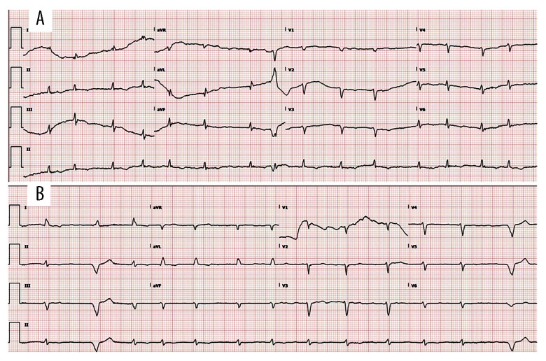
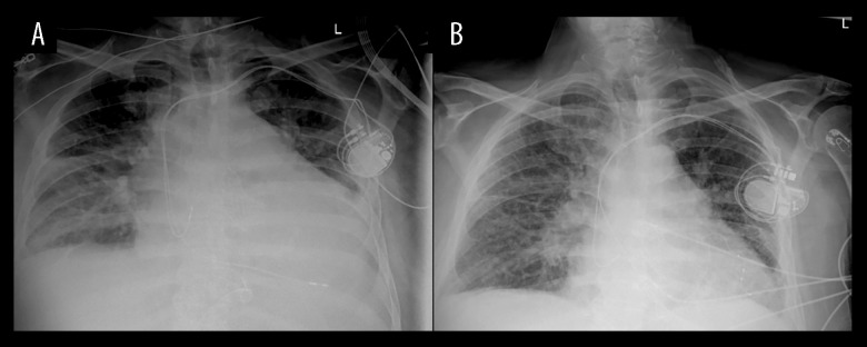
Blood pressure at presentation was 116/96, respiratory rate 20, and heart rate was 91 beats per minute. He was afebrile, and O2 was 98% on room air. Complete blood count and comprehensive metabolic profile were unremarkable. However, the EKG showed atrial fibrillation with a heart rate of 95, as shown in Figure 1, with no ST elevation. Brain natriuretic peptide was 806 pg/mL (normal value is <100 pg/ml), and troponin was not elevated. He required 2 L oxygen via nasal cannula, and a chest X-ray showed probable small left pleural effusion and mild cardiomegaly, suggesting edema, as seen in Figure 2. However, on day 3, he started to become tachycardic with a heart rate above 108 and started desaturating, requiring 5 L of oxygen by nasal cannula.
Figure 1.
(A) EKG on admission, (B) EKG on the day of transfer.
Figure 2.
(A) Chest X-ray prior to echocardiography, (B) Chest X-ray on the day of transfer.
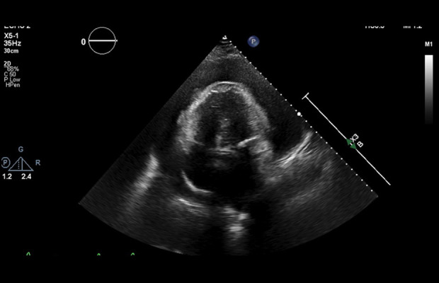
On hospital day 4, an echocardiogram showed reduced left ventricular ejection fraction estimated at 35–40%, severely decreased RV systolic function, right ventricular diastolic collapse, severely increased left atrial size, mildly increased right atrial size, moderate global left ventricular hypokinesis, and, most importantly, a large pericardial effusion suggesting early signs of hemopericardium, with cardiac tamponade as noted in Figure 3. Given his hemodynamic instability and signs of cardiac tamponade on EKG, Cardiothoracic Surgery was consulted and clopidogrel and apixaban were held for a possible pericardial window. Lasix was also held because of the large hemopericardium. The patient was made nil per os and scheduled for an urgent subxiphoid pericardial window.
Figure 3.

Four-chamber view echocardiography illustrating fluid surrounding cardiac structure.
Intraoperative transesophageal echocardiography confirmed a large circumferential hemopericardium, and a total of 1700 cc of bloody pericardial fluid was removed via the pericardial window. A portion of the pericardial tissue itself was excised. A sample was sent to Microbiology, and the remainder was sent to Pathology. A follow-up transesophageal echocardiogram revealed complete resolution. The patient’s hemodynamics improved upon drainage of the fluid. The pathology report of pericardial effusion and tissue showed fragments of fibro-membranous tissue with mild degenerative changes, which concluded with clinical hemopericardium.
A repeat EKG on postoperative day 4 showed left ventricular ejection fraction estimated at 40–45%, mildly increased right ventricular size, mild-to-moderate tricuspid valve regurgitation, moderate pulmonary hypertension, right ventricular systolic pressure of 49.6 mmHg, and improved pericardial effusion. The chest tube was removed, as it was draining minimal fluid. The patient was transferred to the general medical floor following improvement in clinical status and radiological findings on chest X-ray, as illustrated in Figure 2, and EKG, as shown in Figure 1.
Discussion
Bleeding is a possible complication of direct oral anticoagulant (DOACs), including apixaban, with gastro-intestinal problems being of especially high risk [1]. Nevertheless, few cases of bleeding into rarer sites, such as the pericardial space (hemopericardium) caused by different direct oral anticoagulant (DOACs), have been published [8]. Furthermore, hemopericardium associated with rivaroxaban and dabigatran was more often reported than with apixaban [6].
A recent systematic review found only 41 reported cases of DOACs-induced hemopericardium, with apixaban accounting for only 11 of them [6]. Furthermore, several factors were linked to these cases, such as older age, male sex, multiple comorbidities, and renal dysfunction [6–8]. Iatrogenic interventions such as pacemaker implantation can cause pericarditis, which may develop into hemopericardium due to direct oral anticoagulant (DOACs) use [6].
Patients with liver and kidney dysfunction can be prone to a higher risk of complications as direct oral anticoagulant (DOACs) are excreted from the body mainly through bile, both directly and indirectly (after being metabolized via the CYP3A4) [5]. Renal excretion is 80% for dabigatran, 35% for rivaroxaban, and 27% for apixaban [7].
Drud–drug interactions with direct oral anticoagulant (DOACs) include amiodarone, diltiazem, losartan, amlodipine, escitalopram, and saw palmetto [8]. Furthermore, some case reports have also pointed to such interactions with selective serotonin reuptake inhibitors (SSRIs), chemotherapeutic agents, and antiplatelet drugs such as aspirin and clopidogrel [6,8]. Our patient did not have any of the above risk factors other than a pacemaker, and developed a such complication.
The most common presentation of patients was shortness of breath and lower-extremity edema, and most of the patients were hemodynamically unstable, which is different from our case. Furthermore, arrhythmia (atrial fibrillation) was the indication for anticoagulation in most cases [6,7], and most patients developed symptoms within 2–12 weeks [1].
Since hemopericardium is a life-threatening condition that can present acutely as cardiac tamponade or with more subacute, varying, non-specific symptoms (eg, shortness of breath in a hemodynamically stable patient), which was the case in our patient, it is very important to diagnose and manage it once clinical suspicion has raised [8]. Physical examination, electrocardiogram, and imaging findings such as hypotension, pulsus paradoxus, elevated jugular venous pressure, pulsus alternans, or cardiomegaly can greatly aid in the diagnosis [8]. Nevertheless, once hemopericardium is suspected, urgent diagnostic echocardiography (ECHO) must be done to confirm the diagnosis [8].
Pericardiocentesis currently remains the main treatment, but pericardial window could be considered with symptomatic pericardial effusion (either circumferential or loculated) [9]. There are no routine tests done to monitor the effectiveness of direct oral anticoagulant (DOACs) and no reported cases of the use of Andexanet alfa in the management of such cases, although idarucizumab was used in 1 case to reverse the effect of dabigatran, with improvement, which makes the idea promising [5,8]. Considering the above, the use of direct thrombin inhibitors or anti-Factor Xa assays and assessing the effectiveness of reversal agents such as Andexanet alfa will be very important [7,8].
Conclusions
In conclusion, this case sheds light on the uncommon but critical complications associated with direct oral anticoagulant therapy. With only a handful of reported cases, the rarity of this condition underscores the need for heightened awareness among clinicians. Our patient’s intricate medical history accentuates the challenges in managing anticoagulation in individuals with multiple comorbidities. Prompt diagnosis through echocardiography and successful pericardiocentesis highlight the importance of swift intervention, especially in hemodynamically unstable patients. As the use of direct oral anticoagulants continues to rise, further research is essential to establish effective monitoring protocols and explore the role of reversal agents in managing these rare and potentially life-threatening complications.
Footnotes
Publisher’s note: All claims expressed in this article are solely those of the authors and do not necessarily represent those of their affiliated organizations, or those of the publisher, the editors and the reviewers. Any product that may be evaluated in this article, or claim that may be made by its manufacturer, is not guaranteed or endorsed by the publisher
Declaration of Figures’ Authenticity
All figures submitted have been created by the authors who confirm that the images are original with no duplication and have not been previously published in whole or in part.
References:
- 1.Nasir SA, Babu Pokhrel N, Baig A. Hemorrhagic pericardial effusion from apixaban use: Case report and literature review. Cureus. 2022;14(10):e30021. doi: 10.7759/cureus.30021. [DOI] [PMC free article] [PubMed] [Google Scholar]
- 2.Olagunju A, Khatib M, Palermo-Alvarado F. A possible drug–drug interaction between eliquis and amiodarone resulting in hemopericardium. Cureus. 2021;13(2):e13486. doi: 10.7759/cureus.13486. [DOI] [PMC free article] [PubMed] [Google Scholar]
- 3.Sigawy C, Apter S, Vine J, Grossman E. Spontaneous hemopericardium in a patient receiving apixaban therapy: First case report. Pharmacotherapy. 2015;35(7):e115–17. doi: 10.1002/phar.1602. [DOI] [PubMed] [Google Scholar]
- 4.Agrawal A, Kerndt CC, Manna B. StatPearls [Internet] Treasure Island (FL): StatPearls Publishing; 2024. Apixaban. [Updated 2024 Feb 22] [PubMed] [Google Scholar]
- 5.Cinelli M, Uddin A, Duka I, et al. Spontaneous hemorrhagic pericardial and pleural effusion in a patient receiving apixaban. Cardiol Res. 2019;10(4):249–52. doi: 10.14740/cr902. [DOI] [PMC free article] [PubMed] [Google Scholar]
- 6.Sheikh AB, Shah I, Sagheer S, et al. Hemopericardium in the setting of direct oral anticoagulant use: An updated systematic review. Cardiovasc Revasc Med. 2022;39:73–83. doi: 10.1016/j.carrev.2021.09.010. [DOI] [PubMed] [Google Scholar]
- 7.Asad ZUA, Ijaz SH, Chaudhary AMD, et al. Hemorrhagic cardiac tamponade associated with apixaban: A case report and systematic review of literature. Cardiovasc Revasc Med. 2019;20(11S):15–20. doi: 10.1016/j.carrev.2019.04.002. [DOI] [PMC free article] [PubMed] [Google Scholar]
- 8.Aguilar-Gallardo JS, Das S, et al. Clinically ambiguous hemorrhagic cardiac tamponade associated with apixaban. Cureus. 2022;14(4):e24290. doi: 10.7759/cureus.24290. [DOI] [PMC free article] [PubMed] [Google Scholar]
- 9.Imazio M, Adler Y. Management of pericardial effusion. Eur Heart J. 2013;34(16):1186–97. doi: 10.1093/eurheartj/ehs372. [DOI] [PubMed] [Google Scholar]


