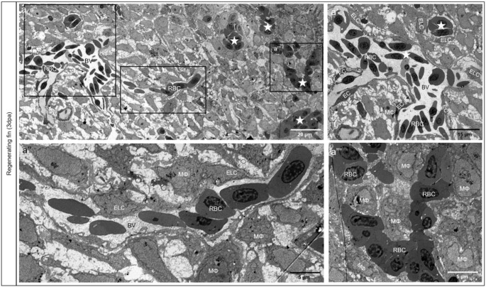
Fig. 1.
Endothelial cell mosaic during neoangiogenesis in the regenerating zebrafish fin. (a) an overview of the regenerative tissue at 3dpa with perfused blood vessels (BV) containing red blood cells (RBC), and extravasal clusters (asterisks) or single RBC; the distal part of the fin is depicted on the right side. The proximal part of the fin with large and perfused blood vessel with well-differentiated endothelial cells (EC) and classical morphological appearance is depicted in a’. At the distal (right) part, first vessels containing a mosaic of typical EC and Endothelial-Like Cells (ELC) are detectible (asterisks). a’’ depicts a transient segment with continuous vascular coverage. The latter consist of ELCs with long sleeves surrounding the RBCs. The morphological appearance of the ELCs closely resembles the macrophage (MΦ) structural phenotype. Different, transient forms are detectible. At the vascular front, the cells surrounding the RBC clusters are classical MΦ (a’’’). Images are acquired by transmission electron microscopy

