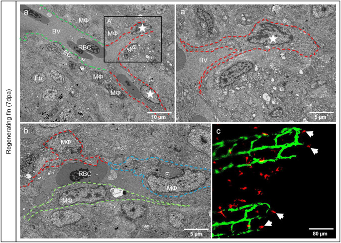
Fig. 2.
The vasculature expands by transformation of the adjacent macrophages into endothelial like cells. a) mosaic blood vessel containing a typical blood vessel segment with well-defined ECs (green dash line), and an atypical one appearing as a channel (red dash line) covered by MΦ (asterisk) extensions. At higher magnification (a’), MΦ at the vascular tip (asterisk) send a cytoplasmic extensions (dotted line) to the stock EC cells. (b) elongated MΦ (traced red, blue and green) is part of the vascular wall with extensions fencing the lumen containing plasma and RBCs. In vivo investigations (ECs appear in green and MΦ in red) confirmed MΦ at the front of the vessel tips (c, arrows) corresponding to that demonstrated in a and a’ by TEM. Images a, a’ and b are acquired by the transmission electron microscope and image c by confocal microscopy

