Abstract
Background
A stent with characteristics of a hybrid design may have advantages in improving the patency of symptomatic iliofemoral vein obstruction. This study assessed the safety and effectiveness of the V-Mixtent Venous Stent in treating symptomatic iliofemoral outflow obstruction.
Methods
Eligible patients had a Clinical-Etiologic-Anatomic-Physiologic (CEAP) C classification of ≥ 3 or a Venous Clinical Severity Score (VCSS) pain score of ≥ 2. The primary safety endpoint was the rate of major adverse events within 30 days. The primary effectiveness endpoint was the 12-month primary patency rate. Secondary endpoints included changes in VCSS from baseline to 6 and 12 months, alterations in CEAP C classification, Chronic Venous Disease Quality of Life Questionnaire (CIVIQ-14) scores at 12 months, and stent durability measures.
Results
Between December 2020 and November 2021, 171 patients were enrolled across 15 institutions. A total of 185 endovenous stents were placed, with 91.81% of subjects receiving one stent and 8.19% receiving 2 stents. Within 30 days, only two major adverse events occurred (1.17%; 95% confidence interval [CI], 0.14–4.16%), below the literature-defined performance goal of 11% (P < .001). The 12-month primary patency rate (91.36%; 95% CI, 85.93–95.19%; P < .001) exceeded the literature-defined performance goal. VCSS changes from baseline demonstrated clinical improvement at 6 months (− 4.30 ± 3.66) and 12 months (− 4.98 ± 3.67) (P < .001). Significant reduction in symptoms, as measured by CEAP C classification and CIVIQ-14, was observed from pre-procedure to 12 months (P < .001).
Conclusions
The 12-month outcomes confirm the safety and effectiveness of the V-Mixtent Venous Stent in managing symptomatic iliofemoral venous outflow obstruction, including clinical symptom improvement compared to before treatment.
Supplementary Information
The online version contains supplementary material available at 10.1186/s12916-024-03545-2.
Keywords: Venous stenting, Iliofemoral venous outflow obstruction, Patency, Multicenter study, Clinical improvement
Background
Deep venous stenting has gained significant popularity as a prevalent approach for managing acute or chronic venous obstruction, including non-thrombotic iliac vein lesions (NIVL) in recent years [1, 2]. This is particularly relevant for individuals classified as Clinical-Etiologic-Anatomic-Physiologic (CEAP) C ≥ 3 or with a Venous Clinical Severity Score (VCSS) score ≥ 2 or both. Patients falling into these categories, exhibiting over 50% stenosis on imaging modalities such as computed tomography venography, magnetic resonance venography, intravascular ultrasound, or venography, should be considered for venous stenting [2]. Similarly, patients with identified stenotic lesions undergoing thrombus removal for acute iliofemoral deep vein thrombosis (DVT) should also be considered eligible candidates for venous stenting [2].
Explorations into venous stenting in pelvic veins commenced in the early 1990s [3–7], with subsequent extensive scrutiny of peer-reviewed publications by Raju in 2013 affirming that “Iliac vein stenting is safe, with negligible morbidity (< 1%), and exhibits patency rates of 90% to 100% for non-thrombotic disease and 74% to 89% for post-thrombotic disease at 3 to 5 years” [8]. The reported outcomes spanning short, mid, and long-term periods over the past 25 years consistently demonstrate promise. Venous stents, as a whole, have undergone meticulous design enhancements aimed at improving their effectiveness and facilitating the endovascular treatment of intricate venous lesions. These advancements include increased flexibility, expandability, and improved visibility [9]. Despite these improvements, the ongoing challenge lies in striking the optimal balance between radial force/crush resistance and high flexibility. Consequently, the selection of a stent should be tailored to suit the specific circumstances, considering factors such as flexibility, the risk of stent fracture (e.g., when crossing the inguinal ligament), and the imperative for heightened crush resistance.
In this context, the V-Mixtent Venous Stent (Shanghai EndoVas Medical Technology Co., Ltd, Shanghai, China), employing a modular design concept with non-uniform stents that combine laser-cut and woven structures, presents a promising solution. The objective of this study is to assess the safety and effectiveness of the V-Mixtent Venous Stent in the management of iliofemoral venous outflow obstruction.
Methods
Study design
This research report adheres to the STROCSS standards [10]. This prospective, single-arm multicenter trial was designed to include adult participants aged ≥ 18 and ≤ 75 years who presented with symptomatic chronic non-malignant obstruction of the iliofemoral venous system. The primary objective was to assess the safety and effectiveness of the V-Mixtent Venous Stent. To ensure a diverse patient population, the study involved large medical centers, with efforts directed towards achieving an equitable distribution of enrollments across centers, where each center enrolled ≤ 30% of the total sample. Consecutive patient screening was employed to minimize selection bias, and treated venous segments included the common iliac vein (CIV), external iliac vein (EIV), and common femoral vein (CFV).
The implementation process of the study can be seen in Additional file 1: Fig. S1. The inclusion and exclusion criteria for the study were detailed in the additional file (Additional file 1: Table S1). Inclusion criteria necessitated patients to have manifested unilateral and clinically significant venous obstruction, defined as clinical classification ≥ C3 of the CEAP classification [11] or a VCSS score [12] ≥ 2, along with luminal diameter reduction ≥ 50% on biplanar venography. Biplanar venography involves visualizing the target blood vessel at its maximum and minimum diameters through naked-eye observation. Exclusion criteria included recent pulmonary embolism, contralateral venous disease, inferior vena cava involvement, limited life expectancy, known allergy to stent components, near-term family planning, uncontrolled or active coagulopathy, bleeding diathesis, or planned concurrent venous procedures within 30 days of stent implantation. Stenting was indicated for acute DVT post-thrombectomy or thrombolysis in cases of residual iliac vein obstruction. Pre-procedure clinical assessments, incorporating CEAP C classification, VCSS, and Chronic Venous Insufficiency Questionnaire (CIVIQ-14) [13], were performed.
Between December 2020 and November 2021, a total of 171 patients underwent implantation at 15 centers in China. The study has a scheduled follow-up period of 5 years. The study protocol received approval from institutional review boards or ethical committees at participating centers. A Clinical Events Committee (CEC) was responsible for adjudicating events related to primary safety and effectiveness endpoints, while a Data and Safety Monitoring Board oversaw safety outcomes. Image data will be evaluated by independent core laboratories, adhering to the principles outlined in the Declaration of Helsinki. The study obtained approval from the Institutional Review Board at each study center, and all patients provided informed consent.
Device description
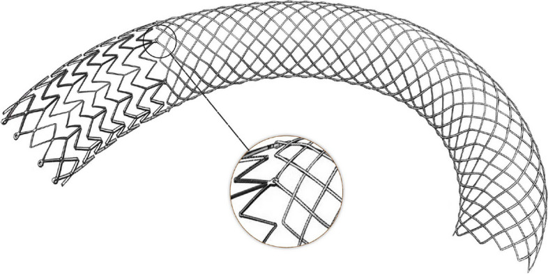
The V-Mixtent Venous Stent was a self-expanding nitinol stent featuring radiopaque markers (Fig. 1). We compared the properties of the V-Mixtent Venous Stent with those of other venous stents (Additional file 1: Table S2). Uniform in diameter, the stent was available in six sizes: 10, 12, 14, 16, 18, and 20 mm. Stent length options included 60, 80, 100, 120, and 150 mm. The stent incorporated etched segments, which came in three lengths: 37, 47, and 57mm. It is noteworthy that not all stent lengths were compatible with the utilization of etched segments.
Fig. 1.

V-Mixtent Venous Stent design
Preloaded within 8F or 10F over-the-wire introducers of 80-cm lengths, the stent was purpose-designed for iliofemoral veins. The common iliac vein segment of the stent displayed a laser-etched structure, ensuring robust radial support and local resistance to compression. During surgical procedures, the etched section was released initially without a rebound effect, ensuring precise positioning and accurate deployment. Additionally, the stent boasted a woven structure designed to conform to the physiological curvature of the iliofemoral vein, providing a high degree of flexibility. The weaving pattern facilitated the smooth downward extension of the distal end upon release, rendering it suitable for placement in tortuous and diametrically varying vessels and enabling joint crossing. A looped structure was strategically employed to establish a flexible connection between the etched and weaving sections. Furthermore, all silk tips were embedded within the stent, a feature aimed at minimizing damage to the vessel wall.
Implant procedure
Baseline assessment of percentage diameter stenosis was conducted using biplanar venography. Stent sizing, placement extent, and evaluation of postprocedural residual stenosis were guided by venography. The procedure recommended both vessel predilation and post-stent dilatation. The stents were deliberately oversized by 2 to 4 mm in consideration of the surrounding vasculature, the expanded balloon diameter used for predilation, or the standard diameter of the vein intended for stenting. Recommendations included achieving comprehensive lesion coverage and extending into healthy tissue, both caudally and cranially, by 5 to 10 mm. For cases involving multiple stents, a recommended stent overlap of ≥ 1 cm was advised. Post-procedural assessment of lesion and stent placement characteristics was performed through two-view venography. Participants underwent a 12-month standardized anticoagulation regimen post-stent implantation, with the specific protocol determined by the research physician based on the participant’s condition [14]. The main anticoagulant drugs used include rivaroxaban, edoxaban, argatroban, troxerutin, low molecular weight heparin, and unfractionated heparin.
Patient follow-up
Treatment success was appraised by gauging enhancements in clinical assessment scales (CEAP, VCSS, and CIVIQ-14) at 6 and 12 months. The evaluation encompassed the assessment of the incidence of stent fracture and clinical migration. Imaging follow-up included duplex ultrasound at 6 and 12 months to assess stent flow, two-view venography (or CTA), and radiography at 12 months for a quantitative appraisal of patency and stent integrity, respectively. Follow-up visits were conducted at 1 month, 6 months, and 1 year to scrutinize adverse events and investigate antithrombotic measures. Taking into account the differences in long-term patency rates between patients with thrombotic and non-thrombotic types after stent implantation [8], we have grouped the results of this section.
Study definitions
The primary safety endpoint consisted of a composite of major adverse events (MAEs) within 30 days post-procedure, encompassing complications related to the device or procedure. The primary effectiveness endpoint was the 12-month primary patency rate, defined as freedom from occlusion or thrombosis within the target vessel, and freedom from surgical or endovascular intervention to maintain patency. Technical success signified the successful delivery and deployment of the stent, while procedural success indicated improved flow through the target lesion (stenosis ≤ 50%) with no MAEs before discharge. Clinical migration was defined as stent movement requiring intervention (Additional file 1: Table S3) [15, 16].
Statistical analysis
The study design was predicated on power calculations derived from prior research on dedicated venous stents [15, 17, 18]. Given a sample size of 171 evaluable patients, the study achieved over 80% power for the primary safety endpoint, assuming a major adverse event (MAE) rate of 4% and a performance goal of 11%. Similarly, the study attained over 80% power for the primary effectiveness endpoint, assuming a primary patency of 84% and a performance goal of 74%. Both analyses were conducted using a two-tailed exact probability method with a significance level of 0.05.
Statistical analyses were performed using SAS for Windows, release 9.4 (SAS Institute, Cary, NC). Continuous variables are expressed as the mean ± standard deviation, while categorical variables are presented as counts and percentages. For patients unable to be observed for the primary effectiveness endpoint, the worst-case imputation method (WOCF) was employed for imputing the primary safety assessment endpoint. Regarding the primary effectiveness endpoint, which might encounter missing data due to imaging follow-up, supplementary clinical indirect effectiveness estimation methods such as Doppler ultrasound and appropriate data imputation methods were utilized for sensitivity analysis. These supplementary approaches serve as supportive evidence for the primary analysis, enhancing the robustness and reliability of the study findings.
Results
Baseline patient characteristics
A total of 171 patients, comprising 73 with primary thrombosis (PT) and 98 with non-thrombotic lesions (NT), from 15 participating sites were deemed eligible and underwent V-Mixtent Venous Stent placement (Additional file 1: Table S4). Baseline demographics and comorbid medical conditions are presented in Table 1. The mean age of the patients was 59 ± 9 years, ranging from 30 to 83 years, and a balanced gender distribution was observed. Common medical histories included peripheral venous disease (48.54%) and hypertension (33.33%). Further details of baseline venous clinical assessments are provided in Table 2, revealing that C3 had the highest representation in the CEAP C classification, accounting for 44.44%.
Table 1.
Demographic and preexisting comorbid medical conditions
| Characteristic | Value |
|---|---|
| Age, years (n = 171) | |
| Mean ± standard deviation | 59 ± 9 |
| Range | 30–83 |
| Sex | |
| Male patients | 47.95 (82/171) |
| Female patients | 52.05 (89/171) |
| BMI, kg/m2 (n = 170) | |
| Mean ± standard deviation | 25.33 ± 3.43 |
| Range | 18.59–35.56 |
| Medical history | |
| DM | 8.77 (15/171) |
| Type 1 | 0 (0/15) |
| Type 2 | 100.00 (15/15) |
| Hypertension | 33.33 (57/171) |
| Smoking status | |
| Never | 85.38 (146/171) |
| Previous | 5.26 (9/171) |
| Current | 9.36 (16/171) |
| Coronary heart disease | 6.43 (11/171) |
| PE (> 6 months) | 2.92 (5/171) |
| Stroke | 6.43 (11/171) |
Data are presented as mean (SD), percentage (no./No.), unless noted otherwise
BMI body mass index, PE pulmonary embolism, DM diabetes mellitus
Table 2.
Baseline venous clinical assessments
| Venous assessment | Value |
|---|---|
| CEAP C classification | |
| C0-2 | 1.17 (2/171) |
| C3 | 44.44 (76/171) |
| C4a | 22.22 (38/171) |
| C4b | 6.43 (11/171) |
| C5 | 14.04 (24/171) |
| C6 | 11.70 (20/171) |
| VCSS (n = 171) | |
| Mean ± standard deviation | 7.43 ± 4.18 |
| Range | 2–21 |
| CIVIQ-14 score (n = 171) | |
| Mean ± standard deviation | 26.94 ± 10.97 |
| Range | 14–70 |
Data are presented as mean (SD), percentage (no./No.), unless noted otherwise
CEAP Clinical, Etiological, Anatomical, Pathophysiological, VCSS venous clinical severity score, CIVIQ-14 Chronic Venous Disease Quality of Life Questionnaire
Procedural results
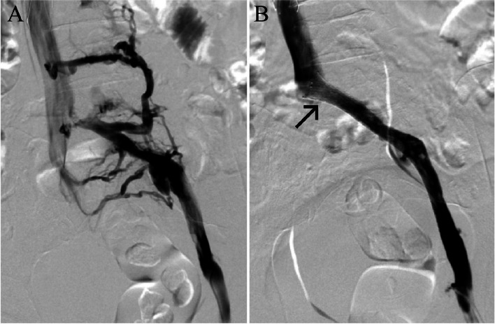
Details regarding patient indications for stent placement, lesion characteristics, and venographic measurements are outlined in Table 3. The left leg was the predominant target limb (91.23%, 156/171). The core laboratory-assessed mean lesion length was 64.59 ± 45.43 mm, and the mean diameter stenosis was 81.33 ± 14.68%, with 13.45% showing total occlusions. Preimplant dilation was performed in the majority of stent placements (98.25%, 168/171). Most patients (96.49%, 165/171) exhibited lesions in the common iliac vein (CIV). A total of 185 endovenous stents were placed, with 91.81% (157/171) of subjects receiving one stent and 8.19% (14/171) receiving 2 stents (Table 3). Predominantly, the implanted stents (51.89%, 96/185) were 14 mm in diameter, followed by 33.51% being 16 mm in diameter, and 10.81% being 12 mm in diameter. The most common stent length was 100 mm. Following stent placement, adjunct balloon dilation was conducted for 66.28% (114/171) of cases. The technical success rate was 100%. Post-procedure diameter stenosis, as assessed by venography, was 24.29% ± 11.12% (Table 3). The procedural success rate was 99.42% (170/171); one patient experienced thrombotic occlusion within the stent, necessitating secondary intervention before discharge. No contralateral deep vein thrombosis (DVT) events were observed within 12 months. The proportion of patients with postoperative antithrombotic therapy duration exceeding 6 months is 85.80% (139/162) [19]. The core laboratory conducted individual diameter stenosis assessments for patients with available venographic data or Doppler ultrasound (DUS), which were utilized to evaluate the primary effectiveness endpoint. A representative subject’s preprocedural and postprocedural venography is displayed in Fig. 2.
Table 3.
Indication and core laboratory-reported baseline lesion characteristics and venographic measurements
| Characteristic | Value |
|---|---|
| Study lesion laterality | |
| Left | 91.23 (156/171) |
| Right | 8.77 (15/171) |
| Lesion etiology | |
| PT | 42.69 (73/171) |
| NT | 57.31 (98/171) |
| Access site | |
| Femoral | 66.08 (113/171) |
| Popliteal | 31.58 (54/171) |
| Superficial vein | 2.34 (4/171) |
| Anesthesia type | |
| Local | 98.83 (169/171) |
| General | 0.58 (1/171) |
| Spinal | 0.58 (1/171) |
| Study lesion locationa | |
| CIV | 96.49 (165/171) |
| EIV | 18.13 (31/171) |
| CFV | 3.51 (6/171) |
| Reference vessel diameter, mm (n = 171) | |
| Mean ± standard deviation | 13.96 ± 3.03 |
| Range | 3.50–31.00 |
| Lesion length, mm (n = 171) | |
| Mean ± standard deviation | 64.59 ± 45.43 |
| Range | 5.00–247.00 |
| Preimplant dilatation performed | 98.25 (168/171) |
| No. of endovenous stents placed | |
| 1 | 91.81 (157/171) |
| 2 | 8.19 (14/171) |
| Diameter stenosis, % | |
| Before procedure (n = 171) | |
| Mean ± standard deviation | 81.23 ± 14.09 |
| Range | 50.00–100.00 |
| Post-procedure (n = 171) | |
| Mean ± standard deviation | 24.29 ± 11.12 |
| Range | 0.54–49.67 |
| At 12 months (n = 162) | |
| Mean ± standard deviation | 31.45 ± 23.58 |
| Range | 0.45–100.00 |
| Total occlusion | 13.45 (23/171) |
Data are presented as mean (SD), percentage (no./No.), unless noted otherwise
PT post-thrombotic, NT non-thrombotic, CIV common iliac vein, EIV external iliac vein, CFV common femoral vein
aLesions could have involved more than one location. Therefore, the number of lesion locations totaled more than the total number of patients enrolled
Fig. 2.
Preprocedural and postprocedural venography of a patient, with the lesion primarily located in the common iliac vein (CIV). The black arrows indicate the stent. A Preprocedural venography shows significant collateral circulation. B The patient received one stent implantation, and venography shows disappearance of vascular stenosis and collateral circulation
Safety outcomes
Data for the primary safety endpoint were available for all 171 patients. A total of 2 subjects (1.17%) experienced major adverse events (MAEs) within 30 days (refer to Table 4). Both patients belonged to the primary thrombosis (PT) group, and these MAEs were clinically driven reinterventions due to stent thrombosis. The upper bound of the two-sided 95% confidence interval (CI) was 4.16%, which was below the 11% performance goal, signifying the successful achievement of the primary safety endpoint (P < 0.001).
Table 4.
Primary effectiveness and safety end points
| Outcomes | Primary cause subgroups | ||
|---|---|---|---|
| Total, P value | NT | PT | |
| Primary safety failures at 30 days (SS) | 1.17 (2/171), < .001 | 0 (0/98) | 2.74 (2/73) |
| Primary patency at 12 months (PPS) | 91.36 (148/162), < .001 | 97.92 (94/96) | 81.82 (54/66) |
| Primary patency at 12 months (FAS) | 86.55 (148/171), < .001 | 95.92 (94/98) | 73.97 (54/73) |
Data are presented as percentage (no./No.), unless noted otherwise
FAS full analysis set, PPS per-protocol set, SS safe analysis set, NT non-thrombotic, PT post-thrombotic
Over the 1-year follow-up period, among the 171 subjects, 27 (15.79%) reported a total of 40 procedure-related adverse events (AEs). Eight patients (4.68%) reported a total of 9 device-related AEs, including 4 cases of occasional lumbar discomfort post-surgery, a sensation of soreness and swelling in the lower back, general lower back pain and discomfort, and left-sided lumbar distension. The remaining cases were related to thrombosis within the stent, thrombosis in the left common iliac vein, exacerbated thrombosis within the stent, and deep vein thrombosis. The occurrence rate of MAEs within 12 months was 5.26% (9/171), and all were clinically driven reinterventions due to stent thrombosis. Additionally, three patients died: two cases were unrelated to the device and procedure, while the cause of death for one patient was unknown due to loss to follow-up. Importantly, no device defects or bleeding events meeting the MAE definition occurred during the study, emphasizing the safety profile of the V-Mixtent Venous Stent.
Effectiveness outcomes
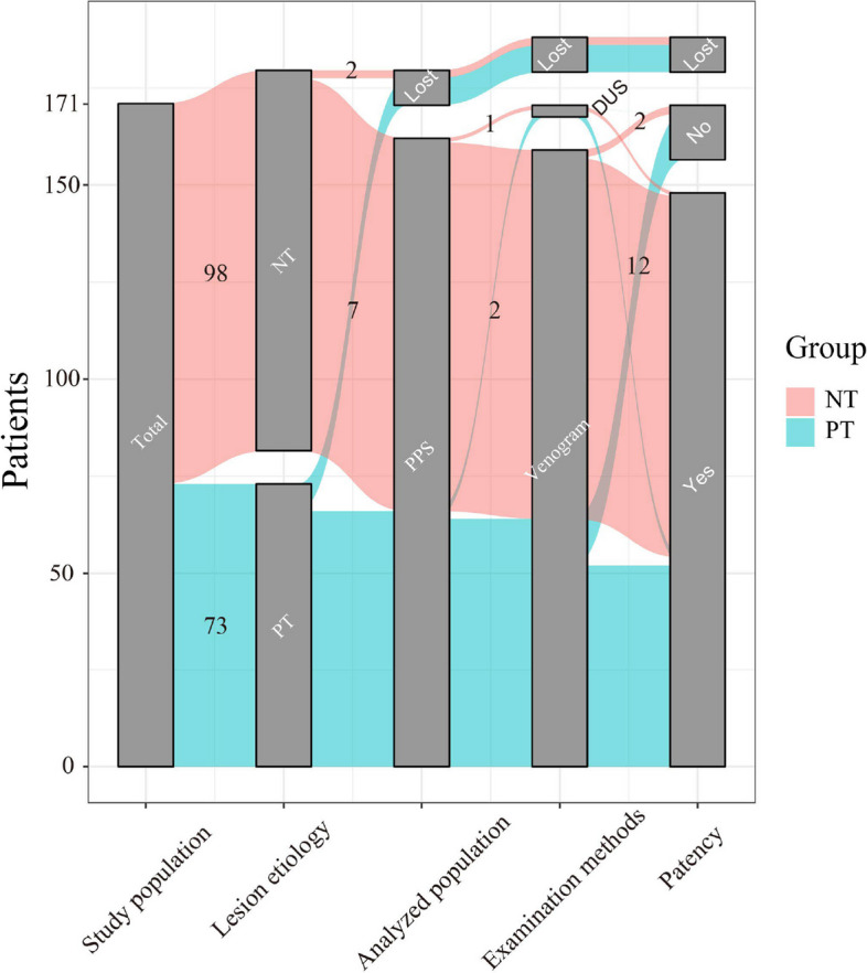
In total, 162 out of 171 patients (94.74%) were eligible for the primary effectiveness endpoint (Fig. 3). Among them, 159 subjects had the imaging component of primary patency assessed through venogram, while the remaining 3 subjects underwent analysis using Doppler ultrasound (DUS). Nine subjects were excluded from the primary patency analysis for various reasons: 3 subjects passed away, 3 were lost to follow-up, and 3 declined further follow-up. Out of the 162 assessable patients, 148 subjects (91.36%) achieved primary patency (refer to Table 4). The lower limit of the two-sided 95% confidence interval (CI) was 85.93%, surpassing the performance goal of 74% (P < 0.001).
Fig. 3.

Sankey diagram displayed the population flow in this study. The numbers on the colored ribbons in the figure indicate the included population. The different lines represent various scenarios of traffic diversion. PT, post-thrombotic; NT, non-thrombotic; PPS, per-protocol set; DUS, doppler ultrasound
Among the 14 patients who experienced a loss of patency during the 12-month follow-up, 12 were primary thrombosis (PT) patients. In this group, two patients underwent clinically driven target vessel revascularization before the conclusion of the one-year follow-up, whereas the remaining 12 patients encountered patency loss by the 1-year follow-up milestone. Sensitivity analyses were performed to evaluate the influence of missing data on the primary patency endpoint. Even when considering up to 9 missing data points as a loss of primary patency, the results remained consistent with the primary analysis. Subgroup analyses indicated a primary patency of 97.92% (94/96) in the non-thrombotic lesions (NT) group and 81.82% (54/66) in the PT group.
Clinical outcomes
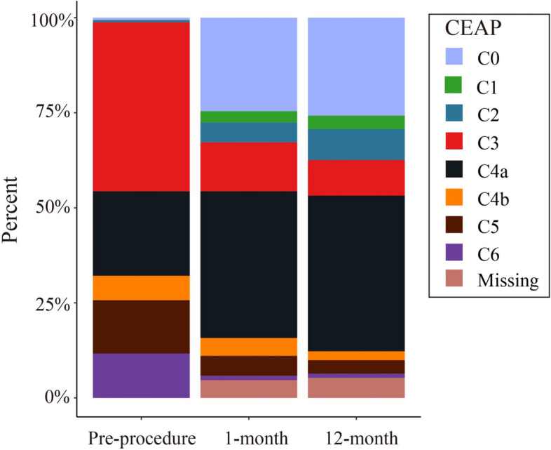
Following stent placement, patients reported a progressive reduction in symptoms, evidenced by improvements in three clinical scales at 6 and 12 months (refer to Table 5 and Fig. 4). Out of 162 patients, 138 (85.19%) experienced symptomatic improvement (VCSS score improvement ≥ 2 points), leading to a mean VCSS score decrease from 7.43 ± 4.18 at baseline to 2.56 ± 2.95 at 12 months. Only three patients (1.85%) exhibited worsened VCSS scores at the 12-month mark. On the Chronic Venous Insufficiency Questionnaire (CIVIQ-14), the mean score decreased from 26.94 ± 10.97 at baseline to 16.04 ± 4.58 at 12 months. Among the 162 patients, over half (58.02%) reported an improvement in quality of life (QOL), demonstrating at least a 6-point decrease in CIVIQ-14 score, while four patients (2.47%) considered themselves worsened at this time point. A significant correlation between follow-up time points and the CEAP C classification was observed (P < 0.001). At 12 months post-procedure, the CEAP C classification improvement rate (C classification improvement ≥ 1) was 66.05%, with 39.51% falling under grade C3 or below.
Table 5.
Change in Venous Clinical Severity Score (VCSS) and Chronic Venous Disease Quality of Life Questionnaire (CIVIQ-14) from baseline through 12 months
| Baseline (N = 171) | 6 months (N = 163), P value | 6 months change from baseline | 12 months (N = 162), P value | 12 months change from baseline | |
|---|---|---|---|---|---|
| VCSS | 7.43 ± 4.18 | 3.18 ± 3.08, < .001 | − 4.30 ± 3.66 | 2.56 ± 2.95, < .001 | − 4.98 ± 3.67 |
| NT | 7.92 ± 3.91 | 3.12 ± 2.63, < .001 | − 4.83 ± 3.65 | 2.43 ± 2.35, < .001 | − 5.50 ± 3.78 |
| PT | 6.77 ± 4.46 | 3.27 ± 3.66, < .001 | − 3.56 ± 3.56 | 2.74 ± 3.66, < .001 | − 4.30 ± 3.73 |
| CIVIQ-14 | 26.9 ± 10.97 | 17.60 ± 6.48, < .001 | − 9.50 ± 10.46 | 16.04 ± 4.58, < .001 | − 10.75 ± 11.04 |
| NT | 24.72 ± 9.86 | 16.85 ± 6.34, < .001 | − 8.05 ± 9.15 | 15.60 ± 3.23, < .001 | − 9.20 ± 9.87 |
| PT | 29.92 ± 11.73 | 18.70 ± 6.57, < .001 | − 12.15 ± 11.92 | 16.71 ± 6.02, < .001 | − 14.03 ± 12.72 |
Data are presented as mean (SD), unless noted otherwise
P value determined using paired t test
VCSS Venous Clinical Severity Score, CIVIQ-14 Chronic Venous Disease Quality of Life Questionnaire
Fig. 4.

Change in Clinical, Etiological, Anatomical, Pathophysiological (CEAP) C classification from baseline through 12 months
Given these results and recognizing potential differences in safety and effectiveness outcomes between the non-thrombotic lesions (NT) and primary thrombosis (PT) groups, a subgroup analysis was conducted. The improvements in CIVIQ-14 scores and VCSS for both the NT and PT groups at various follow-up time points are presented in Table 5. Significant differences in CIVIQ-14 score improvements were observed between the NT and PT groups at the 6-month (P = 0.018) and 12-month (P = 0.014) follow-up periods.
Discussion
This report presents the outcomes of a prospective multicenter feasibility study evaluating a specialized venous stent. The study highlights a noteworthy 12-month patency rate and minimal safety concerns, emphasizing the positive impact of the intervention on clinical outcomes. Comprehensive assessments, including CEAP, VCSS, and CIVIQ-14, indicate significant improvements. Despite challenges in measuring improvements over time without a control dataset and accounting for factors such as patient selection and disease state variations, the study surpassed predefined pretrial performance benchmarks for primary safety and effectiveness rates. This underscores the potential benefits of the stent’s unique design, combining etched and braided structures. Further research is essential to deepen our understanding of these promising outcomes and explore the stent’s potential applications.
Prospective multicenter studies offer a higher degree of representativeness in assessing therapeutic intervention outcomes compared to single-center or retrospective analyses. Despite inherent differences, the clinical and morphological outcomes of the presented study align with prior commendable reports on venous stenting [15, 16, 20–22]. The consistency in results across various studies reinforces the significance of interventional treatments for individuals grappling with symptomatic iliofemoral obstructive venous disease. Notably, the 30-day freedom from MAE in the VIRTUS [15] and VERNACULAR [22] studies was substantial, recording rates of 98.8% and 93.5%, respectively. It is essential to note that the VIRTUS and VERNACULAR trials predominantly included patients with chronic venous disease, excluding those with acute DVT [15, 22]. This selective approach, while potentially optimizing candidates for stent placement, differs from earlier research that validated the safety of stenting in patients after thrombus removal [23]. Additionally, the VIVO study [16] accurately represented a real-world population, and the ABRE study [21] featured a population most akin to that of the VIVO study, encompassing acute DVT, post-thrombotic syndrome, and NIVLs. The corresponding 12-month primary patency outcomes across these studies ranged from 84.0% (VIRTUS study), 88.6% (VERNACULAR study), 89.9% (VIVO study), to 88.0% (ABRE study).
Distinct etiologies of iliofemoral lesions, particularly chronic PT conditions, have been linked to worse outcomes compared to non-thrombotic (NT) or acute DVT cases [20, 24]. In the VIRTUS trial, primary patency was notably higher in NT cases (96.2%) compared to chronic PT cases (79.8%), averaging at 84% for the overall cohort [15]. Nevertheless, providing treatment to patients in the PT group proves valuable, supported by substantial enhancements in Quality of Life (QOL). Consistent with this pattern, the current study demonstrates higher patency in NT patients. Additionally, an interesting observation in this study is that, at both follow-up time points, the improvement in CIVIQ-14 scores in the PT group was significantly higher than that in the NT group. This difference could be attributed to the higher baseline CIVIQ-14 scores in the PT group. Typically, patients with NIVLs exhibit shorter lesions, leading to enhanced primary patency due to the compact, segmental nature of their lesions and the normal status of their inflow and outflow veins [15, 21, 22]. However, whether there exists a clinically significant difference in patency outcomes between patients with acute DVT or those with chronic post-thrombotic disease requires further clarification. Findings from a large retrospective study suggested higher patency rates for patients with chronic post-thrombotic disease [25], while the ABRE study indicated superior patency rates for patients with acute DVT [21]. More extensive data comparing stent patency after treating post-thrombotic disease or following acute DVT removal are necessary to comprehensively understand and characterize the nuances in outcomes based on different etiologies of iliofemoral lesions.
Throughout the 12-month follow-up period of this study, no occurrences of stent migration or integrity damage were observed. We also know that the currently used stents (Abre, Venovo, and Wallstent) have a very low risk of stent fractures associated with crossing the inguinal ligament. It is essential to note, however, that prior studies have documented instances of stent fractures and migrations [15, 16]. Stent migration in the venous system is a rare but potentially consequential complication that could necessitate open surgery. Mitigating the risk of stent migration involves implementing thorough training programs, employing appropriate stent selection practices, ensuring excellent procedural techniques, and maintaining a dedicated follow-up program [26–28].
Our study has several limitations. While outcomes in venous stenting have consistently shown promise, the level of supporting evidence remains modest due to a lack of controlled prospective trials scrutinizing performance and safety aspects. Due to the heterogeneity between research centers, there may be potential adverse biases in the patient selection process. Most studies, including this one, often omit reporting outcomes in specific high-risk cohorts. Merely citing average patency rates may not offer comprehensive insights into the precise mechanisms of patency failure. Factors such as stented length, ensuring full coverage of the diseased vessel, stent inflow/outflow adequacy, and adherence to appropriate anticoagulation protocols are crucial considerations. Multivariate analysis in previous studies has underscored stent length as a significant predictor of late severe in-stent area reduction [29]. There are also viewpoints suggesting that stent patency from healthy vein to healthy vein is more important. Apprehensions about stent durability stem from instances of non-braided stents extending into the CFV. This is akin to arterial stents crossing joints that are susceptible to neointimal hyperplasia and stent fracture, posing a potential risk of premature stent failure [30]. The single-arm study design poses limitations on analyzing longitudinal measures like pain and VCSS, potentially leading to an overestimation of device-attributed improvements. It is noteworthy that the determination of stent dimensions in this study was guided by biplanar venography rather than intravascular ultrasound, which is a limitation in the study design [31]. It is also noteworthy that approximately 13.45% of patients experienced total occlusion of the entire iliofemoral segments, with variations in occlusion rates between the NT and extensively lesioned cases. These demographic characteristics suggest potential differences and challenges compared to other studies [15, 16].
Conclusions
In conclusion, this study firmly establishes the safety and effectiveness of an innovative iliac vein stent for managing symptomatic iliofemoral venous obstruction. The findings can be applied to a broader real-world population grappling with chronic venous insufficiency, given the study’s inclusion of a diverse and challenging patient sample across 15 sites, with standardized data collection and review procedures adhering to a shared protocol. Despite the promising 1-year outcomes observed in venous stenting, a deeper understanding necessitates further investigation into long-term results. Ongoing data collection and analysis on this patient cohort will extend for a period of 5 years, offering a comprehensive view of the stent’s enduring effectiveness and safety profile.
Research registration unique identifying number (UIN)
1. Name of the registry: China Clinical Trial Registration Center.
2. Unique identifying number or registration ID: ChiCTR2300073019.
3. Hyperlink to your specific registration (must be publicly accessible and will be checked): Home—https://www.chictr.org.cn/hvshowprojectEN.html?id=230092&v=1.0.
Supplementary Information
Additional file 1: Table S1. Inclusion and Exclusion Criteria in The Study. Table S2. Comparison of venous stent properties. Table S3. Definitions of Key Study Outcomes. Table S4. List of Enrolling Centers and Investigators in The Study. Fig. S1. Study Implementation Flowchart.
Acknowledgements
The authors would like to thank all the staff and patients in this study.
Abbreviations
- AE
adverse event(s)
- BMI
body mass index
- CEAP
Clinical-Etiologic-Anatomic-Physiologic
- CIV
common iliac vein
- CFV
common femoral vein
- CIVIQ-14
Chronic venous insufficiency questionnaire-14
- CEC
Clinical events committee
- VCSS
Venous clinical severity score
- DVT
Deep vein thrombosis
- DM
Diabetes mellitus
- DUS
Doppler ultrasound
- EIV
External iliac vein
- FAS
Full analysis set
- MAE
Major adverse events(s)
- NIVL
Non-thrombotic iliac vein lesion
- NT
Non-thrombotic
- PPS
Per protocol set
- PE
Pulmonary embolism
- PT
Post-thrombotic
- QOL
Quality of life
- SS
Safe analysis set
- WOCF
Worst-case imputation method
Authors’contributions
C.S.: writing—original draft, methodology, visualization, formal analysis. X.-W.L.: investigation, writing—review and editing. H.-T.S.: investigation, writing—review and editing. L.Z.: investigation, writing—review and editing. S.-Y.W.: investigation, writing—review and editing. H.-P.L.: investigation, writing—review and editing. K.L.: investigation, writing—review and editing. S.S.: investigation, writing—review and editing. Z.-J.L.: investigation, writing—review and editing. Y.-Y.L.: investigation, writing—review and editing. G.-D.C.: investigation, writing—review and editing. M.-R.L.: investigation, writing—review and editing. Y.-G.K.: investigation, writing—review and editing. B.Y.: investigation, writing—review and editing. K.Y.: investigation, writing—review and editing. P.Y.: methodology, supervision, writing—review and editing. W.W.: methodology, supervision, writing—review and editing. Trial investigators: investigation, project administration, data curation. All authors read and approved the final manuscript.
Funding
The clinical study was sponsored by Shanghai EndoVas Medical Technology Co., Ltd.
Availability of data and materials
No datasets were generated or analysed during the current study.
Declarations
Ethics approval and consent to participate
Ethical approval has been obtained for this research study (GCP No. 202009217). This information has already been described in the manuscript.
Consent for publication
Not applicable.
Competing interests
The authors declare no competing interests.
Footnotes
Publisher’s Note
Springer Nature remains neutral with regard to jurisdictional claims in published maps and institutional affiliations.
Wei Wang and Pu Yang contributed equally to this work.
Contributor Information
Pu Yang, Email: puyang@csu.edu.cn.
Wei Wang, Email: weiwangcsu@csu.edu.cn.
References
- 1.Taha MAH, et al. A clinical guide to deep venous stenting for chronic iliofemoral venous obstruction. J Vasc Surg Venous Lymphat Disord. 2022;10(1):258-266.e1. 10.1016/j.jvsv.2020.12.087 [DOI] [PubMed] [Google Scholar]
- 2.Villalba L, et al. RETRACTED: Australian and New Zealand Society for Vascular Surgery clinical practice guidelines on venous outflow Obstruction of the femoral-iliocaval veins. J Vasc Surg Venous Lymphat Disord. 2023;11(4):832–42. 10.1016/j.jvsv.2023.02.002 [DOI] [PubMed] [Google Scholar]
- 3.Berger A, Jaffe JW, York TN. Iliac compression syndrome treated with stent placement. J Vasc Surg. 1995;21(3):510–4. 10.1016/S0741-5214(95)70295-4 [DOI] [PubMed] [Google Scholar]
- 4.Alemany J, Montag H, Wozniak G. Indications for stent implantation in stenoses of the pelvic veins: early results. Vasa Suppl. 1991;33:103. [PubMed] [Google Scholar]
- 5.Carlson JW, et al. Management of pelvic venous stenosis with intravascular stainless steel stents. Gynecol Oncol. 1995;56(3):362–9. 10.1006/gyno.1995.1064 [DOI] [PubMed] [Google Scholar]
- 6.Elson JD, et al. Vena caval and central venous stenoses: management with Palmaz balloon-expandable intraluminal stents. J Vasc Interv Radiol. 1991;2(2):215–23. 10.1016/S1051-0443(91)72285-9 [DOI] [PubMed] [Google Scholar]
- 7.Sawada S, et al. Application of expandable metallic stents to the venous system. Acta Radiol. 1992;33(2):156–9. 10.1177/028418519203300216 [DOI] [PubMed] [Google Scholar]
- 8.Raju S. Best management options for chronic iliac vein stenosis and occlusion. J Vasc Surg. 2013;57(4):1163–9. 10.1016/j.jvs.2012.11.084 [DOI] [PubMed] [Google Scholar]
- 9.Kang CH, et al. Comparison of open-cell stent and closed-cell stent for treatment of central vein stenosis or occlusion in hemodialysis patients. Iran J Radiol. 2016;13(4). 10.5812/iranjradiol.37994 [DOI] [PMC free article] [PubMed] [Google Scholar]
- 10.Mathew G, et al. STROCSS 2021: strengthening the reporting of cohort, cross-sectional and case-control studies in surgery. Int J Surg. 2021;96. 10.1016/j.ijsu.2021.106165 [DOI] [PubMed] [Google Scholar]
- 11.Eklöf B, et al. Revision of the CEAP classification for chronic venous disorders: consensus statement. J Vasc Surg. 2004;40(6):1248–52. 10.1016/j.jvs.2004.09.027 [DOI] [PubMed] [Google Scholar]
- 12.Rutherford RB, et al. Venous severity scoring: an adjunct to venous outcome assessment. J Vasc Surg. 2000;31(6):1307–12. 10.1067/mva.2000.107094 [DOI] [PubMed] [Google Scholar]
- 13.Launois R, et al. Construction and international validation of CIVIQ-14 (a short form of CIVIQ-20), a new questionnaire with a stable factorial structure. Qual Life Res. 2012;21(6):1051–8. 10.1007/s11136-011-0008-3 [DOI] [PubMed] [Google Scholar]
- 14.Milinis K, et al. Antithrombotic therapy following venous stenting: international Delphi consensus. Eur J Vasc Endovasc Surg. 2018;55(4):537–44. 10.1016/j.ejvs.2018.01.007 [DOI] [PubMed] [Google Scholar]
- 15.Razavi MK, et al. Pivotal study of endovenous stent placement for symptomatic iliofemoral venous obstruction. Circ Cardiovasc Interv. 2019;12(12). 10.1161/CIRCINTERVENTIONS.119.008268 [DOI] [PubMed] [Google Scholar]
- 16.Hofmann LR, et al. Twelve-month end point results from the evaluation of the Zilver Vena venous stent in the treatment of symptomatic iliofemoral venous outflow obstruction (VIVO clinical study). J Vasc Surg Venous Lymphat Disord. 2023;11(3):532-541.e4. 10.1016/j.jvsv.2022.12.066 [DOI] [PubMed] [Google Scholar]
- 17.Razavi M, et al. The initial report on 1-year outcomes of the feasibility study of the VENITI VICI VENOUS STENT in symptomatic iliofemoral venous obstruction. J Vasc Surg Venous Lymphat Disord. 2018;6(2):192–200. 10.1016/j.jvsv.2017.10.014 [DOI] [PubMed] [Google Scholar]
- 18.Dake M, O’Sullivan G. A prospective, multicenter evaluation of the Venovo self-expanding vascular stent used to treat obstructive lesions of the iliac and femoral veins: 12-month results from the VERNACULAR trial. J Vasc Interv Radiol. 2019;30(3):S3. 10.1016/j.jvir.2018.12.036 [DOI] [Google Scholar]
- 19.Guo B, et al. Principles of Optimal Antithrombotic Therapy for Iliac VEnous Stenting (POATIVES): A national expert-based Delphi consensus study. J Vasc Surg Venous Lymphat Disord. 2024;12(2):101739. 10.1016/j.jvsv.2023.101739 [DOI] [PubMed] [Google Scholar]
- 20.Razavi MK, Jaff MR, Miller LE. Safety and effectiveness of stent placement for iliofemoral venous outflow obstruction: systematic review and meta-analysis. Circ Cardiovasc Interv. 2015;8(10). 10.1161/CIRCINTERVENTIONS.115.002772 [DOI] [PubMed] [Google Scholar]
- 21.Murphy E, et al. Pivotal study evaluating the safety and effectiveness of the Abre venous self-expanding stent system in patients with symptomatic iliofemoral venous outflow obstruction. Circ Cardiovasc Interv. 2022;15(2). 10.1161/CIRCINTERVENTIONS.121.010960 [DOI] [PMC free article] [PubMed] [Google Scholar]
- 22.Dake MD, et al. Three-year results from the venovo venous stent study for the treatment of iliac and femoral vein obstruction. Cardiovasc Intervent Radiol. 2021;44(12):1918–29. 10.1007/s00270-021-02975-2 [DOI] [PMC free article] [PubMed] [Google Scholar]
- 23.Vedantham S, et al. Pharmacomechanical thrombolysis and early stent placement for iliofemoral deep vein thrombosis. J Vasc Interv Radiol. 2004;15(6):565–74. 10.1097/01.RVI.0000127894.00553.02 [DOI] [PubMed] [Google Scholar]
- 24.Neglén P, et al. Stenting of the venous outflow in chronic venous disease: long-term stent-related outcome, clinical, and hemodynamic result. J Vasc Surg. 2007;46(5):979–90. 10.1016/j.jvs.2007.06.046 [DOI] [PubMed] [Google Scholar]
- 25.Mabud TS, et al. Lower extremity venous stent placement: a large retrospective single-center analysis. J Vasc Interv Radiol. 2020;31(2):251-259.e2. 10.1016/j.jvir.2019.06.011 [DOI] [PubMed] [Google Scholar]
- 26.Sayed MH, et al. A review of the incidence, outcome, and management of venous stent migration. J Vasc Surg Venous Lymphat Disord. 2022;10(2):482–90. 10.1016/j.jvsv.2021.07.015 [DOI] [PubMed] [Google Scholar]
- 27.Badesha AS, et al. A systematic review on the incidence of stent migration in the treatment of acute and chronic iliofemoral disease using dedicated venous stents. Ann Vasc Surg. 2022;83:328–48. 10.1016/j.avsg.2021.12.084 [DOI] [PubMed] [Google Scholar]
- 28.Shammas N. Avoiding iliac vein stent migration: techniques and appropriate sizing. Vasc Dis Manag. 2017;14:E247-248. [Google Scholar]
- 29.Ye K, et al. Technical details and clinical outcomes of transpopliteal venous stent placement for postthrombotic chronic total occlusion of the iliofemoral vein. J Vasc Interv Radiol. 2014;25(6):925–32. 10.1016/j.jvir.2014.02.031 [DOI] [PubMed] [Google Scholar]
- 30.Moeri L, et al. Braided or laser-cut self-expanding nitinol stents for the common femoral vein in patients with post-thrombotic syndrome. J Vasc Surg Venous Lymphat Disord. 2021;9(3):760–9. 10.1016/j.jvsv.2020.08.027 [DOI] [PubMed] [Google Scholar]
- 31.Gagne PJ, et al. Venography versus intravascular ultrasound for diagnosing and treating iliofemoral vein obstruction. J Vasc Surg Venous Lymphat Disord. 2017;5(5):678–87. 10.1016/j.jvsv.2017.04.007 [DOI] [PubMed] [Google Scholar]
Associated Data
This section collects any data citations, data availability statements, or supplementary materials included in this article.
Supplementary Materials
Additional file 1: Table S1. Inclusion and Exclusion Criteria in The Study. Table S2. Comparison of venous stent properties. Table S3. Definitions of Key Study Outcomes. Table S4. List of Enrolling Centers and Investigators in The Study. Fig. S1. Study Implementation Flowchart.
Data Availability Statement
No datasets were generated or analysed during the current study.


