Abstract
Background and study aims Endoscopic resection of appendiceal orifice (AO) polyps extending inside the appendiceal lumen is challenging given the inability to determine polyp lateral margins and risk of appendicitis. Transcecal endoscopic appendectomy (TEA) ensures en bloc resection of these complex polyps.
Patients and methods This case series includes patients who underwent TEA by a single endoscopist in the United States. Technical success was defined as achieving complete removal of the appendix along with AO polyp in an en bloc fashion.
Results In total, nine patients were included (mean age 69.7 ± 9.6 years). The average appendix size was 4.07 ± 2.02 cm. Technical success was achieved in 100% of the patients. The average procedure length was 118.1 ± 44.21 minutes. The en bloc resection rate, R0 resection rate, and curative resection rates were 100%. Patients were observed for an average of 3.1 ± 1.6 days. One patient developed loculated fluid collection 9 days post procedure, which resolved on its own with oral antibiotic therapy. No other adverse events were recorded.
Conclusions This was an early study of the feasibility of TEA in the United States. This novel technique, in early-stage development, is potentially safe and associated with a minimal risk profile in expert hands. Further prospective studies are needed to standardize the technique.
Keywords: Endoscopy Lower GI Tract; Polyps / adenomas / ...; Endoscopic resection (polypectomy, ESD, EMRc, ...)
Introduction
Historically, surgical resection has been the traditional approach for resecting complex appendiceal polyps extending into the appendiceal orifice (AO). The surgical approach mainly entails open or laparoscopic partial cecectomy, ileocecectomy, and right hemicolectomy. Although surgery is the gold standard for management of malignant lesions of the appendix, it is associated with long-term morbidity such as chronic diarrhea and bacterial overgrowth if the ileocecal valve is removed 1 2 . Although endoscopic resection of AO polyps is possible, conventional endoscopic mucosal resection (EMR) would not guarantee polyp-free margins 3 . Endoscopic full thickness resection (EFTR) is an attractive option in this setting. However, it is not feasible for polyps > 2 cm or appendiceal polyps extending to the base of the cecum. In addition, endoscopic resection of these polyps is challenging due to limited maneuverability and it carries the hazard of appendicitis 4 .
The en bloc resection rate for AO polyps via EMR or endoscopic submucosal dissection (ESD) ranges from 59% to 95%, depending on the size and complexity of the polyp as well as the endoscopic technique 5 6 7 8 . However, polyps extending deeply into the appendiceal lumen remain a challenge.
Transcecal endoscopic appendectomy (TEA) aims to address the limitations of standard endoscopic resection techniques. Due to the limited space within the appendiceal lumen, even with advanced resection methods such as ESD and EFTR the peripheral margins may not be accessible due to polyp extension deeply within the appendiceal lumen. TEA is superior not only for en bloc resection of complex appendiceal polyp, but also eliminates the risk of appendicitis associated with other resection modalities such as EFTR.
There have been only a few reports of use of endoscopic appendectomy for management of complex appendiceal polyps, mainly from Asian centers 9 10 11 12 13 . In this case series, we report successful use of TEA for management of complex AO polyps in a practice located in the United States with a focus on use of a stabilization and traction device to expedite the procedure.
Patients and methods
Study design
This was a retrospective case series of nine patients who underwent TEA for management of complex AO polyps. The project was approved by the Baylor College of Medicine Institutional Review Board (H-50836).
Study population
Nine patients who underwent TEA for lateral spreading tumors of the AO were retrospectively enrolled. Our inclusion criteria were polyps > 2 cm extending inside the AO or obstructing the AO completely without the ability to determine the maximum extent of polyps within the appendiceal lumen. Patients with a previous history of appendectomy were excluded.
Technique
Step-by-step video demonstration of transcecal endoscopic appendectomy.
Video 1
All procedures were performed by an experienced advanced endoscopist (MO) who has successfully performed over 650 ESD procedures 14 . All patients were seen in advance by the endoscopist performing the procedure to discuss the risks and benefits of this novel technique in a clinic visit prior to the procedure and verified informed consent was obtained. All procedures were performed under general anesthesia with a long-acting paralyzing agent. Levofloxacin 500 mg was administered intravenously (IV) before the procedure.
A 3.8-mm single-channel colonoscope Pentax (Pentax America, New Jersey, United States) or Olympus CF-HQ190L (Olympus America, Pennsylvania, United States) was used. A transparent disposable plastic cap was fitted to the distal end of the colonoscope. Stabilization during dissection was achieved by advancing the colonoscope to the cecum using an overtube (double balloon endoluminal intervention platform [DiLumen, Lumendi, Connecticut, United States] or Pathfinder Endoscope Overtube [Neptune Medical, California, United States]).
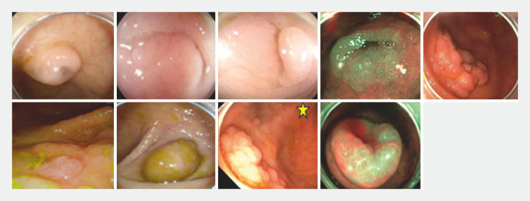
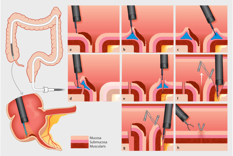
Upon reaching the cecum, the AO was examined to assess polyp extension within the appendiceal lumen ( Fig. 1 ). Demarcation of the border of the appendiceal polyp in the cecum was made with the tip of the dissection knife using ERBE electrocautery (Soft coagulation, effect 5, watts 50) ( Fig. 2 a ). Subsequently, submucosal injection of compound HESPAN solution (6% hetastarch in 0.9% sodium chloride mixed with 3 cc of methylene blue) was used with an injection needle in a circumferential manner around the AO ( Fig. 2 b and Fig. 2 c ). Afterward, a circumferential mucosal incision was made around the AO polyp using an electrocautery knife (1.5-mm Dual Knife [Olympus America Inc., Center Valley, Pennsylvania, United State] or 2-mm Orise Proknife [Boston Scientific Corporation, Marlborough, Massachusetts, United States]) ( Fig. 2 d , Fig. 2 e ). The incision was made using the ERBE setting endocut Q (2-1-1).
Fig. 1.
Appearance of appendiceal polyps upon initial endoscopic evaluation. All lesions were biopsied during the index colonoscopy before TEA and final pathology was remarkable for tubular adenoma or sessile serrated lesion. Only one lesion contained low-grade dysplasia. There was no evidence of high-grade dysplasia or carcinoma in any of the initial histopathological results. The lesion with the star is the only case in which tissue biopsy was not obtained because of the classic adenomatous appearance of the polyp. Before embarking on TEA, this lesion was meticulously examined via light chromoendoscopy to ensure that no cancerous features were present.
Fig. 2.
Schematic diagram of step-by-step demonstration of endoscopic transcecal appendectomy.
After making a circumferential incision around the AO polyp, adequate dissection of the submucosal tissue was performed. Traction was applied in all cases either via Lumendi suture line or rubber-band clip traction ( Fig. 2 f ). When applying rubber-band clip traction, the traction was applied while attaching the anal side of the lesion to the opposite cecal wall.
After achieving circumferential dissection of the submucosal plane in the cecum underneath the polyp and once the appendiceal wall was identified, a full thickness incision was extended around the AO to allow advancement of the endoscope into the peritoneum ( Fig. 2 g ). The attachment of the appendix to the mesoappendix was identified followed by dissection of the appendiceal body from the mesoappendiceal fat plane with careful attention not to dissect into the appendiceal wall. Dissection of the appendiceal lumen from the mesoappendix was continued until the tip of the appendix was reached under direct visualization. The tip of the appendix was then dissected from the remaining mesoappendix and adipose tissue surrounding the tip of the appendix. This is a critical step to ensure that no appendiceal stump is left behind. During this stage, application of another point of traction with a figure-eight rubber-band was occasionally utilized to augment traction force, allowing the base of the appendix to be pulled inside the cecum. It is of high importance to pay special attention to the appendicular artery. The appendicular artery contains two portions: 1) The main branch which runs within the mesoappendix to the tip; and 2) The accessory appendicular artery (from ileocolic artery or its branches), which supplies other parts of the appendix except the tip 15 . Prophylactic coagulation is preferentially applied using coagulation grasper (Olympus America, United States) while dissecting the appendix from the mesoappendix.
During dissection of the mesoappendix from the peritoneum, patients were closely monitored for any increase in intraabdominal pressure with close attention to peak inspiratory airway pressure. A significant increase in peak pressure (> 5 points mm/hg) was managed with abdominal decompression using a 14-gauge angiocath peripheral venous catheter placed two finger widths under the umbilicus to prevent tension pneumoperitoneum. Once the position of the catheter was confirmed in the peritoneum, peak inspiratory airway pressure was monitored to ensure that it gradually returned to baseline. The angiocath peripheral venous catheter was then left in place during dissection.
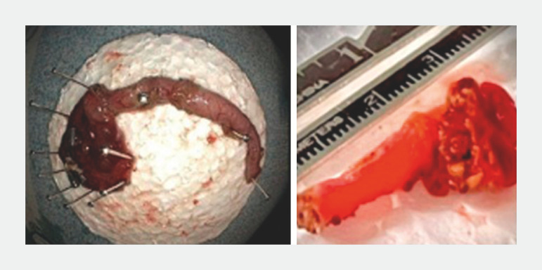
Careful macroscopic examination of the resected specimen is a key step to ensure complete excision of the appendix ( Fig. 3 ). The scope was then advanced to the peritoneum to ensure that any residual fluid adjacent to the appendectomy site and intraperitoneal gas were completely suctioned. A small volume of sterile water (if needed) was used to ensure that all residual liquid and clotted blood particles were aspirated from all gutters of the peritoneal cavity. Complete closure of the full thickness resection site was performed using hemostatic clips ( Fig. 2 h ). For closure, a two-step technique was applied to facilitate closure: tissue approximation followed by full closure with enforcement of additional hemostatic clips. For tissue approximation, we used a variety of commercially available devices, including the X-Tack Endoscopic HeliX Tacking System (Apollo Endosurgery, Austin, Texas, United States), dual-action tissue (DAT) clip (Micro-Tech Endoscopy, USA, Ann Arbor, Michigan, United States), and MANTIS Clip (Boston Scientific, Marlborough, Massachusetts, United States). After closure of the defect was completed, carbon dioxide was used for insufflation to fully expand the cecum and ensure full closure with a gas leak test. A step-by-step demonstration of this technique is available in Video 1 .
Fig. 3.
Careful examination of the resected appendix to ensure complete removal.
Appendix and polyp sizes were collected from the pathology report. Using post formalin fixation size is associated with underestimation of the actual length of the appendix as the result of tissue shrinkage 16 17 . However, because the majority of the polyps extended into the appendiceal lumen, in order to maintain consistency in reporting the diameters, the decision was made to use the post fixation sizes.
Patients were routinely admitted for observation and started on a clear liquid diet immediately post procedure.
Outcome definition
The main aim of this study was technical success, which was defined as achieving complete removal of the appendix and the polyp in en bloc fashion. En bloc resection was achieved if a polyp was removed in one piece. R0 (complete) resection was defined as en bloc resection with negative horizontal and vertical margins. Curative resection entailed complete histological resection with no risk of lymphovascular or perineural invasion.
Patients were monitored closely for adverse events (AEs) post procedure such as abdominal pain, abscess formation, delayed bleeding, and perforation. AEs were classified per the American Society for Gastrointestinal Endoscopy lexicon as mild (hospitalization for 1–3 days), moderate (hospitalization for 4–9 days), or severe (hospitalization for more than 10 days) to define severity 18 .
Results
Patient and procedure characteristics
Nine patients (77.8% female) with a mean age of 69.7 ± 9.6 years underwent TEA. American Society of Anesthesiologists (ASA) scores were 1, 2, and 3 in one, four, and four patients, respectively. One patient was on aspirin and apixiban and two patients were on aspirin only. The average body mass index of the patients was 25.29 ± 3.1 kg/m 2 . Patient and procedure baseline characteristics and outcomes are summarized in Table 1 .
Table 1 Patient, polyp, and procedure characteristics and outcome and follow-up information.
| Age/gender | Appendix size (cm) | Polyp size (cm) | Appearance/Paris | Path | Length (Min) | Knife | Overtube | Traction | # of clips | R0 resection | Curative resection | Length of stay (days) | Follow up |
| AO, appendiceal orifice; DAT, dual-action tissue; DK, Dual Knife; LGD, low-grade dysplasia; OPK: OrisePro Knife; SB, SB knife; SSA, sessile serrated adenoma; TA, tubular adenoma; TVA, tubulovillous adenoma. *This lesion was closed with five clips and one through the scope suturing device. † This patient was admitted for elective inguinal hernia repair 2 weeks after, inguinal hernia repair and found to have some tissue possibly serosal in the right paracolic gutter, which was biopsied. The final pathology showed fibroconnective tissue and adipose tissue without any evidence of residual appendiceal tissue. | |||||||||||||
| 72/F | 3.7 × 2.2 | 1.5 × 0.8 | Entire AO/Is | SSA | 65 | DK | Lumendi | Lumendi double balloon | 5 | R0 | Yes | 3 | Post-procedure ileus, managed conservatively |
| 69/M | 2.8 × 2.0 | 1 × 1 | Entire AO, extending inside /Is | TA | 90 | OPK | None | Rubber-band clip | 8 | R0 | Yes | 1 | None |
| 67/F | 3.9 × 2.8 | 2.1 × 1.9 | 50% of the AO/ IIa | SSA | 169 | OPK | Lumendi | Rubber-band clip | 3 | R0 | Yes | 5 | Multiple walled-off fluid collections on Day 9, management conservatively with complete resolution of collections of Day 17 |
| 51/M | 2.6 × 2.1 | 1.6 × 1.2 | Entire AO, extending inside/Is | SSA | 86 | OPK | Pathfinder | Rubber-band clip | 7 | R0 | Yes | 3 | Mild abdominal pain |
| 69/F | 2.4 × 0.9 | 1.1 × 0.9 | Entire AO, extending inside/IIa | SSA | 83 | OPK | Pathfinder | Rubber-band clip | 6 | R0 | Yes | 3 | Mild abdominal pain |
| 84/M | 8.5 × 2.9 | 2 × 1.9 | Entire AO, extending inside/ IIa+IIC | TA | 136 | OPK, SBK | Pathfinder | Rubber-band clip | 7 | R0 | Yes | 1 | None |
| 61/F | 5.2 × 5 | 1.8 × 1.4 | 80% of the AO expanding inside/ IIA+IIc | TVA | 128 | OPK | Pathfinder | Rubber-band clip | 13 | R0 | Yes | 4 | Mild abdominal pain |
| 83/F | 3 × 3 | 1.8 × 1.4 | Entire AO, extending inside/IIa | SSA w/ LGD | 97 | OPK | Pathfinder | Rubber-band clip | 6 | R0 | Yes | 6 | Mild abdominal pain |
| 71/F | 7 × 0.9 | 5 × 0.9 | 80% of the AO expanding inside/ IIA+IIc | SSA | 209 | OPK, SBK | Pathfinder | Rubber-band clip | 5* | R0 | Yes | 3 | Mild abdominal pain † |
The polyps occupied the entire AO in five cases and 50% to 80% the circumference with deep extension within the appendiceal lumen in the remaining four cases. Scope stabilization with a Lumendi or Pathfinder overtube was used in all cases except one in which severe stricturing diverticular disease was present and there was no need for additional stabilization devices.
The average size of the resected appendix was 4.07 ± 2.02 cm. The mean size of polyps in the resected specimens was 1.96 ± 0.99 cm 2 . The final polyp pathology was remarkable for sessile serrated adenoma (SSA) in five cases, tubular adenoma in two patients, tubulovillous adenoma in one case, and SSA with low-grade dysplasia in one case.
Study outcomes and adverse events
Technical success was achieved in 100% of the cases. All polyps were removed en bloc. Associated R0 resection and curative resection rates were 100%. The average procedure length was 118.1 ± 44.21 minutes. All defects were successfully and completely closed with an average of 6.63 ± 2.8 hemostatic clips. In one case, a through-the-scope endoscopic suturing device was used to facilitate tissue approximation and then full closure was reinforced with through-the-scope endoscopic hemostatic clips.
Patients were observed in the hospital post procedure for an average of 3.1 ± 1.6 days (range 1–6 days). Mild and moderate AEs were noted in six and three patients, respectively. The most common clinical AE was abdominal discomfort/pain (n = 7), which was managed conservatively with empiric antibiotics. One patient developed a loculated fluid collection 9 days post procedure. This patient was observed in the hospital for 5 days post procedure for abdominal pain. During the initial admission, she was managed conservatively with IV antibiotics. In light of her benign abdominal exam, afebrile status, and clinical improvement, no radiological imaging was obtained. She was discharged home after resolution of her symptoms on oral antibiotics on Day 5. However, on Day 9, she developed recurrent abdominal pain. Per patient preferences, knowing the risks, an outpatient computed tomography (CT) scan was obtained, which showed loculated fluid collections without any obvious evidence of a leak. Considering she was doing better overall on oral antibiotics, outpatient percutaneous drainage by interventional radiology was planned. Follow-up CT scan on the day of the planned procedure was remarkable for complete resolution of the fluid collections without any intervention. We hypothesize that this fluid collection was most likely secondary to a local inflammatory reaction because as there was no leak noted on CT scan. No cases delayed bleeding or delayed perforation were observed in any of the patients.
Discussion
In this case series, we reported outcomes of TEA for managing complex appendiceal polyps extending deeply into the appendiceal lumen in a western practice. Technical success, R0 resection, and curative resection were achieved in all cases. No major AEs requiring surgical intervention were observed and all minor AEs were successfully treated with conservative management.
TEA is a novel technique with limited published case series (two case series and four case reports) in the literature and mainly from Asia (China) ( Table 2 ) 9 10 11 12 13 19 . A technique similar to ours was applied for TEA in all these previously published series. Traction with snare was utilized to facilitate dissection in all published series except one in which the authors utilized a side-by-side colonoscope to apply dynamic traction with a snare. In the only western published case report, traction with double balloon traction platform was applied 19 . In this technique, the authors completely avoided entering the peritoneal cavity and TEA purely relied on dissection of the visualized portion of the appendix that was being pulled within the cecal lumen 19 . Compared with the Asian experience, we achieved similar results in our cohort with shorter average procedure times. This could be explained by utilization of stabilization devices and application of different points of traction with rubber-band clips, resulting in faster and efficient dissection.
Table 2 Characteristics of previously published Asian studies regarding TEA.
| Number of patients | Traction method | Procedure length (minutes) | Length of hospital stay | Appendix size (cm) | Pathology | Adverse events | |
| Liu et al, China, 2019 9 | 1 case | Snare traction | 140 | 4 days | 3.5 × 2 | Villous adenoma with low-grade intraepithelial neoplasia | None |
| Chen et al, China, 2021 10 | 4 cases | Snare traction | 112.7 ± 6.1 | 8 ± 1.4 (7–10 days), NPO (4–7) | 1.7 ± 0.25 | Sessile serrated | None |
| Gua et al, China, 2022 11 | 13 cases | Dynamic traction with using a second scope | Median 167 (90–220) | 8 (6–18), NPO 4 (3–13) | 2 (0.8–5) | Adenoma = 4 Sessile serrated = 2 High-grade intraepithelial neoplasia = 2 Low-grade intraepithelial neoplasia = 1 Low-grade appendiceal mucinous neoplasm (Tis) = 1 Appendicitis with abscess or cyst=3 | None |
| Ma et al, China, 2023 12 | 1 case | Snare traction | 50 | 5 days | 4 | Chronic appendicitis | None |
| Yuan et al, China, 2019 13 | 1 case | Snare traction | – | 3 days | 5 × 2 | Chronic appendicitis | None |
| Kantsevoy et al, USA, 2022 19 | I case | DiLumen double balloon | 101 | 0 Day | 4 | Appendicular intussusception | None |
Another major difference between the published eastern experience and our western experience is the length of stay. In our cohort, patients were started on a clear liquid diet immediately after the procedure in contrast to the Asian experience, in which patients were kept NPO for at least 3 days. We believe early oral feeding initiation is safe as long as complete and secured closure was confirmed. Early oral feeding is beneficial for preventing post procedure ileus. The length of hospital stay was comparatively lower and without any major delayed AEs.
There are a few key points to consider while performing this novel method for TEA. First, we advocate for utilizing stabilization devices. Overtubes provide the stability needed to dissect in the cecum and they act as conduits to remove the endoscope for lens cleaning when needed. It is worth mentioning that the high fat concentration within the mesoappendix leads to occasional fogginess of the endoscopic lens, hence the need for colonoscopy lens cleaning.
The second key point is application of traction. Applying step-by-step traction in various locations during the procedure makes the dissection plane accessible at all times and expedites the procedure. The third point is to ensure complete dissection of the entire appendix and remove the tip of the appendix from the surrounding mesoappendix and adipose tissue intact. This step is crucial to prevent stump appendicitis. Stump appendicitis is a rare phenomenon with an estimated prevalence of 0.15% after laparoscopic appendectomy 20 . The appendix is variable in length and can range from 2 to 20 cm based on race, gender, and ethnicity 21 . Peri-endoscopic CT scan can provide additional information about the length and configuration of the appendix and appendiceal artery. CT scan also provides information excluding malignant space-occupying tumors deeper within the appendiceal lumen. Fourth is to pay attention to the appendicular artery. The appendiceal artery is visible while dissecting the appendix from the mesoappendix toward the base. Preemptive coagulation of the artery using a coagulation grasper is recommended to avoid detrimental bleeding. Because of the high fat content of the mesoappendix, dissection of the fat around the root of the appendiceal artery completely before using a coagulation grasper for hemostasis is recommended to ensure complete and safe conduction of the energy for hemostasis. We advocate for multidirectional traction placement to pull the appendiceal body toward the cecal lumen continuously, which facilitates better visualization of the dissection plane and control of any unexpected bleeding during dissection. We suggest early utilization of various endoscopic tools in our armamentarium for preemptive coagulation and control of bleeding, such as injection of epinephrine and prophylactic placement of hemostatic clips at the origin of the appendiceal artery within the visualized portion of the mesoappendix in combination with use of a coagulation grasper for thermal hemostasis. However, a coagulation grasper was sufficient for hemostasis in our experience.
The fifth point is early decompression of the abdomen using an angiocatheter to ensure hemodynamic stability. The sixth point is that it is imperative to advance the scope within the peritoneal cavity to suction residual liquids and smoke generated from cautery to avoid delayed infection or peritoneal irritation. A flexible colonoscope with a working channel of 3.3 mm is effective in reaching all the gutters within the peritoneal cavity and able to adequately suction all the foreign materials from it. In contrast to the Asian experience, early initiation of a clear liquid diet in our cohort was well tolerated by patients, with prevention of postoperative ileus. Accordingly, we advocate for initiation of oral feeding immediately post procedure to expedite hospital discharge and hasten patient recovery. Needless to stay, we advocate for obtaining a CT scan with oral, IV and rectal contrast for patients with worsening abdominal distension and persistent disproportional abdominal pain to rule out a leak or contained perforation. Finally, TEA is a feasible technique in the hands of expert endoscopists for removal of benign adenomatous and sessile serrate lesions involving the AO. We are not advocating use of this novel technique for management of cancerous and mucinous lesions of the appendix because of the high risk of peritoneal dissemination.
Conclusions
Natural orifice transluminal endoscopic surgery (NOTES) is superior to and has more advantages than laparoscopic surgery because it avoids an external incision and its associated AEs, such as wound infection, pain, herniation, and adhesions. Ours is one of the early reports of outcomes with use of such a novel technique in the United States, with results comparable to the described case series from Asian centers. Our method is focused on utilizing traction and stabilization devices to make the procedure feasible and applicable for widespread use. This novel technique, in its developing phase, also is associated with a minimal AE profile in the hands of expert endoscopists. Larger prospective studies are needed to further standardize this technique.
Footnotes
Conflict of Interest Tara Keihanian: Tara Keihanian is a consultant for Neptune Medical, ConMed, and Lumendi. Mai Khalaf: No conflict of interest to disclose. Fuad Zain Aloor: No conflict of interest to disclose. Dina Hani Zamil: No conflict of interest to disclose. Salmaan A. Jawaid: Salmaan Jawaid is a consultant for ConMed, Creo Medical, and Lumendi Mohamed O. Othman: Mohamed O. Othman is a consultant for Olympus, Boston Scientific Corporation, Abbvie, ConMed, Creo Medical, Lumendi and Apollo. Mohamed O. Othman received research grants from Lucid Diagnostics, AbbVie and ConMed.
References
- 1.Park KJ, Choi HJ, Kim SH. Laparoscopic approach to mucocele of appendiceal mucinous cystadenoma: feasibility and short-term outcomes in 24 consecutive cases. Surg Endosc. 2015;29:3179–3183. doi: 10.1007/s00464-014-4050-4. [DOI] [PubMed] [Google Scholar]
- 2.Yang IJ, Seo M, Oh HK et al. Surgical outcomes of single-port laparoscopic surgery compared with conventional laparoscopic surgery for appendiceal mucinous neoplasm. Ann Coloproctol. 2021;37:239–243. doi: 10.3393/ac.2020.11.08. [DOI] [PMC free article] [PubMed] [Google Scholar]
- 3.Huang ES, Chumfong IT, Alkoraishi AS et al. Combined endoscopic mucosal resection and extended laparoscopic appendectomy for the treatment of periappendiceal, cecal, and appendiceal adenomas. J Surg Res. 2020;252:89–95. doi: 10.1016/j.jss.2020.02.006. [DOI] [PubMed] [Google Scholar]
- 4.Fahmawi Y, Hanjar A, Ahmed Y et al. Efficacy and safety of full-thickness resection device (ftrd) for colorectal lesions endoscopic full-thickness resection: A systematic review and meta-analysis. J Clin Gastroenterol. 2021;55:e27–e36. doi: 10.1097/MCG.0000000000001410. [DOI] [PMC free article] [PubMed] [Google Scholar]
- 5.Binmoeller KF, Hamerski CM, Shah JN et al. Underwater EMR of adenomas of the appendiceal orifice (with video) Gastrointest Endosc. 2016;83:638–642. doi: 10.1016/j.gie.2015.08.079. [DOI] [PubMed] [Google Scholar]
- 6.Song EM, Yang HJ, Lee HJ et al. Endoscopic resection of cecal polyps involving the appendiceal orifice: A KASID multicenter study. Dig Dis Sci. 2017;62:3138–3148. doi: 10.1007/s10620-017-4760-2. [DOI] [PubMed] [Google Scholar]
- 7.Jacob H, Toyonaga T, Ohara Y et al. Endoscopic submucosal dissection of cecal lesions in proximity to the appendiceal orifice. Endoscopy. 2016;48:829–836. doi: 10.1055/s-0042-110396. [DOI] [PubMed] [Google Scholar]
- 8.Patel AP, Khalaf MA, Riojas-Barrett M et al. Expanding endoscopic boundaries: Endoscopic resection of large appendiceal orifice polyps with endoscopic mucosal resection and endoscopic submucosal dissection. World J Gastrointest Endosc. 2023;15:386–396. doi: 10.4253/wjge.v15.i5.386. [DOI] [PMC free article] [PubMed] [Google Scholar]
- 9.Liu BR, Ullah S, Ye L et al. Endoscopic transcecal appendectomy: a novel option for the treatment of appendiceal polyps. VideoGIE. 2019;4:271–273. doi: 10.1016/j.vgie.2019.03.004. [DOI] [PMC free article] [PubMed] [Google Scholar]
- 10.Chen T, Xu A, Lian J et al. Transcolonic endoscopic appendectomy: a novel natural orifice transluminal endoscopic surgery (NOTES) technique for the sessile serrated lesions involving the appendiceal orifice. Gut. 2021;70:1812–1814. doi: 10.1136/gutjnl-2020-323018. [DOI] [PMC free article] [PubMed] [Google Scholar]
- 11.Guo L, Ye L, Feng Y et al. Endoscopic transcecal appendectomy: a new endotherapy for appendiceal orifice lesions. Endoscopy. 2022;54:585–590. doi: 10.1055/a-1675-2625. [DOI] [PMC free article] [PubMed] [Google Scholar]
- 12.Ma ZL, Pan HT, Xu JQ et al. Endoscopic transcecal appendectomy for recurrent appendicitis after previous endoscopic mucosal resection. Endoscopy. 2023;55:E1095–E1096. doi: 10.1055/a-2173-7608. [DOI] [PMC free article] [PubMed] [Google Scholar]
- 13.Yuan XL, Cheung O, Du J et al. Endoscopic transcecal appendectomy. Endoscopy. 2019;51:994–995. doi: 10.1055/a-0889-7289. [DOI] [PubMed] [Google Scholar]
- 14.Ahmed Y, Othman M. EMR/ESD: Techniques, complications, and evidence. Curr Gastroenterol Rep. 2020;22:39. doi: 10.1007/s11894-020-00777-z. [DOI] [PubMed] [Google Scholar]
- 15.Swathipriyadarshini C, Rajilarajendran H, Balaji T et al. A comprehensive study of mesoappendix and arterial pattern of appendix. Turk J Surg. 2022;38:55–59. doi: 10.47717/turkjsurg.2022.5502. [DOI] [PMC free article] [PubMed] [Google Scholar]
- 16.Lam D, Kaneko Y, Scarlett A et al. The effect of formalin fixation on resection margins in colorectal cancer. Int J Surg Pathol. 2019;27:700–705. doi: 10.1177/1066896919854159. [DOI] [PubMed] [Google Scholar]
- 17.Song JS, Kim BS, Yang MA et al. Underestimation of endoscopic size in large gastric epithelial neoplasms. Clin Endosc. 2022;55:760–766. doi: 10.5946/ce.2021.269. [DOI] [PMC free article] [PubMed] [Google Scholar]
- 18.Cotton PB, Eisen GM, Aabakken L et al. A lexicon for endoscopic adverse events: report of an ASGE workshop. Gastrointest Endosc. 2010;71:446–454. doi: 10.1016/j.gie.2009.10.027. [DOI] [PubMed] [Google Scholar]
- 19.Kantsevoy SV, Robbins G, Raina A et al. Purely endoscopic appendectomy. VideoGIE. 2022;7:265–267. doi: 10.1016/j.vgie.2022.03.010. [DOI] [PMC free article] [PubMed] [Google Scholar]
- 20.Dikicier E, Altintoprak F, Ozdemir K et al. Stump appendicitis: a retrospective review of 3130 consecutive appendectomy cases. World J Emerg Surg. 2018;13:22. doi: 10.1186/s13017-018-0182-5. [DOI] [PMC free article] [PubMed] [Google Scholar]
- 21.Willekens I, Peeters E, De Maeseneer M et al. The normal appendix on CT: does size matter? PLoS One. 2014;9:e96476. doi: 10.1371/journal.pone.0096476. [DOI] [PMC free article] [PubMed] [Google Scholar]


