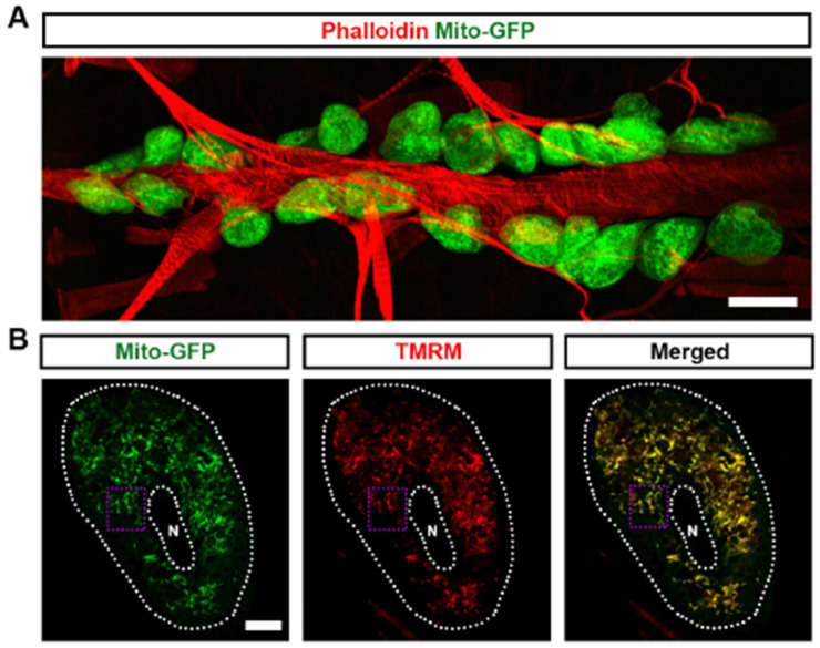
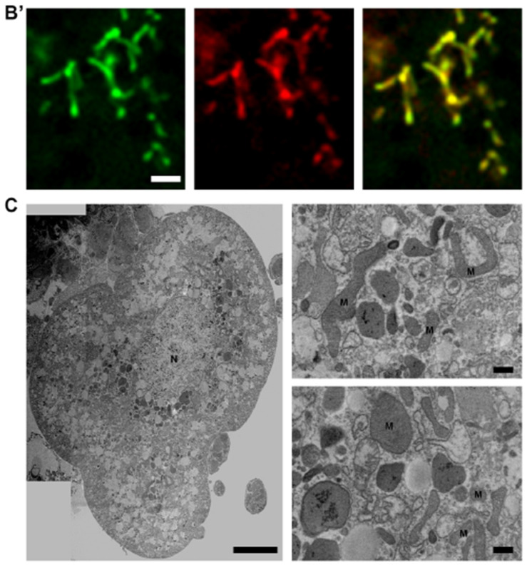
Figure 1.
Mitochondria in Drosophila nephrocytes: (A) UAS-mito-GFP (green) induced by nephrocyte-specific driver Dot-Gal4 to label the mitochondria in Drosophila nephrocytes (Dot > mito-GFP, female, 4-day-old). Phalloidin (red) labels the actin filaments and was used to visualize the fly heart. Scale bar = 20 µm. (B,B’) Dot > mito-GFP (green) was to label the mitochondria in Drosophila nephrocytes (female, 4-day-old). Mitochondrial membrane potential was indicated by tetramethylrhodamine, methyl ester (TMRM; red). White dotted lines, outline the nephrocyte and the nucleus (N); purple dotted box, outlines the area magnified in (B’). Scale bar: (B) = 5 µm; (B’) = 1 µm. (C) Mosaic of transmission electron microscopy (TEM) images showing nephrocyte ultrastructure with nucleus (N) and mitochondria (M). Scale bars: (left) = 5 µm; (right, both) = 2 µm.


