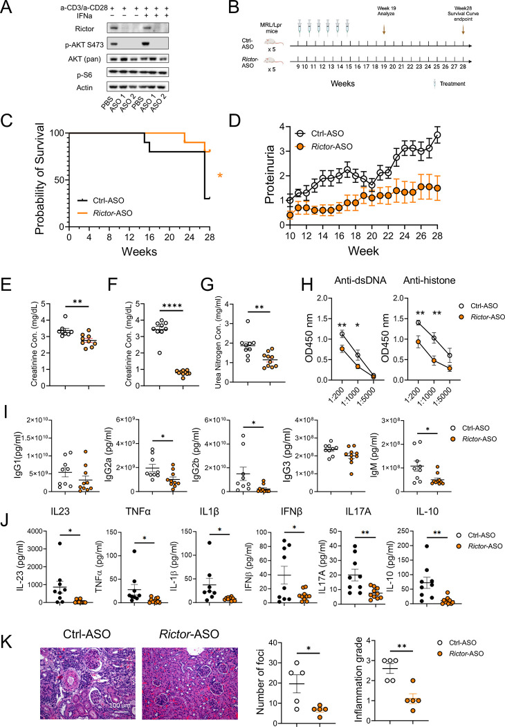
Figure 4: Anti-mouse Rictor-ASO treatment benefits lupus-like symptoms in MRL/lpr mice.
(A) Immunoblot image showing the deletion of RICTOR by Rictor-ASO in mouse CD4+ T cells. (B) Treatment scheme of Rictor-/Ctrl-ASO in MRL/lpr mice. (C) Kaplan–Meier cumulative survival plot of Rictor-ASO and Ctrl-ASO treated MRL/lpr mice. (D) Proteinuria changes of Rictor-ASO and Ctrl-ASO treated MRL/lpr mice over 28 weeks. Urine (E) and serum (F) creatinine concentration of MRL/lpr mice at 19 weeks. (G) Serum urea nitrogen concentration at 19 weeks. (H) Tiers of anti-dsDNA (left) and anti-histone (Right) antibodies in mouse serum at 19 weeks. Serum inflammatory cytokine concentration (I) and Ig isotypes concentration (J) at 19 weeks. (K) Representative H&E staining images of kidneys at 19 weeks; Left: quantitative cortex inflammation grade; right: number of foci. Scale bar: 100 μm. ns, not significant; *p < 0.05, **p < 0.01. The Kaplan–Meier estimator was used for survival curve analysis (C). Unpaired student t-tests were used for two groups comparison (E – K). Error bars represent SEM.

