

Case Presentation
A 5-month-old healthy male presented with a 3-month history of a small nodule on the skin of the trunk. Physical examination revealed a pink nontender roundish domed nodule with a smooth surface and a stretched texture 6 mm in diameter on the midline of the abdomen (Figure 1). The differential diagnosis included Spitz nevus, fibrous tumor, and xanthogranuloma. Dermoscopy revealed a whitish pink non-structured central area with dilated linear irregular unfocused vessels peripherally. Histological examination showed a mesenchymal neoplasm with the WHO 2018/2023 spectrum of histological features and immunophenotypic profile of cellular neurothekeoma.
Figure 1.
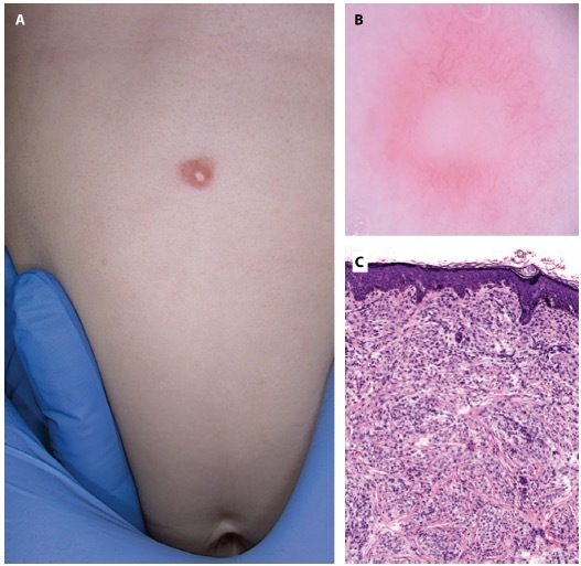
(A) Photo of neurothekeoma. (B) Videodermoscopy scan. (C) Histology.
Teaching Point
Cellular neurothekeoma (CN) is an uncommon benign soft tissue dermal neoplasm. It can occur on any cutaneous location but is commonly seen on the upper limbs of young children and adolescents [1]. CN tumors show increased cellularity, a lack of myxoid stroma, negativity to S100 protein, and often positivity to NK1C3 (CD57). Unlike the classical type of neurothekeoma (nerve sheath myxoma) in the cellular variant of the tumor, a histiocytic or fibrohistiocytic differentiation has been suggested by recent evidence.
Dermatoscopic diagnosis is challenging because CN is not distinctive, featuring arborizing vessels on a reddish background, whitish streak areas, or peripheral pigment network. So, it is often mistaken for basal cell carcinoma or dermatofibroma [2].
In conclusion, although it is rare to see a CN in infancy, it is important to keep in mind this kind of tumor in the differential diagnosis of nodular lesions in pediatric age. It is crucial to recognize this entity in children to prevent misdiagnosis and excessively aggressive treatments.
Footnotes
Funding: None.
Competing Interests: None.
Authorship: All authors have contributed significantly to this publication.
References
- 1.Murphrey M, Huy Nguyen A, White KP, Krol A, Bernert R, Yarbrough K. Pediatric cellular neurothekeoma: Seven cases and systematic review of the literature. Pediatr Dermatol. 2020;37(2):320–325. doi: 10.1111/pde.14043. [DOI] [PubMed] [Google Scholar]
- 2.Choi S, Cho SI, Lee C, Kwak Y, Mun JH. Dermoscopy of multiple cellular neurothekeoma: An analysis of 11 neurothekeomas in a middle-aged woman. Australas J Dermatol. 2020;61(1):e73–e76. doi: 10.1111/ajd.13185. [DOI] [PubMed] [Google Scholar]