



Summary
RAS proteins are conserved guanosine triphosphate (GTP) hydrolases (GTPases) that act as molecular binary switches and play vital roles in numerous cellular processes. Upon GTP binding, RAS GTPases adopt an active conformation and interact with specific proteins termed RAS effectors that contain a conserved ubiquitin-like domain, thereby facilitating downstream signaling. Over 50 effector proteins have been identified in the human proteome, and many have been studied as potential mediators of RAS-dependent signaling pathways. Biochemical and structural analyses have provided mechanistic insights into these effectors, while studies using model organisms have complemented our understanding of their role in physiology and disease. Yet, many critical aspects regarding the dynamics and biological function of RAS-effector complexes remain to be elucidated. In this review, we discuss the mechanisms and functions of known RAS effector proteins, provide structural perspectives on RAS-effector interactions, evaluate their significance in RAS-mediated signaling, and explore their potential as therapeutic targets.
Keywords: RAS, effector, RBD, Ras-binding domain, GTPase
eTOC blurb
RAS proteins act as molecular switches by binding GTP and interacting with effectors to facilitate downstream signaling. This review by Mozzarelli et al. discusses the mechanisms, structures, and functions of RAS effectors, their roles in signaling, and their potential as therapeutic targets, emphasizing the need for deeper insights into RAS-effector interactions.
Introduction
The RAS family of small GTPases, comprising 36 members, exhibits homology in their protein sequence. Advancements in sequencing and cloning technologies have revealed their phylogenetic relationship and highlighted critical differences in protein sequences present within their distinct regulatory motifs1,2. The latter has significant importance in the biological function of RAS GTPases, defining their biochemical properties, interactions with regulatory proteins, subcellular localization, and ability to engage with downstream protein effectors. Most RAS GTPases exhibit high affinity towards guanosine diphosphate and triphosphate (GDP/GTP, respectively) and their intrinsic enzymatic activity promotes GTP hydrolysis to GDP, with RAS proteins being loaded with GTP through a nucleotide exchange reaction. These cyclic reactions are inherently catalytically inefficient, and therefore, guanine exchange factors (GEF) facilitate GTP loading, and GTPase activating proteins (GAP) accelerate the rate of hydrolysis3. The coordinated function of these factors ensures efficient, rapid, and tightly regulated RAS signaling in cells (Figure 1A).
Figure 1. The fundamentals of RAS-effector signaling.
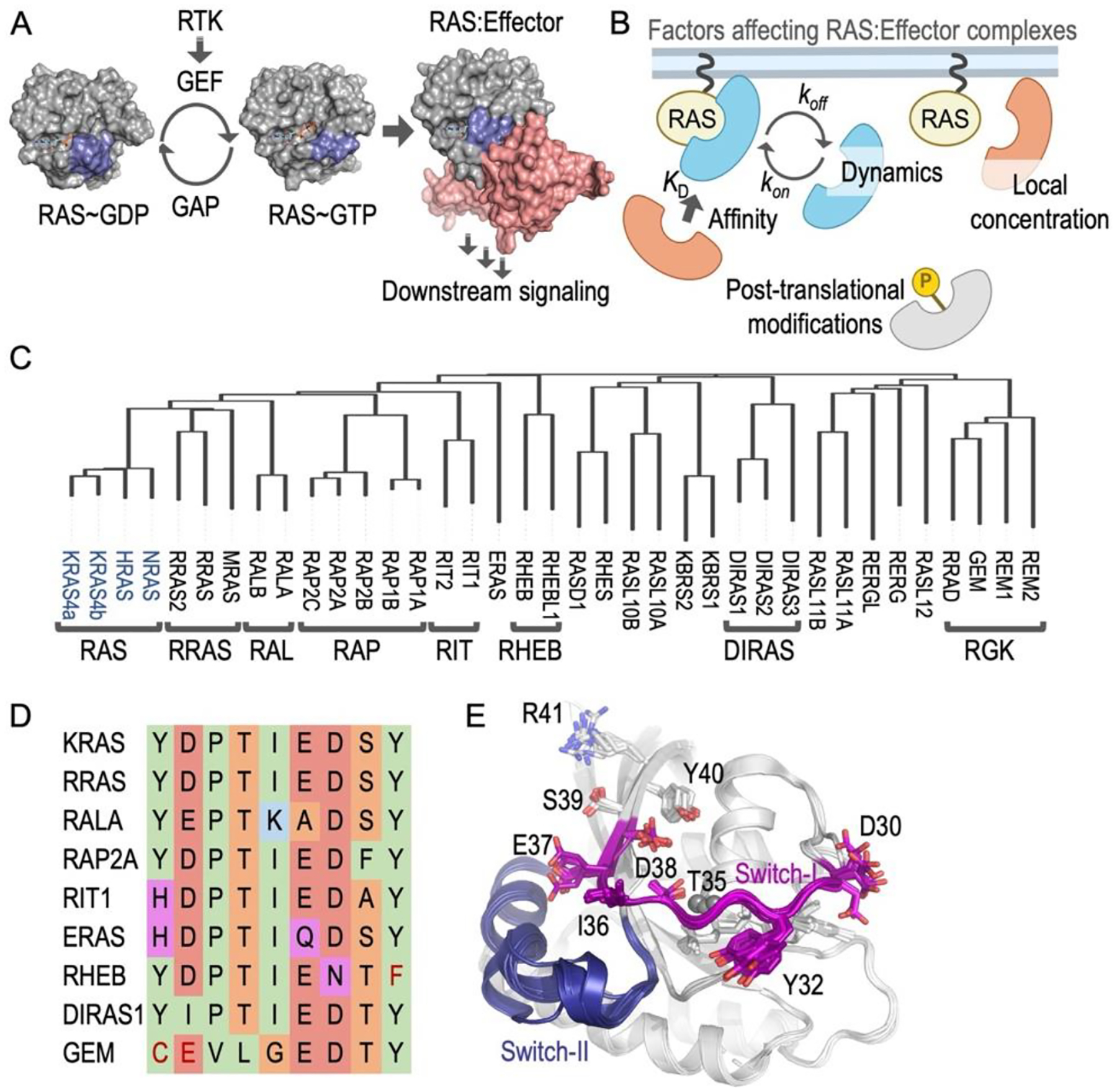
(A) Representative overview of the RAS binary switch. The effector binding domain (highlighted in blue) undergoes extensive conformational changes upon nucleotide exchange facilitating effector complex formation (red). PDB 6MBT (KRAS-GDP), 6MBQ (KRAS-GppNHp), and 6XHB (KRAS and CRAF RBD-CRD). (B) Potential factors affecting the competitive nature of RAS effectors (different effectors depicted as colored shapes). Dissociation constant (KD) is the main driver of effector affinity, and some studies have systematically assessed the interaction (KD) between RAS proteins and effectors. For instance, HRASGTP KD (μM) for CRAF (0.09) < RASSF5 (0.24) < RALGDS (2.5) < PLCε (3.7) < PI3Kα (84.3)40. The dynamics of the interaction are also affected by association (kon) and dissociation (koff) rate constants, affecting the interaction between RAS and different effectors even if their affinity at equilibrium is similar7. Local concentrations188 (or subcellular localization) and post-translational modifications of effectors can also modify their apparent affinity. For instance, RAP1 S11 phosphorylation and H/K/NRAS K147 mono-ubiquitination have been suggested to modify RAF interaction189,190 (C) Dendrogram of the RAS family of GTPases highlights their phylogenetic relationships and main clades, representing important subfamilies like classical RAS proteins (in blue). (D) Protein sequence alignment in the effector binding domain of different RAS GTPases demonstrates similarities and differences in critical residues that contribute to effector specificity. Amino acids are highlighted as follows: small polar (orange), hydrophobic (green), polar (magenta), negatively charged (red), and positively charged (blue). Red font indicates similar properties to the KRAS amino acid, but distinct amino acid (synonymous change). Amino acid sequences include KRAS (32–40), RRAS (58–66), RALA (43–51), RAP2A (32–40), RIT1 (50–58), ERAS (70–78), RHEB (35–43), RHES (48–56), DIRAS1 (36–44) and GEM (106–114). (E) Structural overlay of RAS protein in RAS-effector complexes, highlighting the conformational similarities of key interacting residues in the effector binding domain.
RAS binding to GTP induces conformational changes, creating a unique binding interface for interaction with specific effector proteins, which possess a conserved ubiquitin fold domain known as the RAS-binding domain (RBD). Other effector domains binding to RAS were initially classified as RAS-association (RA) domains due to their limited sequence similarity to RBDs, with both types typically spanning 80–100 amino acids. However, subsequent structural studies have shown that, despite low primary sequence homology, these domains possess the characteristic ubiquitin fold and a similar RAS interaction mechanism, suggesting their reclassification under the RBD category4,5. While the presence of RBD/RA indicates the potential for a specific protein to act as a RAS effector, this is not always the case, making the definition of a RAS effector somewhat ambiguous. At a minimum, a hypothetical RAS effector must bind its cognate RAS protein only when GTP-bound. In addition, this interaction should lead to changes in the activity or function of the effector; although there are also effectors that just act as a scaffold. Therefore, confirming a protein’s role as a RAS effector necessitates experimental validation.
Our current view of RAS-effector signaling predominantly follows a linear model and assumes that each RAS protein has multiple downstream effectors to diversify the signaling output. However, a major question in the field is how we can reconcile a model in which different effectors compete for limited GTP-bound RAS molecules6. In this conundrum, the importance of the RAS effector kinetics is crucial; not only the affinity towards an effector is important, but how such affinity is affected by different factors (Figure 1B). For instance, are there high-affinity effectors that signal first and block available RAS molecules for other low-affinity effectors? More importantly, do these also modify the association between RAS and GAPs through steric hindrance, given the overlapping nature of their binding interface? Conversely, can low-affinity effectors play a significant role downstream of RAS-GTP if their local concentration is high enough? In this context, systems biology approaches are beginning to tackle these important questions7–9.
There are critical concepts regarding the biology of RAS effectors that warrant at least reevaluation and, at most, substantial reconsideration. For example, many structural and biophysical studies carried out using RAS effectors use isolated RBD/RA domains, despite reports showing additional binding contacts outside of these domains10,11. Moreover, isolated RBD/RA do not necessarily capture important functional details of the full-length protein, such as post-translational modifications that could modify affinity, other adjacent domains that might occlude accessibility, or its subcellular localization, among others10,12,13. Another important aspect that remains understudied is the role of RAS effectors in the in vivo context. While many potential effectors have been characterized in vitro or in cell-based assays, their role during development, disease, or at the organismal level remains poorly understood. In fact, there are limited studies using mouse models in which the suppression of an effector can counteract phenotypes associated with a gain-of-function RAS allele.
The diversity of RAS proteins and their binary switch
The RAS superfamily of small GTPases encompasses over 160 members distributed in 5 distinct families: RAS, RHO, RAB, ARF, and RAN2,14. Within the RAS family, there are 36 proteins that are further classified into distinct monophyletic groups based on their amino acid sequence conservation15 (Figure 1C). Notably, the subfamily of RAS GTPases can play dual roles in regulating cellular proliferation, broadly categorized into those promoting cell proliferation or survival and those exhibiting growth-suppressive properties15. Key members such as classical RAS proteins, H/K/NRAS, are frequently mutated in cancers and, hence, have been extensively studied for their roles in cell proliferation, apoptosis, and differentiation. Similar functions appear to be regulated by the closely related members RRAS/RRAS2/MRAS. Other members, like RALA/B and RAP1/2, are implicated in cell migration, adhesion, and cytoskeletal organization, while RHEB activates the mTORC1 signaling pathway, impacting cell growth and metabolism16. However, many RAS proteins remain poorly characterized at the biochemical and cellular levels.
RAS proteins are characterized by a globular domain, known as the G-domain, and N- and C-terminal extensions that provide unique properties to these proteins. The C-terminal extensions are particularly important because they generally contain amino acids responsible for subcellular localization and show low sequence conservation across the family; for this reason, this extension is also referred to as the hypervariable region (HVR). Many RAS proteins contain a C-terminal CAAX box (C: cysteine; A: aliphatic residue; X: terminal residue), which is prenylated at the cysteine residue and promotes plasma membrane anchorage. Additionally, some RAS proteins are palmitoylated at one or two cysteines upstream of the CAAX motif, while others, lacking these motifs, associate with the plasma membrane, or endomembranes through charge complementation17. The role of the HVR is highly relevant in the context of effector signaling because subcellular localization can dictate the ability to bind to specific effectors. Molecular simulations have proposed that different HVRs can position the G-domain in configurations with differing accessibility to certain effectors18,19.
Within the G-domain, there are several regulatory regions that are important for the coordination of the nucleotide, interaction with effectors, and other regulatory mechanisms. The G-domain of most RAS proteins acts as a molecular binary switch that toggles between conformational states bound to GDP/GTP. The GTP-bound state, also known as the active conformer, allows for effector binding and activation of signaling cascades that regulate essential cellular functions. Structural analyses of RAS have identified two critical switch regions—switch-I (aa 30–38 in H/K/NRAS) and switch-II (aa 59–76) that are central to protein-protein interactions and exhibit dynamic conformational shifts between the GDP/GTP-bound states, often compared to a loaded-spring mechanism20,21. In the loaded spring state, RAS presents the interface residues essential for effector engagement (mainly the effector binding domain; aa 32–40). After GTP is hydrolyzed and the γ phosphate is released, RAS reverts to a GDP-bound state, causing the switch regions to adopt ‘open’ conformations, releasing effector proteins. Despite the high conservation of the G-domain, variations in regulatory motifs diversify the properties of RAS proteins, particularly those affecting the effector binding motif. These variations lead to changes in affinity and, thus, effector selectivity (Figure 1D, E).
RAS-GTP binds specific domains in RAS effector
The discovery of RAF and PI3 kinases as RAS effectors accelerated our understanding of the molecular mechanisms of effector association. RAF was the first RAS effector discovered, and its exclusive association with RAS-GTP, validated by several studies, confirmed its role in selectively transmitting signals from active RAS22–26. The N-terminal region of CRAF (RAF1) had been shown to antagonize the transformation effects of oncogenic RAS in cells, and consequently, this region was found to bind RAS-GTP directly through its RBD27. This discovery was integral in understanding the initiation of the RAF-MEK-ERK (MAPK) signaling cascade downstream of RAS. In parallel, PI3K emerged as a second class of validated RAS effector after initial findings linked PI3K activity with RAS28, but the direct interaction between PI3K catalytic subunit with RAS-GTP was only substantiated later29. This interaction was delineated through RAS-GTP binding to p110α, the catalytic subunit of the PI3Kα lipid kinase complex, and subsequently extended to the p110γ isoform, mediated through their respective RBDs30,31.
Following the identification of RAF and PI3K as RAS effectors, several exploratory efforts were launched to identify additional protein effectors. Utilizing cDNA libraries coupled with yeast two-hybrid assays using RAS GTPases as baits revealed a broad range of RAS effectors. Additionally, using the consensus amino acid sequences characteristic of RBD/RA domains, data mining of the human proteome identified over 50 proteins containing similar domains that could act as potential effectors of RAS-like GTPases (Figure 2). Beyond RAF and PI3K kinases, this diverse family of effectors includes several RAL GTPase GEFs (e.g., RALGDS, RGL1–4), AFDN/Afadin, the RASSF family, RIN1, and PLCε1, among others32–39. This diversity underscores the complexity of RAS signaling, extending past the known RAF and PI3K-mediated growth and survival pathways. However, detailed studies focusing on the specificity of effector binding within the extensive RAS family of GTPases have been limited, despite their importance in understanding the potential binding preference. Furthermore, the mechanisms by which binding to RAS family GTPases alters the functions of these effector proteins remain largely unexplored for most effectors.
Figure 2. Proposed human RAS effectors.
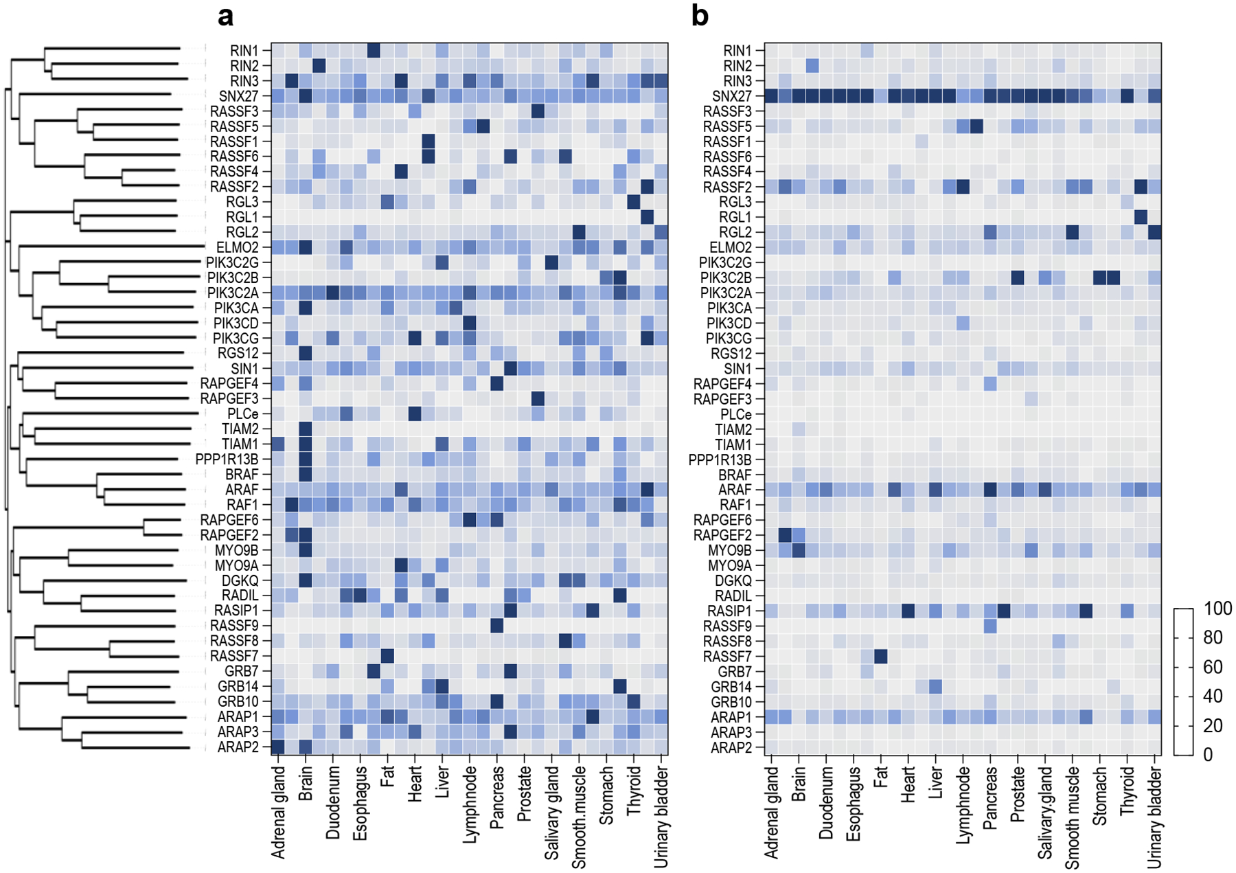
There are over 50 human proteins in the human proteome that contain a prototypical RBD/RA domain in their amino acid sequence and have been proposed as RAS effectors. The figure shows a dendrogram resulting from the amino acid alignment of the RBD/RA domains of the indicated potential RAS effectors. Heat maps represents the protein expression levels of these effectors in the selected tissues (PRIDE Dataset)191. The values represent protein levels normalized to the highest expressing protein across each tissue (a) and to the highest espressing tissue (b) (and given a value of 100).
RAS-effector interactions
Several factors influence the formation of RAS-effector complexes in cells, including affinity, selectivity, competition, and the potential effect of neomorphic mutations (those changing the molecular properties) in RAS GTPases, among others. Classical RAS proteins exhibit a high binding affinity towards RAF kinases, with dissociation constants (KD) ranging from 50–200 nM40. This affinity is in stark contrast to interactions with other effector molecules, which generally fall into the low micromolar range. Structural models of 54 putative RAS effectors have been used to estimate the binding affinity to KRAS, revealing striking differences8. Yet, RAS proteins are tethered to membranes and an effector containing a membrane-targeting domain can enhance affinity by over 100-fold, effectively increasing local concentration and reducing the three-dimensional reaction space to two dimensions41. In addition, previous studies have shown that RAS proteins organize into transient nanoclusters on the plasma membrane, each containing approximately 5–6 molecules, which affect effector recruitment and activation42. Contrary to earlier theories of RAS dimerization influencing RAF kinase activity, current consensus suggests that interactions of HVR with membrane phospholipids drive RAS clustering43. This conclusion is supported by several evidences, which show similar clustering patterns irrespective of the G-domain44. Typically, the affinities between RAS-GTP proteins and effectors are measured using their isolated RBD domains; while this approach provides biophysical insights into RAS-RBD interaction, it fails to consider important aspects of the effector’s regulation. For instance, some RBDs are occluded by adjacent domains through autoinhibitory conformations; in other cases, adjacent domains may directly interact with the RAS protein, modifying the overall binding affinity10,45,46.
Another important aspect is selectivity, the effector’s ability to preferentially bind one RAS protein over another. This selectivity is influenced by specific residues in the effector binding domain and their respective affinities. This is well-exemplified by RAF kinases, which display selective interactions with H/K/NRAS, and exhibit minimal interactions with other RAS proteins. In contrast to the high-affinity binding of RAF kinases, other effectors, such as PI3Kα and RASSF5, demonstrate a more moderate affinity that can lead to a less restricted specificity40. For instance, PI3Kα is known to interact with additional RAS GTPases like the members of the RRAS subfamily47. Broader interaction profiles could indicate a more versatile role in cellular signaling, contrasting highly selective interactions of RAF. One of the few studies that analyzed interaction and downstream effects of both well-characterized and novel RAS effectors in a large panel of RAS proteins, revealed remarkable differences in their selectivity; certain effectors exhibit more selectivity than others48. Importantly, affinity and selectivity are not necessarily interconnected; this is exemplified by the effector Sin1, which shows similar in vitro affinities towards H/K/NRAS, but selectivity for KRAS4a in cells10. This originates from the unique HVR, likely due to subcellular localization or G-domain accessibility. Therefore, affinity and selectivity need to be assessed experimentally both in vitro and in cells.
Effector proteins play a crucial role in directing downstream signals of RAS GTPases, involving multiple direct effectors that are often co-expressed and co-localized, highlighting the intricate interplay and competition initially recognized as the problem of specificity6. Since all effector RBDs target the same switch regions of RAS-GTP, this leads to a competitive dynamic among effectors, especially when the GTPase availability is limited. From a biochemical perspective, the effector with the highest affinity is typically a winner in this competition. However, experimental data in cellular and organismal contexts indicate that lower-affinity effectors also play significant biological roles. Thus, it’s conceivable that additional factors influence this competition. Unfortunately, the complexity of this problem has prevented the development of robust experimental models to test this hypothesis, leaving limited experimental evidence available in this regard other than models from systems biology8,49,50. To date, it is still unknown how effectors’ activity/function is regulated upon RAS-GTP interaction; some proposed hypotheses include localization, changes in effector conformation, or a combination of these. The dynamic nature of the RAS nucleotide cycling, effector binding and inactivation by GAPs has been reviewed by others50 and experimental approaches using in vitro network reconstitution have revealed the intricate relationship between GEFs/GAPs and effectors51.
Pathogenic mutations in RAS genes result in higher levels of RAS-GTP, increasing the total number of RAS-effector complexes and potentially altering their binding profile. This may result in interaction with lower-affinity effectors or force atypical interactions. These mutations not only favor RAS-GTP by disrupting the GTPase cycle but can also act as neomorphs. This is exemplified with RASopathy alleles, where certain RAS proteins, which typically do not bind RAF kinases, show enhanced signaling through this pathway52–54. Because RAS oncogenic mutations have been widely used as a model to increase RAS-GTP in cells, one must consider whether the effects caused by effectors using this model are truly due to their physiological association with RAS-GTP or as a result of the neomorphic mutation. Hence, although many effector proteins seem to play crucial roles in cells with mutant H/K/NRAS, it is likely that most of the >50 effectors discovered so far act as physiological effectors for less explored RAS-like GTPases. This has been well-exemplified in several studies that systematically analyzed the interaction of different effectors with distinct RAS GTPases48,55,56. Thus, a comprehensive understanding of RAS-effector signaling necessitates analyzing all effectors in a biological system, considering the concentrations and affinities of all entities involved for the cellular outcome.
Structural insights into RAS-effector complexes
Structural studies of RAS-GTP in complex with RBD/RA (and adjacent domains) of various effectors have advanced our understanding of RAS-effector interactions (Figure 3).
Figure 3. Structural overview of RAS-effector complexes.
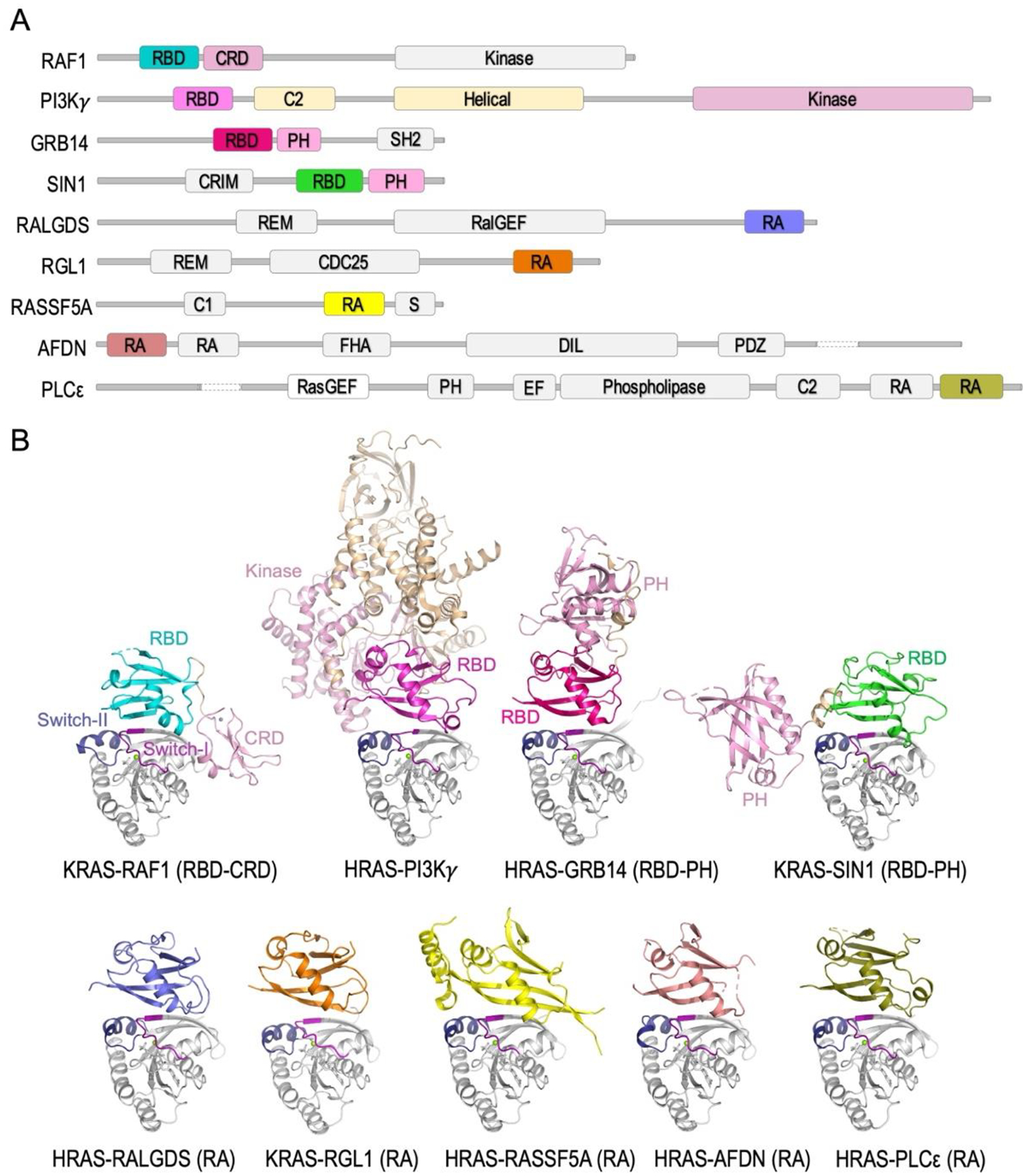
(A) Domain organization of human effector proteins, whose structures have been solved in complex with RAS proteins, is depicted. The effector domain(s), which are part of the RAS-effector complex, are shown in color, while the remaining domains are in gray. (B) The structures of RAS-effector complexes solved so far are shown in cartoon representation. These structures were aligned using RAS proteins, which are depicted in a grey cartoon with switch-I and switch-II regions colored purple and blue, respectively. Nucleotides and Mg ions are represented as sticks and spheres and are colored gray and green, respectively. RBD and adjacent domains are color-coded following the same scheme as in panel A. The structures of CRAF (PDB: 6XI7), PI3Kγ (PDB: 1HE8), GRB14 (PDB: 4K81), and SIN1 (PDB: 7LC1) in complex with H/KRAS were solved with RBD and adjacent domains, whereas the structures of RALGDS (PDB: 1LFD), RGL1 (PDB: 7SCW), RASSF5 (PDB: 3DDC), AFDN (PDB: 6AMB), and PLCε (PDB: 2C5L) in complex with HRAS were primarily solved using RBD/RA domains.
RAF kinases (A/B/CRAF) feature an N-terminal regulatory domain containing RBD and CRD (cysteine-rich domain) and a C-terminal kinase domain (Figure 3A). The RAS-RAF interaction was first elucidated through the crystal structure of RAP1A, a RAS-like GTPase, in complex with the CRAF-RBD, revealing that the RBD adopts a β-sandwich ubiquitin-like fold and forms a continuous β-sheet at the interface57. Subsequent research with H/KRAS validated this interaction pattern11,58. The CRD not only aids RAF in attaching to membranes but also strengthens its binding to RAS11. Crystallographic data reveals that the RBD and CRD form a single elongated entity, with both domains extensively interacting with RAS (Figure 3B). Distinct from the switch-I region-mediated RAS-RBD interface, the RAS-CRD interaction mainly involves the interswitch region and the α5-helix in RAS proteins11,59. Alterations in the RAS-CRD interface markedly diminish CRAF activation with minimal effect on the binding strength, underscoring the criticality of RAS-CRD interactions for complete RAF activation. Cryo-electron microscopy studies examining BRAF complexes have enhanced our knowledge of RAF’s autoinhibited and active states60. These investigations underscore that the autoinhibited state is stabilized through interactions between the kinase domain, the CRD, and 14-3-3 proteins, with pivotal phosphoserines located at both ends of the kinase domain61–64. Activation of RAF by RAS involves RAS binding to the RBD, leading to the disruption of 14-3-3 interactions and the subsequent release of the CRD. This series of events results in RAF dimerization at the plasma membrane, culminating in its full activation. The full-length, active BRAF–14-3-3 complex in the active state yielded no structural details beyond the BRAF kinase domain, suggesting the necessity of incorporating membrane mimetics in future studies to gain a more comprehensive understanding of the RAF activation mechanism.
The four class-I PI3K isoforms, namely PI3Kα, PI3Kβ, PI3Kγ, and PI3Kδ, each contain an RBD. Active RAS proteins bind to the p110α, p110γ, and p110δ catalytic subunits of the PI3K lipid kinase complex via their respective RBDs. RAS exhibits a GTP-dependent yet comparatively weaker binding affinity to PI3K than RAF40. A crystallographic study demonstrated that HRAS binds to the PI3Kγ RBD through its switch-I and II regions, while only the switch-II region interacts with PI3Kγ’s catalytic domain, emphasizing RAS’s dual role in membrane attachment and allosteric activation65 (Figure 3B). Unlike the other class-I PI3K isoforms, PI3Kβ does not bind to RAS-like GTPases; instead, it is activated by the RHO GTPases CDC42 and RAC147.
Utilizing yeast two-hybrid, RALGDS, a GEF for RAL GTPases, was identified as RAS effector38. RALGDS links RAS and RAL signaling, thereby regulating cellular functions like vesicular trafficking and migration38,66. The crystal structure of the HRAS and RALGDS-RA complex revealed a heterotetrameric arrangement, where each HRAS molecule interacts with two RALGDS-RA molecules, one via the switch-I region and another through the switch-II region67. However, later studies suggested that the heterotetrameric arrangement was an artifact of crystal packing, with the physiological interaction primarily involving the switch-I region of RAS68. In mammals, three paralogues of RALGDS have been identified: RGL1 (RALGDS Like 1), RGL2, and RGL3. Recent study suggested that KRAS complexed with RGL1-RA predominantly forms 1:1 dimers in solution, with tetramerization occurring through a very slow association69 (Figure 3B). The interaction of PI3Kγ and RALGDS with RAS is facilitated by forming an antiparallel β-sheet between the RBD and RAS, similar to that observed with RAF. This supports a conserved mode of RBD-RAS recognition, although there are distinctive differences in the relative orientation between the RAS and RBD65 (Figure 3B).
The RAS Association Domain Family (RASSF), comprising ten members (RASSF1–10), represents a large group of RBD-containing effector proteins. Recent research revealed that only RASSF5 directly interacts with classical RAS proteins, distinguishing it from other RASSFs that bind non-classical GTPases55,56. The crystal structure of RAS in complex with the RA domain of RASSF5, containing an additional N-terminal helix, displays the typical RA domain interaction with RAS’ switch-I region seen in other RAS-effector complexes70. However, the N-terminal helix of RASSF5-RA uniquely forms a distinct hydrophobic interaction with the switch-II region of activated RAS (Figure 3B).
AFDN (also known as AF6/MLLT4/Afadin) is characterized by two RBDs and plays a crucial role in the formation and maintenance of cell-cell adherens junctions71 (Figure 3A). The crystal structure of RAS with AFDN’s first RBD shows a classical interaction72. However, similar to RASSF5, AFDN’s first RBD is predicted to feature an N-terminal helix that interacts with the switch-II region of RAS. The NMR structure of AFDN’s second RBD showed a typical ubiquitin-like fold, and alone, it has a very weak affinity for RAS or RAP173. Recent work has shown the interaction of AFDN’s second RBD with RAP2C and, to a lesser extent, HRAS in cells. Interaction of RAS GTPases with AFDN promotes SCRIB interaction, which likely participates in cell contacts and polarity regulation74.
Phospholipase Cε (PLCε), containing two RA domains (RA1/2), is activated by RAP1 and RAS and regulates endocytosis, exocytosis, and cytoskeletal reorganization75,76. NMR studies revealed that both RA domains adopt a ubiquitin fold similar to the RAF-RBD77. However, only RA2 binds to RAS, engaging with both switch regions akin to PI3Kγ. Recent studies revealed that the RA1 domain forms extensive interactions with other PLCε domains to maintain structural and functional integrity78.
GRB14, belonging to the GRB7–10-14 family of multi-domain cytoplasmic adaptor proteins, acts as a tissue-specific regulator of insulin signaling79. GRB14 has been shown to regulate negative feedback on the insulin receptor80. The complex structure of GRB14’s RA and pleckstrin homology (PH) domains with RAS reveals that only the RA domain directly interacts with RAS, forming the typical RAS-RBD interaction surface, while the PH domain does not participate in this interaction81 (Figure 3B). It has been suggested that the PH domain may enhance the RA domain’s positioning for effective interaction with plasma membrane-tethered GTPases, such as RAS and RAP1.
Sin1, encoded by MAPKAP1, is an essential component of the mTORC2 and contains a RBD; in cells, it preferentially binds KRAS4a10,82. Unlike the RAS-GRB14 (RBD-PH) complex, structural analyses of KRAS-Sin1 (RBD-PH) revealed an expanded interaction interface that encompasses the typical RAS-RBD interaction and an additional interaction interface between the RAS switch-II region and the Sin1 linker-PH domain10 (Figure 3B). The KRAS-Sin1 interface is notably more extensive than other RAS-effector complexes, highlighting the importance of both the PH domain in Sin1 and the switch-II region in RAS in facilitating RAS-effector interactions.
In general terms, most RAS-effector complexes are formed by antiparallel β-sheet formations between the RBD β1/β2 and the RAS GTPase β2/β3. Most RAS residues engaging with effectors are conserved, particularly notable hotspots in and around the switch-I region, including Q25, D33, I36, E37, D38, S39, Y40, R41, and in the switch-II region, Y64 (H/K/NRAS numbering)40. Conversely, the majority of residues in RBD/RA domains interacting with RAS display variability. A comparison of CRAF-RBD and RASSF5-RA revealed only two distantly conserved residues between them56. A study analyzing specificity in binding interfaces of RAS with RBDs of CRAF, PI3Kγ, and RALGDS suggested significantly shared binding contacts83. Some RAS residues are crucial for binding all three effectors, while others alter specificity. Mutations in RAS residues D33, D38, I36, and Y40 typically disrupt binding across all three effectors. Specific hydrophobic mutations at I36 impede binding to PI3Kγ and RALGDS but not CRAF, whereas certain changes at L56 enhance CRAF binding and variably affect the others83. Alterations at E37 tend to decrease binding to CRAF and RALGDS but increase it for PI3Kγ. Y64 mutations selectively diminish PI3Kγ and RALGDS binding while maintaining CRAF interaction, and certain mutations at S39 specifically hinder binding to PI3Kγ and CRAF, sparing RALGDS83.
Structural studies on the H/KRAS-CRAF(RBD-CRD) and KRAS-Sin1(RBD-PH) complexes have highlighted crucial interactions with RAS proteins beyond the RBD10,11,65. Given that most RAS-effector complex structures have been obtained primarily using the RBD alone, it is possible that adjacent domains of the RBD/RA in these multi-domain effector proteins may also engage with RAS proteins.
Atypical RAS effectors and isoform-specific effectors
Given the low protein sequence homology among RBD and RA domains, it is also tempting to speculate that many RAS effectors have not yet been discovered, as these will only be able to be identified by experimental means. This notion is supported by the recent identification of “atypical” effectors lacking classical RBD/RA domains. For instance, like Sin1, Hexokinase 1 (HK1) is a KRAS4a effector13. The interaction between HK1 and KRAS4a was attributed to the palmitoylation-depalmitoylation cycle of KRAS4a, facilitating its colocalization with HK1 on the outer mitochondrial membrane. Intriguingly, HK1 lacks the typical RBD/RA sequence or structure. Yet, the full-length HK1 structure reveals a surface-exposed helix–loop–sheet motif that partially resembles the RBD, enabling it to interact with KRAS4a. These findings underscore the possibility that certain RAS GTPases may interact with effectors that do not contain typical RBD. Although there are isoform-specific interactors that have been shown to affect the biology of RAS isoforms, many of these do not satisfy the definition of effector. The identification of novel isoform-specific effectors requires proteomic approaches using different isoforms as baits in both GDP/GTP bound forms, as shown by many recent studies84–87. Future work will be necessary to assess whether some of these interactors bind in a GTP-dependent manner and if they contain atypical RBD domains.
Signaling through RAS effectors at the organismal level
The function of RAS effectors has been studied across model organisms, from yeast to mammals, given the evolutionary conservation of RAS GTPases and their protein effectors (Figure 4A). Notably, high conservation is observed in the switch-I motif, crucial for interactions with downstream effectors88. The budding yeast Saccharomyces cerevisiae has two RAS genes, Ras1 and Ras2, with comparable functions but distinct regulatory mechanisms governing their expression89–91. Ras1/2 bind and activate their downstream effector Cyr1, an ortholog of adenylate cyclase that contains an RBD-like domain, catalyzing cAMP synthesis. This, in turn, activates Bcy1, a Protein Kinase A subunit, driving cell cycle progression, stress response, and spore morphogenesis91–93. Unlike S. cerevisiae, the fission yeast Schizosaccharomyces pombe features a single Ras protein (Ras1) and two main effectors, Byr2 and Scd1. Byr2, part of a kinase cascade including Byr1 and Spk1 activated by Ras1 through its RBD-like domain, regulates pheromone signaling, akin to the mammalian MAPK pathway91,94. While Scd1, a guanine exchange factor (GEF) that regulates Cdc42 and contains a distinct type of RBD, is involved in spindle formation and cytokinesis and is similar to mammalian RALGDS91,95–97.
Figure 4. Signaling pathways of RAS-effector complexes in model organisms and humans.
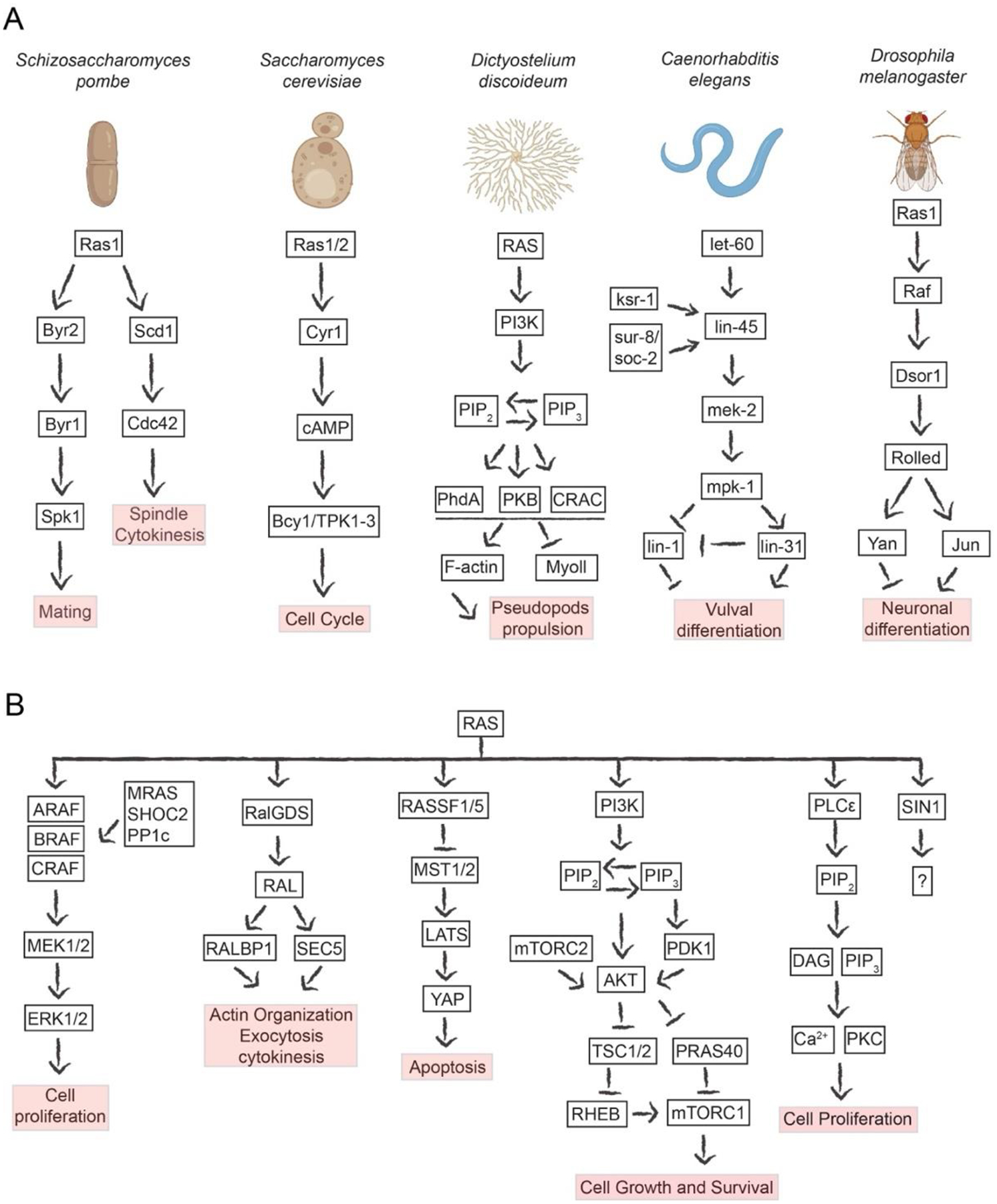
(A) Biochemical pathways regulated by RAS effectors in model organisms, including fission and baker’s yeast, slime mold, roundworms and fruit flies. Note the conserved modes of signal transduction, despite the diverse phenotypic outputs. (B) Overview of some of the different RAS effector signaling pathways that have been characterized in humans. Although many of these have been studied in the context of oncogenic KRAS signaling, they might represent pathways physiologically regulated by non-classical RAS GTPases.
In the slime mold Dictyostelium discoideum, ten RAS proteins have been identified, with RasD and RasG akin to human classical RAS proteins98–100,101. Moreover, the orthologs of several mammalian RAS effectors (PI3K and MAPK pathways) have been identified in this organism100–102. Dictyostelium’s PI3K pathway regulates chemotaxis upon RasG binding, promoting downstream activation of AKT. Similarly, RasC and Rap1 can bind directly to the Sin1 ortholog RIP3 and modulate the activity of the TorC2 complex. In addition to AKT, activation of PI3K in this organism leads to the recruitment of other PH domain-containing proteins such as cytosolic regulator of adenylyl cyclase (CRAC) and PhdA, which in turn promote pseudopod propulsion through F-actin polymerization103.
The genetic characterization of the roundworm Caenorhabditis elegans was pivotal in discovering the role of RAS proteins in larval development, specifically, vulval differentiation104,105. LET-60 is considered as the main RAS ortholog106,107. In addition, screenings for vulval development defects have revealed downstream effectors in this organism, underscoring the RAS-MAPK pathway’s role in regulating this phenotype in worms105,108,109. Indeed, lin-45, mek-2 and mpk-1 were identified as the RAF, MEK and ERK orthologs110–114. However, other genes have also been identified downstream of LET-60, including Sur-8/soc-2 and ksr-1, which positively regulate the RAS-mediated signaling pathway105,115–117. These genes are conserved in humans as SHOC2 and KSR1 and are now known to play critical roles in this signaling pathway.
The discovery of a RAS ortholog (Ras1) in Drosophila melanogaster has advanced our understanding of RAS signaling in fruit flies. Ras1 is responsible for differentiating progenitor cells into neuronal cells, namely eye photoreceptor cells118,119. Initiation of the signal transduction cascade through Ras1 is modulated upstream by the receptor tyrosine kinase Sevenless (Sev) and the GEF Son-of-sevenless (Sos), while Gap1 terminates the signaling118,119–121. Drk (GRB2) serves as a scaffold of Sev and Sos, leading to the activation of Ras1 and its subsequent binding to Raf, in a process that is highly conserved to the mammalian pathway118,122–124. Activated RAS primarily facilitates RAF’s plasma membrane localization through its conserved RBD, where RAF activation occurs125. Once active, RAF initiates the MAPK module through Dsor and Rolled118,126–128.
The insights gained from studies in model organisms played a pivotal role in uncovering the intricacies of RAS signaling in mammals (Figure 4B). While the precise outcomes of the RAS pathway may differ among organisms, the fundamental mechanisms of effector signaling remain conserved across species, using domains that bind only to RAS-GTP. While the evolutionary conservation of the RAS-MAPK cascade is remarkable, the complexity of the RAS signaling pathway increases in mammals, highlighting the significance of studying RAS-effector signaling pathways in less-complex model organisms. In humans, there are >50 putative RAS effectors, compared to the more limited number of effectors in model organisms5,72,129. Moreover, the larger number of RAS proteins in humans contributes to the complexity of the signaling network. In mammals, the best-studied downstream pathways of classical RAS proteins are the MAPK and PI3K pathways. In the MAPK pathway, RAS-GTP recruits the RAF kinases, initiating a complex activation mechanism130. A critical step in this process is the dephosphorylation of RAF proteins at a site N-terminal to the kinase domain by the MRAS-GTP/SHOC2/PP1c complex, underscoring the regulatory role of non-classical RAS proteins in various signaling pathways. Upon dimerization and activation, RAF kinases phosphorylate downstream kinases MEK1/2, which subsequently phosphorylate ERK1/2. This MAPK cascade ultimately stimulates the phosphorylation of many substrates, including transcription factors that promote cellular proliferation. Moreover, ERK1/2 can also negatively regulate the MAPK pathway by phosphorylating and inhibiting SOS1 or CRAF131,132.
In the context of PI3K signaling, alongside classical RAS proteins, RAS-like GTPases such as RRAS, RRAS2, MRAS, and ERAS have been shown to interact with PI3Kα, indicating their potential function as physiological activators of the enzyme47. Upon binding to RAS-GTP, PI3K increases its catalytic activity synergistically with RTK stimulation, phosphorylating phosphatidylinositol 4,5-biphosphate (PIP2) to promote the formation of phosphatidylinositol (3,4,5)-triphosphate (PIP3)131, a second messenger that leads to the recruitment of AKT to the plasma membrane via its PH domain. Membrane-localized AKT is phosphorylated and activated by the kinases PDK1 and mTORC2, enabling it to phosphorylate numerous substrates involved in cell growth and metabolism, including TSC2, PRAS40, FOXO transcription factors, and others133.
Activation of RALGDS by RAS-GTP facilitates its translocation to the plasma membrane and potentially exposes its REM domain, which possesses GEF activity for RALA/B GTPases. RALA/B proteins also have numerous downstream RAS effectors, including RALBP1 and Sec5, to regulate various cellular phenotypes. RALBP1 acts as a GAP for RAC GTPases134, and Sec5 is a critical component of the exocyst complex135. The activation of these pathways promotes actin organization, exocytosis and cytokinesis136. Like RALGDS, other RAL GEF proteins, such as RGL1–3, contain RA domains that interact with different GTP-bound RAS-like proteins with different degrees of specificity48.
RASSF5, also known as NORE1A, was initially identified as the first member of the RASSF family through a yeast two-hybrid screen using mutant RAS as bait35,137. Recent investigations have revealed that RAS proteins can modulate the Hippo pathway by utilizing RASSF family proteins as effectors138. MST kinases were identified as direct interacting partners of RASSF proteins, facilitated by a SARAH domain139. Given their pro-apoptotic characteristics140, these kinases have emerged as promising candidates for mediating RASSF-driven tumor suppression141. MST kinases activate the LATS kinases through phosphorylation, leading to subsequent phosphorylation of the transcriptional regulators YAP and TAZ142. In addition, although NORE1A plays a role in modulating apoptosis, its primary function may lie in acting as a potent effector of RAS-induced senescence137,143–148, but the precise mechanisms by which RAS regulates these pathways, and the contribution of RASSF proteins, remain poorly understood.
Potential therapeutic strategies targeting RAS effectors
Understanding the importance of RAS effectors is also crucial for its therapeutic implications. Identifying critical effectors downstream of activated RAS proteins can provide novel therapeutic targets for cancer and congenital disorders such as RASopathies. Mutations in genes encoding for classical RAS proteins are found in several cancer types; KRAS is mutated in lung, pancreatic and colorectal adenocarcinomas131,149–151, while N/HRAS mutations are frequent in melanoma, head and neck squamous carcinomas and bladder cancer131,152–154. In RASopathies, activating variants in several RAS genes are also frequent155. In all these conditions, the diseased phenotype solely depends on downstream effector signaling.
Given the longstanding challenges of direct RAS inhibition, many therapeutic efforts for RAS-driven cancers have focused on inhibiting downstream effectors. Using animal models of KRAS-driven cancer, it was shown that genetic ablation of the MAPK pathway suppressed tumor onset156. Although ATP-competitive RAF inhibitors have been developed for several tumor types, these inhibitors do not seem to work in the context of RAS-mutant tumors. Indeed, these compounds induce paradoxical activation of the MAPK pathway that results from feedback relief157–159. To overcome this compensatory mechanism, combinations with MEK inhibitors have been developed, although these have been mostly beneficial in BRAF-mutant tumors, including melanoma, lung and thyroid cancers, but not in RAS-mutant tumors. Studies using next-generation pan-RAF inhibitors show less paradoxical activation, opening new therapeutic strategies for the outcomes of patients with RAS-mutated tumors160–163. Based on preclinical evidence, a more targeted approach could involve the specific inhibition of CRAF, particularly through targeted degradation, since this effector appears essential for oncogenic KRAS-driven tumors156. In RASopathies driven by activated RAS, the MAPK pathway seems to be the most important downstream target, and the use of MEK inhibitors in this setting is currently being tested with early reports of efficacy164.
Using mouse models of KRAS and EGFR mutant-driven lung tumors, it was demonstrated that PI3K is required for tumor onset and maintenance165. PI3K inhibitors that specifically target this pathway have been tested in KRAS cancers; however, as monotherapy, these inhibitors do not work in RAS-driven preclinical models nor in clinical trials. Although the combination of MEK and PI3K inhibitors seemed more promising in mouse models166, clinical trials testing this combination showed significant toxicity and questionable efficacy131,167,168.
Targeting other effectors has been tested preclinically as potential therapies for RAS-driven cancers, although no drugs have been developed yet. For example, depleting RALGDS in tumor models reduces cancer incidence, size, and malignant progression in a multistage skin carcinogenesis model169. Similarly, TIAM1 knockout mice were more resistant to HRAS-driven transformation in such skin models and embryonic fibroblasts formed less foci upon KRAS G12V expression170. Mice in which PLCε had been knocked out exhibited opposing results regarding susceptibility to tumor formation upon the skin carcinogenesis protocol, so its role remains questionable171,172. Screenings in different cell lines driven by RAS have uncovered synthetic lethal interactors173. Of these, many are not direct effectors, but CRAF and SHOC2 appear to be essential in these models and are common hits in these screenings173. Although SHOC2 has not been considered a RAS effector, its binding specificity to RAS-GTP forms and recruitment of catalytic activity via protein phosphatase 1 (PP1) proteins makes it act like a RAS effector. Another independent study validated the crucial role of SHOC2 in a KRAS-driven lung cancer model, as the ablation of SHOC2 inhibited tumor development and prevented resistance to MEK inhibitors174. A potential strategy for targeting all RAS effectors involves disrupting the RAS-RBD interaction. Preclinical studies indicated that rigosertib, mimicking activated RAS, disrupts the binding of RAF, PI3K, and RALGDS to RAS-GTP, thereby impeding downstream signaling175,176. However, clinical trials failed to show its efficacy, and subsequent studies raised concerns regarding its specificity177,178. An interesting development in targeting effector RBDs is exemplified by a small molecule (BBO-10203) that binds the PI3Kα-RBD near the RBD-RAS interface, effectively blocking its interaction with classical RAS proteins179. These compounds lay the groundwork for developing similar inhibitors that target the RBDs of other effector proteins, thereby inhibiting their activation by RAS-like GTPases.
Directly targeting allosteric pockets in RAS or effector RBDs near the interface is currently the most promising strategy for treating RAS-driven cancers. Recent advancements include the development of KRAS G12C (GDP) covalent inhibitors180,181, currently approved for treating KRAS G12C mutant lung cancer. Targeting only the inactive KRAS G12C may induce resistance by increasing active GTP-bound KRAS levels; thus, inhibitors targeting both active and inactive states may overcome this resistance182. Recently, inhibitors targeting both nucleotide states of KRAS G12C have been reported183,184. Similarly, multiple efforts have also focused on targeting other RAS mutant alleles and pan-RAS or pan-KRAS inhibitors182,185. These inhibitors either lock RAS in its inactive state or prevent RAS-effector interactions. In this context, a recent chemical strategy that leverages the formation of a tri-complex between the inhibitor, RAS, and cyclophilin A has been reported186; these molecular glues prevent the association with RAS effectors due to steric hindrance. Given that RAS-GTP utilizes overlapping surfaces for interacting with effectors and GAP proteins, approaches aimed at stabilizing mutant RAS-GAP interactions via molecular glues187, preferably also increasing GTPase activity, could effectively block RAS-effector interactions.
Future directions and challenges
Despite the extensive work done to date, a comprehensive and multifaceted experimental approach will be essential to enhance our understanding of RAS-effector interactions. There are several aspects of their biology that will need to be reevaluated: (1) Structural analysis of RAS complexed with full-length effector proteins could reveal the interaction between RAS and RBD-adjacent domains and their collective influence on downstream signaling pathways. (2) The structural aspects of RAS-effector complexes in membrane-mimicking environments are critical, as this aligns closely with physiological conditions, offering a more accurate depiction of these interactions in cellular membranes. (3) Understanding the specific biological roles of classical RAS isoforms and RAS-like GTPases in effector activation, since many effectors are likely activated by non-classical RAS proteins. (4) Study whether oncogenic RAS proteins might activate some of these less-characterized effectors in a neomorphic manner as the active RAS population becomes significantly higher. (5) Discovery of potential isoform-specific interactions with effectors by mechanisms that extend beyond the G-domain binding. (6) Assessing the dynamics of RAS-effector complex formation at the organismal level. Exploring innovative experimental strategies could significantly advance our understanding of RAS-effector interactions. Techniques such as proximity ligation and in vivo tagging of RAS GTPases (and their constitutively GTP-bound mutants) offer promising avenues to address current limitations. Integration of these techniques with robust biochemical and structural analyses will help address key open questions in the field.
Acknowledgments
PC’s laboratory is supported by grants from the NCI (R00CA245122 and R01CA279171). This project was funded in part with federal funds from the National Cancer Institute, NIH contract 75N91019D00024. We thank Tania Gonzalez-Robles for her assistance in generating the protein expression level graph. The content of this publication does not necessarily reflect the views or policies of the Department of Health and Human Services, and the mention of trade names, commercial products, or organizations does not imply endorsement by the U.S. government.
Footnotes
Publisher's Disclaimer: This is a PDF file of an unedited manuscript that has been accepted for publication. As a service to our customers we are providing this early version of the manuscript. The manuscript will undergo copyediting, typesetting, and review of the resulting proof before it is published in its final form. Please note that during the production process errors may be discovered which could affect the content, and all legal disclaimers that apply to the journal pertain.
Declarations of interests
The authors declare no competing interests.
References
- 1.Simanshu DK, Nissley DV, and McCormick F (2017). RAS Proteins and Their Regulators in Human Disease. Cell 170, 17–33. 10.1016/j.cell.2017.06.009. [DOI] [PMC free article] [PubMed] [Google Scholar]
- 2.Wennerberg K, Rossman KL, and Der CJ (2005). The Ras superfamily at a glance. J Cell Sci 118, 843–846. 10.1242/jcs.01660. [DOI] [PubMed] [Google Scholar]
- 3.Bos JL, Rehmann H, and Wittinghofer A (2007). GEFs and GAPs: critical elements in the control of small G proteins. Cell 129, 865–877. 10.1016/j.cell.2007.05.018. [DOI] [PubMed] [Google Scholar]
- 4.Kiel C, Matallanas D, and Kolch W (2021). The Ins and Outs of RAS Effector Complexes. Biomolecules 11, 236. 10.3390/biom11020236. [DOI] [PMC free article] [PubMed] [Google Scholar]
- 5.Smith MJ (2023). Defining bone fide effectors of RAS GTPases. BioEssays 45, 2300088. 10.1002/bies.202300088. [DOI] [PubMed] [Google Scholar]
- 6.Wittinghofer A, and Herrmann C (1995). Ras-effector interactions, the problem of specificity. FEBS Lett 369, 52–56. 10.1016/0014-5793(95)00667-x. [DOI] [PubMed] [Google Scholar]
- 7.Kiel C, Aydin D, and Serrano L (2008). Association Rate Constants of Ras-Effector Interactions Are Evolutionarily Conserved. PLoS Comput Biol 4, e1000245. 10.1371/journal.pcbi.1000245. [DOI] [PMC free article] [PubMed] [Google Scholar]
- 8.Junk P, and Kiel C (2023). Structure-based prediction of Ras-effector binding affinities and design of “branchegetic” interface mutations. Structure 31, 870–883.e5. 10.1016/j.str.2023.04.007. [DOI] [PubMed] [Google Scholar]
- 9.Catozzi S, Ternet C, Gourrege A, Wynne K, Oliviero G, and Kiel C (2022). Reconstruction and analysis of a large-scale binary Ras-effector signaling network. Cell Commun Signal 20, 24. 10.1186/s12964-022-00823-5. [DOI] [PMC free article] [PubMed] [Google Scholar]
- 10.Castel P, Dharmaiah S, Sale MJ, Messing S, Rizzuto G, Cuevas-Navarro A, Cheng A, Trnka MJ, Urisman A, Esposito D, et al. (2021). RAS interaction with Sin1 is dispensable for mTORC2 assembly and activity. Proc Natl Acad Sci U S A 118, e2103261118. 10.1073/pnas.2103261118. [DOI] [PMC free article] [PubMed] [Google Scholar]
- 11.Tran TH, Chan AH, Young LC, Bindu L, Neale C, Messing S, Dharmaiah S, Taylor T, Denson J-P, Esposito D, et al. (2021). KRAS interaction with RAF1 RAS-binding domain and cysteine-rich domain provides insights into RAS-mediated RAF activation. Nat Commun 12, 1176. 10.1038/s41467-021-21422-x. [DOI] [PMC free article] [PubMed] [Google Scholar]
- 12.Thurman R, Siraliev-Perez E, and Campbell SL (2017). RAS ubiquitylation modulates effector interactions. Small GTPases, 1–6. 10.1080/21541248.2017.1371267. [DOI] [PMC free article] [PubMed] [Google Scholar]
- 13.Amendola CR, Mahaffey JP, Parker SJ, Ahearn IM, Chen W-C, Zhou M, Court H, Shi J, Mendoza SL, Morten MJ, et al. (2019). KRAS4A directly regulates hexokinase 1. Nature 576, 482–486. 10.1038/s41586-019-1832-9. [DOI] [PMC free article] [PubMed] [Google Scholar]
- 14.Colicelli J (2004). Human RAS superfamily proteins and related GTPases. Sci STKE 2004, RE13. 10.1126/stke.2502004re13. [DOI] [PMC free article] [PubMed] [Google Scholar]
- 15.Bernal Astrain G, Nikolova M, and Smith MJ (2022). Functional diversity in the RAS subfamily of small GTPases. Biochem Soc Trans 50, 921–933. 10.1042/BST20211166. [DOI] [PubMed] [Google Scholar]
- 16.Singh S, and Smith MJ (2020). RAS GTPase signalling to alternative effector pathways. Biochem Soc Trans 48, 2241–2252. 10.1042/BST20200506. [DOI] [PubMed] [Google Scholar]
- 17.Campbell SL, and Philips MR (2021). Post-translational modification of RAS proteins. Curr Opin Struct Biol 71, 180–192. 10.1016/j.sbi.2021.06.015. [DOI] [PMC free article] [PubMed] [Google Scholar]
- 18.Li Z-L, and Buck M (2017). Computational Modeling Reveals that Signaling Lipids Modulate the Orientation of K-Ras4A at the Membrane Reflecting Protein Topology. Structure 25, 679–689.e2. 10.1016/j.str.2017.02.007. [DOI] [PMC free article] [PubMed] [Google Scholar]
- 19.Ingólfsson HI, Neale C, Carpenter TS, Shrestha R, López CA, Tran TH, Oppelstrup T, Bhatia H, Stanton LG, Zhang X, et al. (2022). Machine learning-driven multiscale modeling reveals lipid-dependent dynamics of RAS signaling proteins. Proc Natl Acad Sci U S A 119, e2113297119. 10.1073/pnas.2113297119. [DOI] [PMC free article] [PubMed] [Google Scholar]
- 20.Milburn MV, Tong L, deVos AM, Brünger A, Yamaizumi Z, Nishimura S, and Kim SH (1990). Molecular switch for signal transduction: structural differences between active and inactive forms of protooncogenic ras proteins. Science 247, 939–945. 10.1126/science.2406906. [DOI] [PubMed] [Google Scholar]
- 21.Gasper R, and Wittinghofer F (2019). The Ras switch in structural and historical perspective. Biol Chem 401, 143–163. 10.1515/hsz-2019-0330. [DOI] [PubMed] [Google Scholar]
- 22.Warne PH, Viciana PR, and Downward J (1993). Direct interaction of Ras and the amino-terminal region of Raf-1 in vitro. Nature 364, 352–355. 10.1038/364352a0. [DOI] [PubMed] [Google Scholar]
- 23.Zhang XF, Settleman J, Kyriakis JM, Takeuchi-Suzuki E, Elledge SJ, Marshall MS, Bruder JT, Rapp UR, and Avruch J (1993). Normal and oncogenic p21ras proteins bind to the amino-terminal regulatory domain of c-Raf-1. Nature 364, 308–313. 10.1038/364308a0. [DOI] [PubMed] [Google Scholar]
- 24.Vojtek AB, Hollenberg SM, and Cooper JA (1993). Mammalian Ras interacts directly with the serine/threonine kinase Raf. Cell 74, 205–214. 10.1016/0092-8674(93)90307-c. [DOI] [PubMed] [Google Scholar]
- 25.Van Aelst L, Barr M, Marcus S, Polverino A, and Wigler M (1993). Complex formation between RAS and RAF and other protein kinases. Proc Natl Acad Sci U S A 90, 6213–6217. 10.1073/pnas.90.13.6213. [DOI] [PMC free article] [PubMed] [Google Scholar]
- 26.Moodie SA, Willumsen BM, Weber MJ, and Wolfman A (1993). Complexes of Ras.GTP with Raf-1 and mitogen-activated protein kinase kinase. Science 260, 1658–1661. 10.1126/science.8503013. [DOI] [PubMed] [Google Scholar]
- 27.Emerson SD, Madison VS, Palermo RE, Waugh DS, Scheffler JE, Tsao KL, Kiefer SE, Liu SP, and Fry DC (1995). Solution structure of the Ras-binding domain of c-Raf-1 and identification of its Ras interaction surface. Biochemistry 34, 6911–6918. 10.1021/bi00021a001. [DOI] [PubMed] [Google Scholar]
- 28.Sjölander A, Yamamoto K, Huber BE, and Lapetina EG (1991). Association of p21ras with phosphatidylinositol 3-kinase. Proc Natl Acad Sci U S A 88, 7908–7912. 10.1073/pnas.88.18.7908. [DOI] [PMC free article] [PubMed] [Google Scholar]
- 29.Kodaki T, Woscholski R, Hallberg B, Rodriguez-Viciana P, Downward J, and Parker PJ (1994). The activation of phosphatidylinositol 3-kinase by Ras. Curr Biol 4, 798–806. 10.1016/s0960-9822(00)00177-9. [DOI] [PubMed] [Google Scholar]
- 30.Rodriguez-Viciana P, Warne PH, Vanhaesebroeck B, Waterfield MD, and Downward J (1996). Activation of phosphoinositide 3-kinase by interaction with Ras and by point mutation. EMBO J 15, 2442–2451. [PMC free article] [PubMed] [Google Scholar]
- 31.Rubio I, Rodriguez-Viciana P, Downward J, and Wetzker R (1997). Interaction of Ras with phosphoinositide 3-kinase gamma. Biochem J 326 (Pt 3), 891–895. 10.1042/bj3260891. [DOI] [PMC free article] [PubMed] [Google Scholar]
- 32.Kikuchi A, Demo SD, Ye ZH, Chen YW, and Williams LT (1994). ralGDS family members interact with the effector loop of ras p21. Mol Cell Biol 14, 7483–7491. 10.1128/mcb.14.11.7483-7491.1994. [DOI] [PMC free article] [PubMed] [Google Scholar]
- 33.Han L, and Colicelli J (1995). A human protein selected for interference with Ras function interacts directly with Ras and competes with Raf1. Mol Cell Biol 15, 1318–1323. 10.1128/MCB.15.3.1318. [DOI] [PMC free article] [PubMed] [Google Scholar]
- 34.Shibatohge M, Kariya K. i, Liao Y, Hu CD, Watari Y, Goshima M, Shima F, and Kataoka T (1998). Identification of PLC210, a Caenorhabditis elegans phospholipase C, as a putative effector of Ras. J Biol Chem 273, 6218–6222. 10.1074/jbc.273.11.6218. [DOI] [PubMed] [Google Scholar]
- 35.Vavvas D, Li X, Avruch J, and Zhang X-F (1998). Identification of Nore1 as a Potential Ras Effector. Journal of Biological Chemistry 273, 5439–5442. 10.1074/jbc.273.10.5439. [DOI] [PubMed] [Google Scholar]
- 36.Boettner B, Herrmann C, and Van Aelst L (2001). Ras and Rap1 interaction with AF-6 effector target. Methods Enzymol 332, 151–168. 10.1016/s0076-6879(01)32199-7. [DOI] [PubMed] [Google Scholar]
- 37.Shao H, and Andres DA (2000). A novel RalGEF-like protein, RGL3, as a candidate effector for rit and Ras. J Biol Chem 275, 26914–26924. 10.1074/jbc.M002241200. [DOI] [PubMed] [Google Scholar]
- 38.Albright CF, Giddings BW, Liu J, Vito M, and Weinberg RA (1993). Characterization of a guanine nucleotide dissociation stimulator for a ras-related GTPase. EMBO J 12, 339–347. 10.1002/j.1460-2075.1993.tb05662.x. [DOI] [PMC free article] [PubMed] [Google Scholar]
- 39.Isomura M, Okui K, Fujiwara T, Shin S, and Nakamura Y (1996). Isolation and mapping of RAB2L, a human cDNA that encodes a protein homologous to RalGDS. Cytogenet Cell Genet 74, 263–265. 10.1159/000134431. [DOI] [PubMed] [Google Scholar]
- 40.Nakhaeizadeh H, Amin E, Nakhaei-Rad S, Dvorsky R, and Ahmadian MR (2016). The RAS-Effector Interface: Isoform-Specific Differences in the Effector Binding Regions. PLoS One 11, e0167145. 10.1371/journal.pone.0167145. [DOI] [PMC free article] [PubMed] [Google Scholar]
- 41.Kholodenko BN, Hoek JB, and Westerhoff HV (2000). Why cytoplasmic signalling proteins should be recruited to cell membranes. Trends Cell Biol 10, 173–178. 10.1016/s0962-8924(00)01741-4. [DOI] [PubMed] [Google Scholar]
- 42.Zhou Y, Prakash P, Gorfe AA, and Hancock JF (2018). Ras and the Plasma Membrane: A Complicated Relationship. Cold Spring Harb Perspect Med 8, a031831. 10.1101/cshperspect.a031831. [DOI] [PMC free article] [PubMed] [Google Scholar]
- 43.Simanshu DK, Philips MR, and Hancock JF (2023). Consensus on the RAS dimerization hypothesis: Strong evidence for lipid-mediated clustering but not for G-domain-mediated interactions. Mol Cell 83, 1210–1215. 10.1016/j.molcel.2023.03.008. [DOI] [PMC free article] [PubMed] [Google Scholar]
- 44.Nan X, Tamgüney TM, Collisson EA, Lin L-J, Pitt C, Galeas J, Lewis S, Gray JW, McCormick F, and Chu S (2015). Ras-GTP dimers activate the Mitogen-Activated Protein Kinase (MAPK) pathway. Proc Natl Acad Sci U S A 112, 7996–8001. 10.1073/pnas.1509123112. [DOI] [PMC free article] [PubMed] [Google Scholar]
- 45.Okada T, Hu CD, Jin TG, Kariya K, Yamawaki-Kataoka Y, and Kataoka T (1999). The strength of interaction at the Raf cysteine-rich domain is a critical determinant of response of Raf to Ras family small GTPases. Mol Cell Biol 19, 6057–6064. 10.1128/MCB.19.9.6057. [DOI] [PMC free article] [PubMed] [Google Scholar]
- 46.Harjes E, Harjes S, Wohlgemuth S, Müller K-H, Krieger E, Herrmann C, and Bayer P (2006). GTP-Ras Disrupts the Intramolecular Complex of C1 and RA Domains of Nore1. Structure 14, 881–888. 10.1016/j.str.2006.03.008. [DOI] [PubMed] [Google Scholar]
- 47.Fritsch R, de Krijger I, Fritsch K, George R, Reason B, Kumar MS, Diefenbacher M, Stamp G, and Downward J (2013). RAS and RHO families of GTPases directly regulate distinct phosphoinositide 3-kinase isoforms. Cell 153, 1050–1063. 10.1016/j.cell.2013.04.031. [DOI] [PMC free article] [PubMed] [Google Scholar]
- 48.Rodriguez-Viciana P, Sabatier C, and McCormick F (2004). Signaling specificity by Ras family GTPases is determined by the full spectrum of effectors they regulate. Mol Cell Biol 24, 4943–4954. 10.1128/MCB.24.11.4943-4954.2004. [DOI] [PMC free article] [PubMed] [Google Scholar]
- 49.Catozzi S, Halasz M, and Kiel C (2021). Predicted ‘wiring landscape’ of Ras-effector interactions in 29 human tissues. npj Syst Biol Appl 7, 10. 10.1038/s41540-021-00170-0. [DOI] [PMC free article] [PubMed] [Google Scholar]
- 50.Kolch W, Berta D, and Rosta E (2023). Dynamic regulation of RAS and RAS signaling. Biochem J 480, 1–23. 10.1042/BCJ20220234. [DOI] [PMC free article] [PubMed] [Google Scholar]
- 51.Coyle SM, and Lim WA (2016). Mapping the functional versatility and fragility of Ras GTPase signaling circuits through in vitro network reconstitution. Elife 5, e12435. 10.7554/eLife.12435. [DOI] [PMC free article] [PubMed] [Google Scholar]
- 52.Cuevas-Navarro A, Wagner M, Van R, Swain M, Mo S, Columbus J, Allison MR, Cheng A, Messing S, Turbyville TJ, et al. (2023). RAS-dependent RAF-MAPK hyperactivation by pathogenic RIT1 is a therapeutic target in Noonan syndrome–associated cardiac hypertrophy. Sci. Adv 9, eadf4766. 10.1126/sciadv.adf4766. [DOI] [PMC free article] [PubMed] [Google Scholar]
- 53.Flex E, Jaiswal M, Pantaleoni F, Martinelli S, Strullu M, Fansa EK, Caye A, De Luca A, Lepri F, Dvorsky R, et al. (2014). Activating mutations in RRAS underlie a phenotype within the RASopathy spectrum and contribute to leukaemogenesis. Hum Mol Genet 23, 4315–4327. 10.1093/hmg/ddu148. [DOI] [PMC free article] [PubMed] [Google Scholar]
- 54.Niihori T, Nagai K, Fujita A, Ohashi H, Okamoto N, Okada S, Harada A, Kihara H, Arbogast T, Funayama R, et al. (2019). Germline-Activating RRAS2 Mutations Cause Noonan Syndrome. Am J Hum Genet 104, 1233–1240. 10.1016/j.ajhg.2019.04.014. [DOI] [PMC free article] [PubMed] [Google Scholar]
- 55.Dhanaraman T, Singh S, Killoran RC, Singh A, Xu X, Shifman JM, and Smith MJ (2020). RASSF effectors couple diverse RAS subfamily GTPases to the Hippo pathway. Sci Signal 13, eabb4778. 10.1126/scisignal.abb4778. [DOI] [PubMed] [Google Scholar]
- 56.Rezaei Adariani S, Kazemein Jasemi NS, Bazgir F, Wittich C, Amin E, Seidel CAM, Dvorsky R, and Ahmadian MR (2021). A comprehensive analysis of RAS-effector interactions reveals interaction hotspots and new binding partners. J Biol Chem 296, 100626. 10.1016/j.jbc.2021.100626. [DOI] [PMC free article] [PubMed] [Google Scholar]
- 57.Nassar N, Horn G, Herrmann C, Scherer A, McCormick F, and Wittinghofer A (1995). The 2.2 A crystal structure of the Ras-binding domain of the serine/threonine kinase c-Raf1 in complex with Rap1A and a GTP analogue. Nature 375, 554–560. 10.1038/375554a0. [DOI] [PubMed] [Google Scholar]
- 58.Fetics SK, Guterres H, Kearney BM, Buhrman G, Ma B, Nussinov R, and Mattos C (2015). Allosteric effects of the oncogenic RasQ61L mutant on Raf-RBD. Structure 23, 505–516. 10.1016/j.str.2014.12.017. [DOI] [PMC free article] [PubMed] [Google Scholar]
- 59.Cookis T, and Mattos C (2021). Crystal Structure Reveals the Full Ras-Raf Interface and Advances Mechanistic Understanding of Raf Activation. Biomolecules 11, 996. 10.3390/biom11070996. [DOI] [PMC free article] [PubMed] [Google Scholar]
- 60.Simanshu DK, and Morrison DK (2022). A Structure is Worth a Thousand Words: New Insights for RAS and RAF Regulation. Cancer Discov 12, 899–912. 10.1158/2159-8290.CD-21-1494. [DOI] [PMC free article] [PubMed] [Google Scholar]
- 61.Kondo Y, Ognjenović J, Banerjee S, Karandur D, Merk A, Kulhanek K, Wong K, Roose JP, Subramaniam S, and Kuriyan J (2019). Cryo-EM structure of a dimeric B-Raf:14-3-3 complex reveals asymmetry in the active sites of B-Raf kinases. Science 366, 109–115. 10.1126/science.aay0543. [DOI] [PMC free article] [PubMed] [Google Scholar]
- 62.Park E, Rawson S, Li K, Kim B-W, Ficarro SB, Pino GG-D, Sharif H, Marto JA, Jeon H, and Eck MJ (2019). Architecture of autoinhibited and active BRAF-MEK1–14-3-3 complexes. Nature 575, 545–550. 10.1038/s41586-019-1660-y. [DOI] [PMC free article] [PubMed] [Google Scholar]
- 63.Martinez Fiesco JA, Durrant DE, Morrison DK, and Zhang P (2022). Structural insights into the BRAF monomer-to-dimer transition mediated by RAS binding. Nat Commun 13, 486. 10.1038/s41467-022-28084-3. [DOI] [PMC free article] [PubMed] [Google Scholar]
- 64.Liau NPD, Wendorff TJ, Quinn JG, Steffek M, Phung W, Liu P, Tang J, Irudayanathan FJ, Izadi S, Shaw AS, et al. (2020). Negative regulation of RAF kinase activity by ATP is overcome by 14-3-3-induced dimerization. Nat Struct Mol Biol 27, 134–141. 10.1038/s41594-019-0365-0. [DOI] [PubMed] [Google Scholar]
- 65.Pacold ME, Suire S, Perisic O, Lara-Gonzalez S, Davis CT, Walker EH, Hawkins PT, Stephens L, Eccleston JF, and Williams RL (2000). Crystal structure and functional analysis of Ras binding to its effector phosphoinositide 3-kinase gamma. Cell 103, 931–943. 10.1016/s0092-8674(00)00196-3. [DOI] [PubMed] [Google Scholar]
- 66.Ferro E, and Trabalzini L (2010). RalGDS family members couple Ras to Ral signalling and that’s not all. Cellular Signalling 22, 1804–1810. 10.1016/j.cellsig.2010.05.010. [DOI] [PubMed] [Google Scholar]
- 67.Huang L, Hofer F, Martin GS, and Kim SH (1998). Structural basis for the interaction of Ras with RalGDS. Nat Struct Biol 5, 422–426. 10.1038/nsb0698-422. [DOI] [PubMed] [Google Scholar]
- 68.Vetter IR, Linnemann T, Wohlgemuth S, Geyer M, Kalbitzer HR, Herrmann C, and Wittinghofer A (1999). Structural and biochemical analysis of Ras-effector signaling via RalGDS. FEBS Lett 451, 175–180. 10.1016/s0014-5793(99)00555-4. [DOI] [PubMed] [Google Scholar]
- 69.Eves BJ, Gebregiworgis T, Gasmi-Seabrook GMC, Kuntz DA, Privé GG, Marshall CB, and Ikura M (2022). Structures of RGL1 RAS-Association Domain in Complex with KRAS and the Oncogenic G12V Mutant. Journal of Molecular Biology 434, 167527. 10.1016/j.jmb.2022.167527. [DOI] [PubMed] [Google Scholar]
- 70.Stieglitz B, Bee C, Schwarz D, Yildiz O, Moshnikova A, Khokhlatchev A, and Herrmann C (2008). Novel type of Ras effector interaction established between tumour suppressor NORE1A and Ras switch II. EMBO J 27, 1995–2005. 10.1038/emboj.2008.125. [DOI] [PMC free article] [PubMed] [Google Scholar]
- 71.Sawyer JK, Harris NJ, Slep KC, Gaul U, and Peifer M (2009). The Drosophila afadin homologue Canoe regulates linkage of the actin cytoskeleton to adherens junctions during apical constriction. J Cell Biol 186, 57–73. 10.1083/jcb.200904001. [DOI] [PMC free article] [PubMed] [Google Scholar]
- 72.Smith MJ, Ottoni E, Ishiyama N, Goudreault M, Haman A, Meyer C, Tucholska M, Gasmi-Seabrook G, Menezes S, Laister RC, et al. (2017). Evolution of AF6-RAS association and its implications in mixed-lineage leukemia. Nat Commun 8, 1099. 10.1038/s41467-017-01326-5. [DOI] [PMC free article] [PubMed] [Google Scholar]
- 73.Wohlgemuth S, Kiel C, Krämer A, Serrano L, Wittinghofer F, and Herrmann C (2005). Recognizing and defining true Ras binding domains I: biochemical analysis. J Mol Biol 348, 741–758. 10.1016/j.jmb.2005.02.048. [DOI] [PubMed] [Google Scholar]
- 74.Goudreault M, Gagné V, Jo CH, Singh S, Killoran RC, Gingras A-C, and Smith MJ (2022). Afadin couples RAS GTPases to the polarity rheostat Scribble. Nat Commun 13, 4562. 10.1038/s41467-022-32335-8. [DOI] [PMC free article] [PubMed] [Google Scholar]
- 75.Smrcka AV, Brown JH, and Holz GG (2012). Role of phospholipase Cε in physiological phosphoinositide signaling networks. Cell Signal 24, 1333–1343. 10.1016/j.cellsig.2012.01.009. [DOI] [PMC free article] [PubMed] [Google Scholar]
- 76.Song C, Satoh T, Edamatsu H, Wu D, Tadano M, Gao X, and Kataoka T (2002). Differential roles of Ras and Rap1 in growth factor-dependent activation of phospholipase C epsilon. Oncogene 21, 8105–8113. 10.1038/sj.onc.1206003. [DOI] [PubMed] [Google Scholar]
- 77.Bunney TD, Harris R, Gandarillas NL, Josephs MB, Roe SM, Sorli SC, Paterson HF, Rodrigues-Lima F, Esposito D, Ponting CP, et al. (2006). Structural and mechanistic insights into ras association domains of phospholipase C epsilon. Mol Cell 21, 495–507. 10.1016/j.molcel.2006.01.008. [DOI] [PubMed] [Google Scholar]
- 78.Rugema NY, Garland-Kuntz EE, Sieng M, Muralidharan K, Van Camp MM, O’Neill H, Mbongo W, Selvia AF, Marti AT, Everly A, et al. (2020). Structure of phospholipase Cε reveals an integrated RA1 domain and previously unidentified regulatory elements. Commun Biol 3, 445. 10.1038/s42003-020-01178-8. [DOI] [PMC free article] [PubMed] [Google Scholar]
- 79.Holt LJ, and Siddle K (2005). Grb10 and Grb14: enigmatic regulators of insulin action--and more? Biochem J 388, 393–406. 10.1042/BJ20050216. [DOI] [PMC free article] [PubMed] [Google Scholar]
- 80.Cooney GJ, Lyons RJ, Crew AJ, Jensen TE, Molero JC, Mitchell CJ, Biden TJ, Ormandy CJ, James DE, and Daly RJ (2004). Improved glucose homeostasis and enhanced insulin signalling in Grb14-deficient mice. EMBO J 23, 582–593. 10.1038/sj.emboj.7600082. [DOI] [PMC free article] [PubMed] [Google Scholar]
- 81.Qamra R, and Hubbard SR (2013). Structural basis for the interaction of the adaptor protein grb14 with activated ras. PLoS One 8, e72473. 10.1371/journal.pone.0072473. [DOI] [PMC free article] [PubMed] [Google Scholar]
- 82.Saxton RA, and Sabatini DM (2017). mTOR Signaling in Growth, Metabolism, and Disease. Cell 168, 960–976. 10.1016/j.cell.2017.02.004. [DOI] [PMC free article] [PubMed] [Google Scholar]
- 83.Weng C, Faure AJ, Escobedo A, and Lehner B (2024). The energetic and allosteric landscape for KRAS inhibition. Nature 626, 643–652. 10.1038/s41586-023-06954-0. [DOI] [PMC free article] [PubMed] [Google Scholar]
- 84.Adhikari H, and Counter CM (2018). Interrogating the protein interactomes of RAS isoforms identifies PIP5K1A as a KRAS-specific vulnerability. Nat Commun 9, 3646. 10.1038/s41467-018-05692-6. [DOI] [PMC free article] [PubMed] [Google Scholar]
- 85.Ternet C, Junk P, Sevrin T, Catozzi S, Wåhlén E, Heldin J, Oliviero G, Wynne K, and Kiel C (2023). Analysis of context-specific KRAS-effector (sub)complexes in Caco-2 cells. Life Sci Alliance 6, e202201670. 10.26508/lsa.202201670. [DOI] [PMC free article] [PubMed] [Google Scholar]
- 86.Kovalski JR, Bhaduri A, Zehnder AM, Neela PH, Che Y, Wozniak GG, and Khavari PA (2019). The Functional Proximal Proteome of Oncogenic Ras Includes mTORC2. Mol Cell 73, 830–844.e12. 10.1016/j.molcel.2018.12.001. [DOI] [PMC free article] [PubMed] [Google Scholar]
- 87.Tang R, Shuldiner EG, Kelly M, Murray CW, Hebert JD, Andrejka L, Tsai MK, Hughes NW, Parker MI, Cai H, et al. (2023). Multiplexed screens identify RAS paralogues HRAS and NRAS as suppressors of KRAS-driven lung cancer growth. Nat Cell Biol 25, 159–169. 10.1038/s41556-022-01049-w. [DOI] [PMC free article] [PubMed] [Google Scholar]
- 88.Parmar N, and Tamanoi F (2010). Rheb G-Proteins and the Activation of mTORC1. In The Enzymes (Elsevier), pp. 39–56. 10.1016/S1874-6047(10)27003-8. [DOI] [PMC free article] [PubMed] [Google Scholar]
- 89.Breviario D, Hinnebusch A, Cannon J, Tatchell K, and Dhar R (1986). Carbon source regulation of RAS1 expression in Saccharomyces cerevisiae and the phenotypes of ras2- cells. Proc. Natl. Acad. Sci. U.S.A 83, 4152–4156. 10.1073/pnas.83.12.4152. [DOI] [PMC free article] [PubMed] [Google Scholar]
- 90.Breviario D, Hinnebusch AG, and Dhar R (1988). Multiple regulatory mechanisms control the expression of the RAS1 and RAS2 genes of Saccharomyces cerevisiae. The EMBO Journal 7, 1805–1813. 10.1002/j.1460-2075.1988.tb03012.x. [DOI] [PMC free article] [PubMed] [Google Scholar]
- 91.Tamanoi F (2011). Ras Signaling in Yeast. Genes & Cancer 2, 210–215. 10.1177/1947601911407322. [DOI] [PMC free article] [PubMed] [Google Scholar]
- 92.Ho J, and Bretscher A (2001). Ras Regulates the Polarity of the Yeast Actin Cytoskeleton through the Stress Response Pathway. MBoC 12, 1541–1555. 10.1091/mbc.12.6.1541. [DOI] [PMC free article] [PubMed] [Google Scholar]
- 93.McDonald CM, Wagner M, Dunham MJ, Shin ME, Ahmed NT, and Winter E (2009). The Ras/cAMP Pathway and the CDK-Like Kinase Ime2 Regulate the MAPK Smk1 and Spore Morphogenesis in Saccharomyces cerevisiae. Genetics 181, 511–523. 10.1534/genetics.108.098434. [DOI] [PMC free article] [PubMed] [Google Scholar]
- 94.Wang Y, Xu HP, Riggs M, Rodgers L, and Wigler M (1991). byr2, a Schizosaccharomyces pombe gene encoding a protein kinase capable of partial suppression of the ras1 mutant phenotype. Mol. Cell. Biol 11, 3554–3563. 10.1128/MCB.11.7.3554. [DOI] [PMC free article] [PubMed] [Google Scholar]
- 95.Wheatley E, and Rittinger K (2005). Interactions between Cdc42 and the scaffold protein Scd2: requirement of SH3 domains for GTPase binding. Biochemical Journal 388, 177–184. 10.1042/BJ20041838. [DOI] [PMC free article] [PubMed] [Google Scholar]
- 96.Li Y, Chen C, and Chang EC (2000). Fission Yeast Ras1 Effector Scd1 Interacts With the Spindle and Affects Its Proper Formation. Genetics 156, 995–1004. 10.1093/genetics/156.3.995. [DOI] [PMC free article] [PubMed] [Google Scholar]
- 97.Li Y, and Chang EC (2003). Schizosaccharomyces pombe Ras1 Effector, Scd1, Interacts With Klp5 and Klp6 Kinesins to Mediate Cytokinesis. Genetics 165, 477–488. 10.1093/genetics/165.2.477. [DOI] [PMC free article] [PubMed] [Google Scholar]
- 98.Robbins SM, Suttorp VV, Weeks G, and Spiegelman GB (1990). A ras -related gene from the lower eukaryote Dictyostelium that is highly conserved relative to the human rap genes. Nucl Acids Res 18, 5265–5269. 10.1093/nar/18.17.5265. [DOI] [PMC free article] [PubMed] [Google Scholar]
- 99.Robbins SM, Williams JG, Jermyn KA, Spiegelman GB, and Weeks G (1989). Growing and developing Dictyostelium cells express different ras genes. Proc. Natl. Acad. Sci. U.S.A 86, 938–942. 10.1073/pnas.86.3.938. [DOI] [PMC free article] [PubMed] [Google Scholar]
- 100.Chubb JR, and Insall RH (2001). Dictyostelium: an ideal organism for genetic dissection of Ras signalling networks. Biochimica et Biophysica Acta (BBA) - General Subjects 1525, 262–271. 10.1016/S0304-4165(01)00111-8. [DOI] [PubMed] [Google Scholar]
- 101.Jaffer ZM, Khosla M, Spiegelman GB, and Weeks G (2001). Expression of activated Ras during Dictyostelium development alters cell localization and changes cell fate. Development 128, 907–916. 10.1242/dev.128.6.907. [DOI] [PubMed] [Google Scholar]
- 102.Meili R, Ellsworth C, and Firtel RA (2000). A novel Akt/PKB-related kinase is essential for morphogenesis in Dictyostelium. Current Biology 10, 708–717. 10.1016/S0960-9822(00)00536-4. [DOI] [PubMed] [Google Scholar]
- 103.Kimmel AR, and Parent CA (2003). The Signal to Move: D. discoideum Go Orienteering. Science 300, 1525–1527. 10.1126/science.1085439. [DOI] [PubMed] [Google Scholar]
- 104.Horvitz HR, and Sternberg PW (1991). Multiple intercellular signalling systems control the development of the Caenorhabditis elegans vulva. Nature 351, 535–541. 10.1038/351535a0. [DOI] [PubMed] [Google Scholar]
- 105.Sternberg PW, and Han M (1998). Genetics of RAS signaling in C. elegans. Trends in Genetics 14, 466–472. 10.1016/S0168-9525(98)01592-3. [DOI] [PubMed] [Google Scholar]
- 106.Rogalski TM, Moerman DG, and Baillie DL (1982). ESSENTIAL GENES AND DEFICIENCIES IN THE UNC-22 IV REGION OF CAENORHABDITIS ELEGANS. Genetics 102, 725–736. 10.1093/genetics/102.4.725. [DOI] [PMC free article] [PubMed] [Google Scholar]
- 107.Clark DV, Rogalski TM, Donati LM, and Baillie DL (1988). The unc-22(IV) region of Caenorhabditis elegans: genetic analysis of lethal mutations. Genetics 119, 345–353. 10.1093/genetics/119.2.345. [DOI] [PMC free article] [PubMed] [Google Scholar]
- 108.Ferguson EL, and Horvitz HR (1985). IDENTIFICATION AND CHARACTERIZATION OF 22 GENES THAT AFFECT THE VULVAL CELL LINEAGES OF THE NEMATODE CAENORHABDITIS ELEGANS. Genetics 110, 17–72. 10.1093/genetics/110.1.17. [DOI] [PMC free article] [PubMed] [Google Scholar]
- 109.Ferguson EL, Sternberg PW, and Horvitz HR (1987). A genetic pathway for the specification of the vulval cell lineages of Caenorhabditis elegans. Nature 326, 259–267. 10.1038/326259a0. [DOI] [PubMed] [Google Scholar]
- 110.Kornfeld K, Guan KL, and Horvitz HR (1995). The Caenorhabditis elegans gene mek-2 is required for vulval induction and encodes a protein similar to the protein kinase MEK. Genes Dev 9, 756–768. 10.1101/gad.9.6.756. [DOI] [PubMed] [Google Scholar]
- 111.Wu Y, and Han M (1994). Suppression of activated Let-60 ras protein defines a role of Caenorhabditis elegans Sur-1 MAP kinase in vulval differentiation. Genes Dev 8, 147–159. 10.1101/gad.8.2.147. [DOI] [PubMed] [Google Scholar]
- 112.Shin H, and Reiner D (2018). The Signaling Network Controlling C. elegans Vulval Cell Fate Patterning. JDB 6, 30. 10.3390/jdb6040030. [DOI] [PMC free article] [PubMed] [Google Scholar]
- 113.Lee M-H, Ohmachi M, Arur S, Nayak S, Francis R, Church D, Lambie E, and Schedl T (2007). Multiple Functions and Dynamic Activation of MPK-1 Extracellular Signal-Regulated Kinase Signaling in Caenorhabditis elegans Germline Development. Genetics 177, 2039–2062. 10.1534/genetics.107.081356. [DOI] [PMC free article] [PubMed] [Google Scholar]
- 114.Singh N, and Han M (1995). sur-2, a novel gene, functions late in the let-60 ras-mediated signaling pathway during Caenorhabditis elegans vulval induction. Genes Dev 9, 2251–2265. 10.1101/gad.9.18.2251. [DOI] [PubMed] [Google Scholar]
- 115.Sieburth DS, Sun Q, and Han M (1998). SUR-8, a Conserved Ras-Binding Protein with Leucine-Rich Repeats, Positively Regulates Ras-Mediated Signaling in C. elegans. Cell 94, 119–130. 10.1016/S0092-8674(00)81227-1. [DOI] [PubMed] [Google Scholar]
- 116.Sundaram M, and Han M (1995). The C. elegans ksr-1 gene encodes a novel raf-related kinase involved in Ras-mediated signal transduction. Cell 83, 889–901. 10.1016/0092-8674(95)90205-8. [DOI] [PubMed] [Google Scholar]
- 117.Kornfeld K, Hom DB, and Horvitz HR (1995). The ksr-1 gene encodes a novel protein kinase involved in Ras-mediated signaling in C. elegans. Cell 83, 903–913. 10.1016/0092-8674(95)90206-6. [DOI] [PubMed] [Google Scholar]
- 118.Wassarman DA, Therrien M, and Rubin GM (1995). The Ras signaling pathway in Drosophila. Current Opinion in Genetics & Development 5, 44–50. 10.1016/S0959-437X(95)90052-7. [DOI] [PubMed] [Google Scholar]
- 119.Simon MA, Bowtell DDL, Dodson GS, Laverty TR, and Rubin GM (1991). Ras1 and a putative guanine nucleotide exchange factor perform crucial steps in signaling by the sevenless protein tyrosine kinase. Cell 67, 701–716. 10.1016/0092-8674(91)90065-7. [DOI] [PubMed] [Google Scholar]
- 120.Rogge RD, Karlovich CA, and Banerjee U (1991). Genetic dissection of a neurodevelopmental pathway: Son of sevenless functions downstream of the sevenless and EGF receptor tyrosine kinases. Cell 64, 39–48. 10.1016/0092-8674(91)90207-F. [DOI] [PubMed] [Google Scholar]
- 121.Gaul U, Mardon G, and Rubin GM (1992). A putative Ras GTPase activating protein acts as a negative regulator of signaling by the Sevenless receptor tyrosine kinase. Cell 68, 1007–1019. 10.1016/0092-8674(92)90073-L. [DOI] [PubMed] [Google Scholar]
- 122.Moodie SA, and Wolfman A (1994). The 3Rs of life: Ras, Raf and growth regulation. Trends in Genetics 10, 44–48. 10.1016/0168-9525(94)90147-3. [DOI] [PubMed] [Google Scholar]
- 123.Stokoe D, Macdonald SG, Cadwallader K, Symons M, and Hancock JF (1994). Activation of Raf as a Result of Recruitment to the Plasma Membrane. Science 264, 1463–1467. 10.1126/science.7811320. [DOI] [PubMed] [Google Scholar]
- 124.Leevers SJ, Paterson HF, and Marshall CJ (1994). Requirement for Ras in Raf activation is overcome by targeting Raf to the plasma membrane. Nature 369, 411–414. 10.1038/369411a0. [DOI] [PubMed] [Google Scholar]
- 125.Lusk J, Lam V, and Tolwinski N (2017). Epidermal Growth Factor Pathway Signaling in Drosophila Embryogenesis: Tools for Understanding Cancer. Cancers 9, 16. 10.3390/cancers9020016. [DOI] [PMC free article] [PubMed] [Google Scholar]
- 126.Marshall CJ (1994). MAP kinase kinase kinase, MAP kinase kinase and MAP kinase. Current Opinion in Genetics & Development 4, 82–89. 10.1016/0959-437X(94)90095-7. [DOI] [PubMed] [Google Scholar]
- 127.O’Neill EM, Rebay I, Tjian R, and Rubin GM (1994). The activities of two Ets-related transcription factors required for drosophila eye development are modulated by the Ras/MAPK pathway. Cell 78, 137–147. 10.1016/0092-8674(94)90580-0. [DOI] [PubMed] [Google Scholar]
- 128.Brunner D, Dücker K, Oellers N, Hafen E, Scholzi H, and Klambt C (1994). The ETS domain protein Pointed-P2 is a target of MAP kinase in the Sevenless signal transduction pathway. Nature 370, 386–389. 10.1038/370386a0. [DOI] [PubMed] [Google Scholar]
- 129.Smith MJ, and Ikura M (2014). Integrated RAS signaling defined by parallel NMR detection of effectors and regulators. Nat Chem Biol 10, 223–230. 10.1038/nchembio.1435. [DOI] [PubMed] [Google Scholar]
- 130.Bonsor DA, and Simanshu DK (2024). RAS and SHOC2 Roles in RAF Activation and Therapeutic Considerations. Annu. Rev. Cancer Biol 8, annurev-cancerbio-062822-030450. 10.1146/annurev-cancerbio-062822-030450. [DOI] [PMC free article] [PubMed] [Google Scholar]
- 131.Moore AR, Rosenberg SC, McCormick F, and Malek S (2020). RAS-targeted therapies: is the undruggable drugged? Nat Rev Drug Discov 19, 533–552. 10.1038/s41573-020-0068-6. [DOI] [PMC free article] [PubMed] [Google Scholar]
- 132.Xie Y, Cao Z, Wong EWP, Guan Y, Ma W, Zhang JQ, Walczak EG, Murphy D, Ran L, Sirota I, et al. (2018). COP1/DET1/ETS axis regulates ERK transcriptome and sensitivity to MAPK inhibitors. Journal of Clinical Investigation 128, 1442–1457. 10.1172/JCI94840. [DOI] [PMC free article] [PubMed] [Google Scholar]
- 133.Hoxhaj G, and Manning BD (2020). The PI3K-AKT network at the interface of oncogenic signalling and cancer metabolism. Nat Rev Cancer 20, 74–88. 10.1038/s41568-019-0216-7. [DOI] [PMC free article] [PubMed] [Google Scholar]
- 134.Park SH, and Weinberg RA (1995). A putative effector of Ral has homology to Rho/Rac GTPase activating proteins. Oncogene 11, 2349–2355. [PubMed] [Google Scholar]
- 135.Murthy M, and Schwarz TL (2004). The exocyst component Sec5 is required for membrane traffic and polarity in the Drosophila ovary. Development 131, 377–388. 10.1242/dev.00931. [DOI] [PubMed] [Google Scholar]
- 136.Cooper JM, Bodemann BO, and White MA (2013). The RalGEF/Ral pathway: evaluating an intervention opportunity for Ras cancers. Enzymes 34 Pt. B, 137–156. 10.1016/B978-0-12-420146-0.00006-8. [DOI] [PubMed] [Google Scholar]
- 137.Donninger H, Schmidt ML, Mezzanotte J, Barnoud T, and Clark GJ (2016). Ras signaling through RASSF proteins. Seminars in Cell & Developmental Biology 58, 86–95. 10.1016/j.semcdb.2016.06.007. [DOI] [PMC free article] [PubMed] [Google Scholar]
- 138.Vos MD, Ellis CA, Bell A, Birrer MJ, and Clark GJ (2000). Ras Uses the Novel Tumor Suppressor RASSF1 as an Effector to Mediate Apoptosis. Journal of Biological Chemistry 275, 35669–35672. 10.1074/jbc.C000463200. [DOI] [PubMed] [Google Scholar]
- 139.Khokhlatchev A, Rabizadeh S, Xavier R, Nedwidek M, Chen T, Zhang X, Seed B, and Avruch J (2002). Identification of a Novel Ras-Regulated Proapoptotic Pathway. Current Biology 12, 253–265. 10.1016/S0960-9822(02)00683-8. [DOI] [PubMed] [Google Scholar]
- 140.Taylor LK, Wang HC, and Erikson RL (1996). Newly identified stress-responsive protein kinases, Krs-1 and Krs-2. Proc. Natl. Acad. Sci. U.S.A 93, 10099–10104. 10.1073/pnas.93.19.10099. [DOI] [PMC free article] [PubMed] [Google Scholar]
- 141.Oh HJ, Lee K-K, Song SJ, Jin MS, Song MS, Lee JH, Im CR, Lee J-O, Yonehara S, and Lim D-S (2006). Role of the Tumor Suppressor RASSF1A in Mst1-Mediated Apoptosis. Cancer Research 66, 2562–2569. 10.1158/0008-5472.CAN-05-2951. [DOI] [PubMed] [Google Scholar]
- 142.Pan D (2010). The Hippo Signaling Pathway in Development and Cancer. Developmental Cell 19, 491–505. 10.1016/j.devcel.2010.09.011. [DOI] [PMC free article] [PubMed] [Google Scholar]
- 143.Serrano M, Lin AW, McCurrach ME, Beach D, and Lowe SW (1997). Oncogenic ras Provokes Premature Cell Senescence Associated with Accumulation of p53 and p16INK4a. Cell 88, 593–602. 10.1016/S0092-8674(00)81902-9. [DOI] [PubMed] [Google Scholar]
- 144.Ferbeyre G, De Stanchina E, Lin AW, Querido E, McCurrach ME, Hannon GJ, and Lowe SW (2002). Oncogenic ras and p53 Cooperate To Induce Cellular Senescence. Molecular and Cellular Biology 22, 3497–3508. 10.1128/MCB.22.10.3497-3508.2002. [DOI] [PMC free article] [PubMed] [Google Scholar]
- 145.Collado M, Gil J, Efeyan A, Guerra C, Schuhmacher AJ, Barradas M, Benguría A, Zaballos A, Flores JM, Barbacid M, et al. (2005). Senescence in premalignant tumours. Nature 436, 642–642. 10.1038/436642a. [DOI] [PubMed] [Google Scholar]
- 146.Collado M, and Serrano M (2010). Senescence in tumours: evidence from mice and humans. Nat Rev Cancer 10, 51–57. 10.1038/nrc2772. [DOI] [PMC free article] [PubMed] [Google Scholar]
- 147.Kennedy AL, Morton JP, Manoharan I, Nelson DM, Jamieson NB, Pawlikowski JS, McBryan T, Doyle B, McKay C, Oien KA, et al. (2011). Activation of the PIK3CA/AKT Pathway Suppresses Senescence Induced by an Activated RAS Oncogene to Promote Tumorigenesis. Molecular Cell 42, 36–49. 10.1016/j.molcel.2011.02.020. [DOI] [PMC free article] [PubMed] [Google Scholar]
- 148.Morton JP, Timpson P, Karim SA, Ridgway RA, Athineos D, Doyle B, Jamieson NB, Oien KA, Lowy AM, Brunton VG, et al. (2010). Mutant p53 drives metastasis and overcomes growth arrest/senescence in pancreatic cancer. Proc. Natl. Acad. Sci. U.S.A 107, 246–251. 10.1073/pnas.0908428107. [DOI] [PMC free article] [PubMed] [Google Scholar]
- 149.The Cancer Genome Atlas Research Network (2014). Comprehensive molecular profiling of lung adenocarcinoma. Nature 511, 543–550. 10.1038/nature13385. [DOI] [PMC free article] [PubMed] [Google Scholar]
- 150.Raphael BJ, Hruban RH, Aguirre AJ, Moffitt RA, Yeh JJ, Stewart C, Robertson AG, Cherniack AD, Gupta M, Getz G, et al. (2017). Integrated Genomic Characterization of Pancreatic Ductal Adenocarcinoma. Cancer Cell 32, 185–203.e13. 10.1016/j.ccell.2017.07.007. [DOI] [PMC free article] [PubMed] [Google Scholar]
- 151.The Cancer Genome Atlas Network (2012). Comprehensive molecular characterization of human colon and rectal cancer. Nature 487, 330–337. 10.1038/nature11252. [DOI] [PMC free article] [PubMed] [Google Scholar]
- 152.Akbani R, Akdemir KC, Aksoy BA, Albert M, Ally A, Amin SB, Arachchi H, Arora A, Auman JT, Ayala B, et al. (2015). Genomic Classification of Cutaneous Melanoma. Cell 161, 1681–1696. 10.1016/j.cell.2015.05.044. [DOI] [PMC free article] [PubMed] [Google Scholar]
- 153.The Cancer Genome Atlas Network (2015). Comprehensive genomic characterization of head and neck squamous cell carcinomas. Nature 517, 576–582. 10.1038/nature14129. [DOI] [PMC free article] [PubMed] [Google Scholar]
- 154.Robertson AG, Kim J, Al-Ahmadie H, Bellmunt J, Guo G, Cherniack AD, Hinoue T, Laird PW, Hoadley KA, Akbani R, et al. (2017). Comprehensive Molecular Characterization of Muscle-Invasive Bladder Cancer. Cell 171, 540–556.e25. 10.1016/j.cell.2017.09.007. [DOI] [PMC free article] [PubMed] [Google Scholar]
- 155.Hebron KE, Hernandez ER, and Yohe ME (2022). The RASopathies: from pathogenetics to therapeutics. Dis Model Mech 15, dmm049107. 10.1242/dmm.049107. [DOI] [PMC free article] [PubMed] [Google Scholar]
- 156.Blasco RB, Francoz S, Santamaría D, Cañamero M, Dubus P, Charron J, Baccarini M, and Barbacid M (2011). c-Raf, but Not B-Raf, Is Essential for Development of K-Ras Oncogene-Driven Non-Small Cell Lung Carcinoma. Cancer Cell 19, 652–663. 10.1016/j.ccr.2011.04.002. [DOI] [PMC free article] [PubMed] [Google Scholar]
- 157.Hatzivassiliou G, Song K, Yen I, Brandhuber BJ, Anderson DJ, Alvarado R, Ludlam MJC, Stokoe D, Gloor SL, Vigers G, et al. (2010). RAF inhibitors prime wild-type RAF to activate the MAPK pathway and enhance growth. Nature 464, 431–435. 10.1038/nature08833. [DOI] [PubMed] [Google Scholar]
- 158.Poulikakos PI, Zhang C, Bollag G, Shokat KM, and Rosen N (2010). RAF inhibitors transactivate RAF dimers and ERK signalling in cells with wild-type BRAF. Nature 464, 427–430. 10.1038/nature08902. [DOI] [PMC free article] [PubMed] [Google Scholar]
- 159.Holderfield M, Nagel TE, and Stuart DD (2014). Mechanism and consequences of RAF kinase activation by small-molecule inhibitors. Br J Cancer 111, 640–645. 10.1038/bjc.2014.139. [DOI] [PMC free article] [PubMed] [Google Scholar]
- 160.Yen I, Shanahan F, Merchant M, Orr C, Hunsaker T, Durk M, La H, Zhang X, Martin SE, Lin E, et al. (2018). Pharmacological Induction of RAS-GTP Confers RAF Inhibitor Sensitivity in KRAS Mutant Tumors. Cancer Cell 34, 611–625.e7. 10.1016/j.ccell.2018.09.002. [DOI] [PubMed] [Google Scholar]
- 161.Peng S-B, Henry JR, Kaufman MD, Lu W-P, Smith BD, Vogeti S, Rutkoski TJ, Wise S, Chun L, Zhang Y, et al. (2015). Inhibition of RAF Isoforms and Active Dimers by LY3009120 Leads to Anti-tumor Activities in RAS or BRAF Mutant Cancers. Cancer Cell 28, 384–398. 10.1016/j.ccell.2015.08.002. [DOI] [PubMed] [Google Scholar]
- 162.Nakamura A, Arita T, Tsuchiya S, Donelan J, Chouitar J, Carideo E, Galvin K, Okaniwa M, Ishikawa T, and Yoshida S (2013). Antitumor Activity of the Selective Pan-RAF Inhibitor TAK-632 in BRAF Inhibitor-Resistant Melanoma. Cancer Research 73, 7043–7055. 10.1158/0008-5472.CAN-13-1825. [DOI] [PubMed] [Google Scholar]
- 163.Wang X, and Kim J (2012). Conformation-Specific Effects of Raf Kinase Inhibitors: Miniperspective. J. Med. Chem 55, 7332–7341. 10.1021/jm300613w. [DOI] [PubMed] [Google Scholar]
- 164.Saint-Laurent C, Mazeyrie L, Yart A, and Edouard T (2024). Novel therapeutic perspectives in Noonan syndrome and RASopathies. Eur J Pediatr 183, 1011–1019. 10.1007/s00431-023-05263-y. [DOI] [PMC free article] [PubMed] [Google Scholar]
- 165.Murillo MM, Rana S, Spencer-Dene B, Nye E, Stamp G, and Downward J (2018). Disruption of the Interaction of RAS with PI 3-Kinase Induces Regression of EGFR-Mutant-Driven Lung Cancer. Cell Reports 25, 3545–3553.e2. 10.1016/j.celrep.2018.12.003. [DOI] [PMC free article] [PubMed] [Google Scholar]
- 166.Engelman JA, Chen L, Tan X, Crosby K, Guimaraes AR, Upadhyay R, Maira M, McNamara K, Perera SA, Song Y, et al. (2008). Effective use of PI3K and MEK inhibitors to treat mutant Kras G12D and PIK3CA H1047R murine lung cancers. Nat Med 14, 1351–1356. 10.1038/nm.1890. [DOI] [PMC free article] [PubMed] [Google Scholar]
- 167.Bedard PL, Tabernero J, Janku F, Wainberg ZA, Paz-Ares L, Vansteenkiste J, Van Cutsem E, Pérez-García J, Stathis A, Britten CD, et al. (2015). A Phase Ib Dose-Escalation Study of the Oral Pan-PI3K Inhibitor Buparlisib (BKM120) in Combination with the Oral MEK1/2 Inhibitor Trametinib (GSK1120212) in Patients with Selected Advanced Solid Tumors. Clinical Cancer Research 21, 730–738. 10.1158/1078-0432.CCR-14-1814. [DOI] [PubMed] [Google Scholar]
- 168.Shapiro GI, LoRusso P, Kwak E, Pandya S, Rudin CM, Kurkjian C, Cleary JM, Pilat MJ, Jones S, De Crespigny A, et al. (2020). Phase Ib study of the MEK inhibitor cobimetinib (GDC-0973) in combination with the PI3K inhibitor pictilisib (GDC-0941) in patients with advanced solid tumors. Invest New Drugs 38, 419–432. 10.1007/s10637-019-00776-6. [DOI] [PubMed] [Google Scholar]
- 169.González-García A, Pritchard CA, Paterson HF, Mavria G, Stamp G, and Marshall CJ (2005). RalGDS is required for tumor formation in a model of skin carcinogenesis. Cancer Cell 7, 219–226. 10.1016/j.ccr.2005.01.029. [DOI] [PubMed] [Google Scholar]
- 170.Malliri A, van der Kammen RA, Clark K, van der Valk M, Michiels F, and Collard JG (2002). Mice deficient in the Rac activator Tiam1 are resistant to Ras-induced skin tumours. Nature 417, 867–871. 10.1038/nature00848. [DOI] [PubMed] [Google Scholar]
- 171.Bai Y, Edamatsu H, Maeda S, Saito H, Suzuki N, Satoh T, and Kataoka T (2004). Crucial role of phospholipase Cepsilon in chemical carcinogen-induced skin tumor development. Cancer Res 64, 8808–8810. 10.1158/0008-5472.CAN-04-3143. [DOI] [PubMed] [Google Scholar]
- 172.Martins M, McCarthy A, Baxendale R, Guichard S, Magno L, Kessaris N, El-Bahrawy M, Yu P, and Katan M (2014). Tumor suppressor role of phospholipase Cε in Ras-triggered cancers. Proc. Natl. Acad. Sci. U.S.A 111, 4239–4244. 10.1073/pnas.1311500111. [DOI] [PMC free article] [PubMed] [Google Scholar]
- 173.Wang T, Yu H, Hughes NW, Liu B, Kendirli A, Klein K, Chen WW, Lander ES, and Sabatini DM (2017). Gene Essentiality Profiling Reveals Gene Networks and Synthetic Lethal Interactions with Oncogenic Ras. Cell 168, 890–903.e15. 10.1016/j.cell.2017.01.013. [DOI] [PMC free article] [PubMed] [Google Scholar]
- 174.Jones GG, Del Río IB, Sari S, Sekerim A, Young LC, Hartig N, Areso Zubiaur I, El-Bahrawy MA, Hynds RE, Lei W, et al. (2019). SHOC2 phosphatase-dependent RAF dimerization mediates resistance to MEK inhibition in RAS-mutant cancers. Nat Commun 10, 2532. 10.1038/s41467-019-10367-x. [DOI] [PMC free article] [PubMed] [Google Scholar]
- 175.Athuluri-Divakar SK, Vasquez-Del Carpio R, Dutta K, Baker SJ, Cosenza SC, Basu I, Gupta YK, Reddy MVR, Ueno L, Hart JR, et al. (2016). A Small Molecule RAS-Mimetic Disrupts RAS Association with Effector Proteins to Block Signaling. Cell 165, 643–655. 10.1016/j.cell.2016.03.045. [DOI] [PMC free article] [PubMed] [Google Scholar]
- 176.Reddy EP, Divakar SK, Vasquez-Del Carpio R, Dutta K, Baker SJ, Reddy R, and Aggarwal AK (2014). Rigosertib Blocks RAS Signaling By Acting As a Small Molecule RAS Mimetic That Binds to the RAS-Binding Domains of RAS Effector Proteins. Blood 124, 5616–5616. 10.1182/blood.V124.21.5616.5616. [DOI] [Google Scholar]
- 177.Ritt DA, Abreu-Blanco MT, Bindu L, Durrant DE, Zhou M, Specht SI, Stephen AG, Holderfield M, and Morrison DK (2016). Inhibition of Ras/Raf/MEK/ERK Pathway Signaling by a Stress-Induced Phospho-Regulatory Circuit. Mol Cell 64, 875–887. 10.1016/j.molcel.2016.10.029. [DOI] [PMC free article] [PubMed] [Google Scholar]
- 178.Jost M, Chen Y, Gilbert LA, Horlbeck MA, Krenning L, Menchon G, Rai A, Cho MY, Stern JJ, Prota AE, et al. (2020). Pharmaceutical-Grade Rigosertib Is a Microtubule-Destabilizing Agent. Mol Cell 79, 191–198.e3. 10.1016/j.molcel.2020.06.008. [DOI] [PMC free article] [PubMed] [Google Scholar]
- 179.Beltran P, Dhirendra S, Xu R, Chen M, Czyzyk D, Donovan S, Feng S, Feng C, Fu L, Lightstone F, et al. (2024). Abstract RF02–02: BBO-10203, a first-in-class, orally bioavailable, selective covalent small molecule that inhibits RAS-driven PI3Kalpha activity without affecting glucose metabolism. Cancer Research 84, RF02–02–RF02–02. 10.1158/1538-7445.SABCS23-RF02-02. [DOI] [Google Scholar]
- 180.Canon J, Rex K, Saiki AY, Mohr C, Cooke K, Bagal D, Gaida K, Holt T, Knutson CG, Koppada N, et al. (2019). The clinical KRAS(G12C) inhibitor AMG 510 drives anti-tumour immunity. Nature 575, 217–223. 10.1038/s41586-019-1694-1. [DOI] [PubMed] [Google Scholar]
- 181.Hallin J, Engstrom LD, Hargis L, Calinisan A, Aranda R, Briere DM, Sudhakar N, Bowcut V, Baer BR, Ballard JA, et al. (2020). The KRASG12C Inhibitor MRTX849 Provides Insight toward Therapeutic Susceptibility of KRAS-Mutant Cancers in Mouse Models and Patients. Cancer Discov 10, 54–71. 10.1158/2159-8290.CD-19-1167. [DOI] [PMC free article] [PubMed] [Google Scholar]
- 182.Holderfield M, Lee BJ, Jiang J, Tomlinson A, Seamon KJ, Mira A, Patrucco E, Goodhart G, Dilly J, Gindin Y, et al. (2024). Concurrent inhibition of oncogenic and wild-type RAS-GTP for cancer therapy. Nature 10.1038/s41586-024-07205-6. [DOI] [PMC free article] [PubMed] [Google Scholar]
- 183.Maciag AE, Stice J, Wang B, Sharma A, Chan A, Lin K, Singh D, Dyba M, Yang Y, Setoodeh S, et al. (2024). Abstract ND07: BBO-8520, a first-in-class, direct inhibitor of KRASG12C (ON), locks GTP-bound KRASG12C in the state 1 conformation resulting in rapid and complete blockade of effector binding. Cancer Research 84, ND07–ND07. 10.1158/1538-7445.AM2024-ND07. [DOI] [Google Scholar]
- 184.Patel S, Bhhatarai B, Calses P, Erlanson D, Everley R, Fong S, Gerken P, Hermann JC, Le T, Liu L, et al. (2023). Abstract 1142: Discovery of FMC-376 a novel orally bioavailable inhibitor of activated KRASG12C. Cancer Research 83, 1142–1142. 10.1158/1538-7445.AM2023-1142. [DOI] [Google Scholar]
- 185.Kim D, Herdeis L, Rudolph D, Zhao Y, Böttcher J, Vides A, Ayala-Santos CI, Pourfarjam Y, Cuevas-Navarro A, Xue JY, et al. (2023). Pan-KRAS inhibitor disables oncogenic signalling and tumour growth. Nature 619, 160–166. 10.1038/s41586-023-06123-3. [DOI] [PMC free article] [PubMed] [Google Scholar]
- 186.Schulze CJ, Seamon KJ, Zhao Y, Yang YC, Cregg J, Kim D, Tomlinson A, Choy TJ, Wang Z, Sang B, et al. (2023). Chemical remodeling of a cellular chaperone to target the active state of mutant KRAS. Science 381, 794–799. 10.1126/science.adg9652. [DOI] [PMC free article] [PubMed] [Google Scholar]
- 187.Ranđelović I, Nyíri K, Koppány G, Baranyi M, Tóvári J, Kigyós A, Tímár J, Vértessy BG, and Grolmusz V (2024). Gluing GAP to RAS Mutants: A New Approach to an Old Problem in Cancer Drug Development. Int J Mol Sci 25, 2572. 10.3390/ijms25052572. [DOI] [PMC free article] [PubMed] [Google Scholar]
- 188.Erlendsson S, and Teilum K (2021). Binding Revisited—Avidity in Cellular Function and Signaling. Front. Mol. Biosci 7, 615565. 10.3389/fmolb.2020.615565. [DOI] [PMC free article] [PubMed] [Google Scholar]
- 189.T D, Venkatraman P, and Vemparala S (2018). Phosphorylation promotes binding affinity of Rap-Raf complex by allosteric modulation of switch loop dynamics. Sci Rep 8, 12976. 10.1038/s41598-018-31234-7. [DOI] [PMC free article] [PubMed] [Google Scholar]
- 190.Sasaki AT, Carracedo A, Locasale JW, Anastasiou D, Takeuchi K, Kahoud ER, Haviv S, Asara JM, Pandolfi PP, and Cantley LC (2011). Ubiquitination of K-Ras enhances activation and facilitates binding to select downstream effectors. Sci Signal 4, ra13. 10.1126/scisignal.2001518. [DOI] [PMC free article] [PubMed] [Google Scholar]
- 191.Wang D, Eraslan B, Wieland T, Hallström B, Hopf T, Zolg DP, Zecha J, Asplund A, Li L, Meng C, et al. (2019). A deep proteome and transcriptome abundance atlas of 29 healthy human tissues. Molecular Systems Biology 15, e8503. 10.15252/msb.20188503. [DOI] [PMC free article] [PubMed] [Google Scholar]