Abstract
Purpose
Liver was one of the most common distant metastatic sites in breast cancer. Patients with distant metastasis were identified as American Joint Committee on Cancer (AJCC) stage IV indicating poor prognosis. However, few studies have predicted the survival in females with T1-2N0-1 breast cancer who developed liver metastasis. This study aimed to explore the clinical features of these patients and establish a nomogram to predict their overall survival.
Results
1923 patients were randomly divided into training (n = 1154) and validation (n = 769) cohorts. Univariate and multivariate analysis showed that age, marital status, race, estrogen receptor (ER), progesterone receptor (PR), human epidermal growth factor receptor-2 (HER2), chemotherapy, surgery and bone metastasis, brain metastasis were considered the independent prognostic indicators. We developed a nomogram according to these ten parameters. The consistency index (c-index) was 0.72 (95% confidence interval CI 0.70–0.74) in the training cohort, 0.72 (95% CI 0.69–0.74) in the validation cohort. Calibration plots indicated that the nomogram-predicted survival was consistent with the recorded 1-, 3- and 5-year prognoses. Decision curve analysis curves in both the training and validation cohorts demonstrated that the nomogram showed better prediction than the AJCC TNM (8th) staging system. Kaplan Meier curve based on the risk stratification system showed that the low-risk group had a better prognosis than the high-risk group (P < 0.001).
Conclusions
A predictive nomogram and risk stratification system were constructed to assess prognosis in T1-2N0-1 breast cancer patients with liver metastasis in females. The risk model established in this study had good predictive performance and could provide personalized clinical decision-making for future clinical work.
Keywords: Liver metastasis, Breast cancer, Nomogram, Prognosis, SEER program
Introduction
Breast cancer is the most common type of cancer affecting women worldwide [1]. The vast majority of cases occurred in women, and male breast cancer accounted for about 1% of all breast cancer cases [2]. Some patients were diagnosed at an early, localized stage and the 5-year survival rate for them was more than 90% [3]. Large local mass and distant organ metastasis were the main causes of treatment failure and death in breast cancer patients [4]. Breast cancer predominantly metastasized to the bones, liver, lungs and brain [5]. Compared to bone and lung metastases, the prognosis was generally poorer once liver metastasis occurred, even in patients who responded to systemic therapy [6]. Some patients were found to have early local lesions with liver metastases and few previous population-based studies on prognostic evaluation existed. For instance, the benefit of removing the primary lesion in patients with breast cancer with liver metastasis (BCLM) remained contentious [7].
American Joint Committee on Cancer (AJCC) TNM staging system was widely regarded as the gold standard for prognostic prediction in tumor patients [8]. However, the AJCC staging system ignored individual characteristics such as histological classification and was not precise enough to predict the individualized survival probability of breast cancer patients. Previous studies have found that age, tumor size, grade, surgery, chemotherapy, radiotherapy, and distant metastasis are related to the prognosis and survival of breast cancer [9]. Recently, the nomogram has been used frequently in the clinical practice of cancer prognosis [10]. In this study, data of patients who were diagnosed as T1-2N0-1 BCLM were collected from the Surveillance, Epidemiology, and End Results (SEER) database to evaluate prognosis, develop an effective prediction model, enable physicians to identify high-risk patients, optimize therapeutic strategies and provide guidance for clinical practice.
Results
Patient demographic and clinical characteristics
In total, we included 1923 patients which were divided into two cohorts randomly: 1154 patients in the training cohort, and 769 patients in the validation cohort. The majority of patients were Caucasian, and the numbers of married and unmarried individuals were roughly equivalent. Grade III represented the largest proportion among the known grades, with 80.8% of patients having an ICD-O-3 code of 8500. The majority of patients underwent chemotherapy, while surgical or radiation interventions were less common. A comparison between the two cohorts identified significant differences in laterality (p < 0.05). Age, race, marital status, pathological grade, T stage, N stage, pathological stage, surgery, radiotherapy, chemotherapy, molecular subtype, pathological type, tumor size, brain metastasis, bone metastasis and lung metastasis were found to not be significantly different (p > 0.05), as shown in Table 1.
Table 1.
Demographics and clinical characteristics of T1-2N0-1 BCLM at diagnosis
| Total | Training cohort | Validation cohort | P value | |
|---|---|---|---|---|
| Age, median [range] | 58 [48,69] | 58 [48,68] | 59 [49,70] | 0.084 |
| Race, n (%) | ||||
| White | 1470 (76.4) | 894 (77.5) | 576 (74.9) | 0.066 |
| Black | 287 (14.9) | 155 (13.4) | 132 (17.2) | |
| Others | 166 (8.6) | 105 (9.1) | 61 (7.9) | |
| Marital status, n (%) | 0.744 | |||
| Married | 944 (49.1) | 570 (49.4) | 374 (48.6) | |
| Unmarried | 979 (50.9) | 584 (50.6) | 395 (51.4) | |
| Grades, n (%) | 0.065 | |||
| I | 88 (4.6) | 43 (3.7) | 45 (5.9) | |
| II | 715 (37.2) | 441 (38.2) | 274 (35.6) | |
| III | 827 (43.0) | 485 (42.0) | 342 (44.5) | |
| Unknown | 293 (15.2) | 185 (16.0) | 108 (14.0) | |
| Tumor size | 10 [1, 30] | 10 [1, 30] | 10 [1, 30] | 0.914 |
| T Stage, n (%) | 0.228 | |||
| T1 | 557 (29.0) | 346 (30.0) | 211 (27.4) | |
| T2 | 1366 (71.0) | 808 (70.0) | 558 (72.6) | |
| N Stage, n (%) | 0.611 | |||
| N0 | 712(37.0) | 422(36.6) | 290(37.7) | |
| N1 | 1211(63.0) | 732(63.4) | 479(62.3) | |
| Laterality, n (%) | 0.001 | |||
| Left | 986 (51.3) | 628 (54.4) | 358 (46.6) | |
| Right | 937 (48.7) | 526 (45.6) | 411 (53.4) | |
| ER, n (%) | 0.089 | |||
| Negative | 593 (30.8) | 339 (29.4) | 254 (33.0) | |
| Positive | 1330 (69.2) | 815 (70.6) | 515 (67.0) | |
| PR, n (%) | 0.716 | |||
| Negative | 883 (45.9) | 526 (45.6) | 357 (46.4) | |
| Positive | 1040 (54.1) | 628 (54.4) | 412 (53.6) | |
| HER2, n (%) | 0.451 | |||
| Negative | 1163 (60.5) | 690 (59.8) | 473 (61.5) | |
| Positive | 760 (39.5) | 464 (40.2) | 296 (38.5) | |
| Surgery, n (%) | 0.473 | |||
| No | 1489 (77.4) | 900 (78.0) | 589 (76.6) | |
| Yes | 434 (22.6) | 254 (22.0) | 180 (23.4) | |
| Chemotherapy, n (%) | 0.886 | |||
| No | 501 (26.1) | 302 (26.2) | 199 (25.9) | |
| Yes | 1422 (73.9) | 852 (73.8) | 570 (74.1) | |
| Radiotherapy, n (%) | 0.978 | |||
| No | 1491 (77.5) | 895 (77.6) | 596 (77.5) | |
| Yes | 432 (22.5) | 259 (22.4) | 173 (22.5) | |
| Brain metastatic, n (%) | 0.053 | |||
| No | 1778 (92.5) | 1056 (91.5) | 722 (93.9) | |
| Yes | 145 (7.5) | 98 (8.5) | 47 (6.1) | |
| Lung metastatic, n (%) | 0.407 | |||
| No | 1415 (73.6) | 857 (74.3) | 558 (72.6) | |
| Yes | 508 (26.4) | 297(25.7) | 211 (27.4) | |
| Bone metastatic, n (%) | 0.801 | |||
| No | 907 (47.2) | 547 (47.4) | 360 (46.8) | |
| Yes | 1016 (52.8) | 607 (52.6) | 409 (53.2) | |
| ICD-O-3, n (%) | 0.996 | |||
| 8500 | 1553 (80.8) | 932 (80.8) | 621 (80.8) | |
| Others | 370 (19.2) | 222 (19.2) | 148 (19.2) | |
Establishment of the overall survival (OS) nomogram
We used univariate and multivariate Cox proportional hazards regression analyses to screen prognostic factors of OS. Univariate Cox regression analysis found that there were statistically significant differences in age, race, marital status, T stage, N stage, estrogen receptor (ER), progesterone receptor (PR), human epidermal growth factor receptor-2 (HER2), surgery, chemotherapy, brain metastasis, bone metastasis and lung metastasis (p < 0.05) (Table 2).
Table 2.
The results of univariate and multivariate Cox regression analyses on variables
| Variables | Univariate analysis | Multivariate analysis | ||
|---|---|---|---|---|
| Hazard ratio (95% CI) | P value | Hazard ratio (95% CI) | P value | |
| Age | 1.030 (1.024–1.035) | < 0.001 | 1.021 (1.015–1.028) | < 0.001 |
| Race | ||||
| White | Reference | Reference | ||
| Black | 1.416 (1.153–1.739) | < 0.001 | 1.476 (1.191–1.828) | < 0.001 |
| Others | 1.002 (0.760–1.320) | 0.989 | 1.190 (0.900–1.574) | 0.222 |
| Marital status | ||||
| Married | Reference | Reference | ||
| Unmarried | 1.512 (1.301–1.757) | < 0.001 | 1.267 (1.084–1.479) | 0.003 |
| Grades | ||||
| I | Reference | |||
| II | 0.782 (0.526–1.163) | 0.225 | ||
| III | 0.873 (0.588–1.294) | 0.498 | ||
| Unknown | 1.124 (0.738–1.711) | 0.586 | ||
| Tumor size | 0.9991 (0.9932–1.005) | 0.778 | ||
| T Stage | ||||
| T1 | Reference | |||
| T2 | 1.072 (0.910–1.264) | 0.403 | ||
| N stage | ||||
| N0 | Reference | Reference | ||
| N1 | 0.756 (0.649–0.881) | < 0.001 | 0.921 (0.787–1.079) | 0.309 |
| Laterality | ||||
| Left | Reference | |||
| Right | 1.021 (0.879–1.186) | 0.785 | ||
| ER | ||||
| Negative | Reference | Reference | ||
| Positive | 0.650 (0.554–0.762) | < 0.001 | 0.595 (0.480–0.739) | < 0.001 |
| PR | ||||
| Negative | Reference | Reference | ||
| Positive | 0.683 (0.588–0.793) | < 0.001 | 0.725 (0.593–0.888) | 0.002 |
| HER2 | ||||
| Negative | Reference | Reference | ||
| Positive | 0.531 (0.453–0.623) | < 0.001 | 0.534 (0.448–0.636) | < 0.001 |
| Surgery | ||||
| No | Reference | Reference | ||
| Yes | 0.627 (0.517–0.759) | < 0.001 | 0.740 (0.608–0.900) | 0.003 |
| Chemotherapy | ||||
| No | Reference | Reference | ||
| Yes | 0.489 (0.417–0.574) | < 0.001 | 0.628 (0.522–0.755) | < 0.001 |
| Radiotherapy | ||||
| No | Reference | |||
| Yes | 1.174 (0.988–1.396) | 0.068 | ||
| Brain metastatic | ||||
| No | Reference | Reference | ||
| Yes | 2.298 (1.810–2.917) | < 0.001 | 2.061 (1.603–2.650) | < 0.001 |
| Lung metastatic | ||||
| No | Reference | Reference | ||
| Yes | 1.560 (1.324–1.838) | < 0.001 | 1.158 (0.972–1.378) | 0.100 |
| Bone metastatic | ||||
| No | Reference | Reference | ||
| Yes | 1.350 (1.161–1.570) | < 0.001 | 1.235 (1.054–1.447) | 0.009 |
| ICD-O-3 | ||||
| 8500 | Reference | |||
| Others | 1.134 (0.942–1.365) | 0.183 | ||
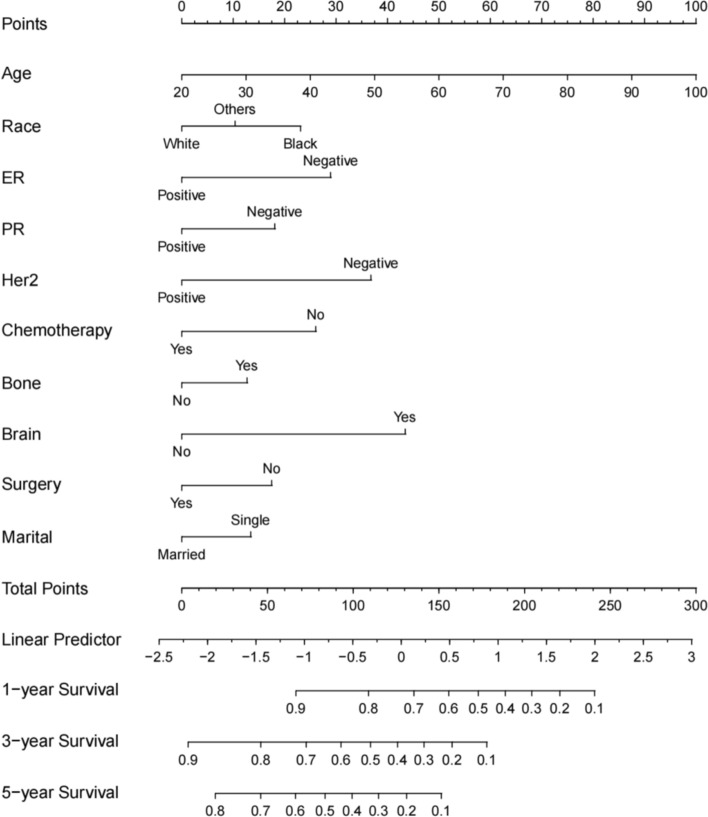
Thirteen statistically significant variables from the aforementioned univariate Cox regression analysis were included in the multivariate analysis. Age, race, marital status, ER, PR, HER2, surgery, brain metastasis, bone metastasis and chemotherapy were proven to be independent risk factors for T1-2N0-1 BCLM and these variables were ultimately used to construct the nomogram (Fig. 1). Risk scores for each variable were calculated to predict the 1-, 3-, and 5-year OS of patients.
Fig. 1.
A nomogram for T1-2N0-1 BCLM patients
Validation of the OS nomogram
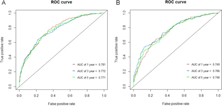
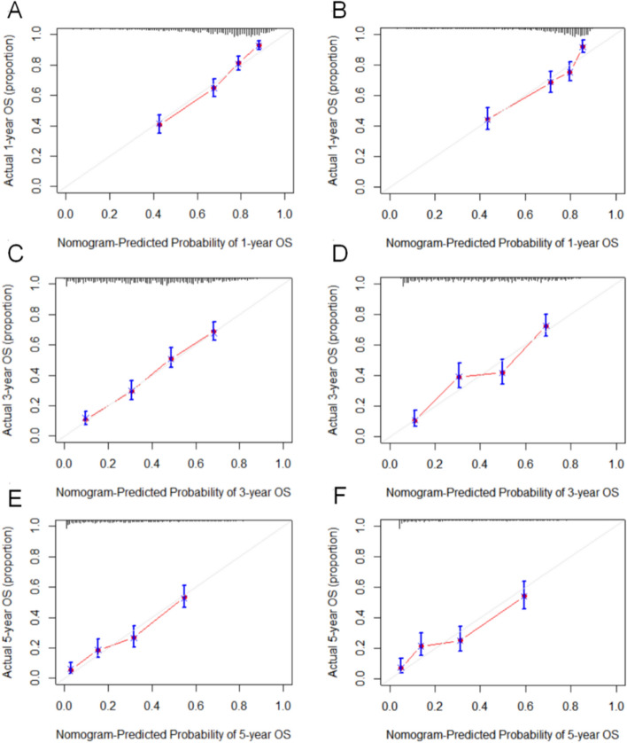
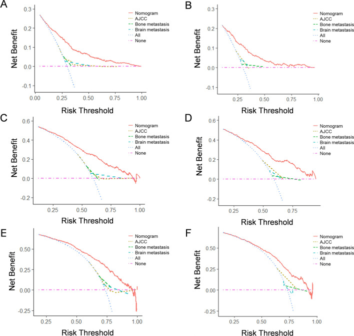
The c-index was 0.72 (95% CI 0.70–0.74) in the training cohort and 0.72 (95% CI 0.69–0.74) in the validation cohort. For receiver operating characteristic (ROC) curves, the area under the receiver operating characteristic curve (AUC) values at 1-, 3-, and 5-year in the training cohort were 0.781, 0.772 and 0.771. The AUC values at 1-, 3-, and 5-year in the validation cohort were 0.749, 0.766 and 0.748 (Fig. 2). Calibration curves for internal validation of the nomogram showed that the predicted OS was approximated to the actual OS (Fig. 3). Besides, decision curve analysis (DCA) showed that the nomogram was more effective in increasing the net clinical benefit compared to AJCC TNM staging, bone metastasis and brain metastasis variables (Fig. 4).
Fig. 2.
ROC of the nomogram for 1-, 3-, and 5-year prediction. A Training cohort based on the nomogram. B Validation cohort based on the nomogram
Fig. 3.
Calibration plots of 1-, 3-, and 5-year in the training cohort (A, C, E) and the validation cohort (B, D, F)
Fig. 4.
DCA curves of 1-, 3-, and 5-year in the training cohort (A, C, E) and the validation cohort (B, D, F)
Risk stratification system
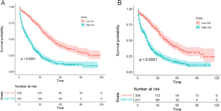
Because these results showed excellent prediction efficiency in survival of the nomogram, we calculated total points based on the predicted score calculated by the nomogram. According to the cutoff value (median points), all the patients were separated into low-risk (total points < 126) and high-risk (total points ≥ 126) groups. The low-risk patients had significantly better OS than the high-risk patients (p < 0.0001) by Kaplan–Meier analyses (Fig. 5) which demonstrated the validity of the risk stratification system.
Fig. 5.
Kaplan curves of low- and high-risk groups in the training cohort A and validation cohort B
Discussion
As breast cancer incidence rates increased continuously, oncologists were faced with the critical task of making accurate prognostic judgments for various breast cancer types [11]. It was important to note that BCLM was a complex and heterogeneous disease, and the different pathological subtypes and treatment modalities employed could have a significant impact on patient survival [12]. According to the AJCC TNM staging system, BCLM were classified as stage IV. T1-2N0-1 BCLM in females was only a minority, and consequently, there have been few studies conducted on the prognosis of such patients.
In the present study, we conducted a retrospective analysis to investigate the demographic, clinicopathological, and prognostic features of female patients with T1-2N0-1 breast cancer who developed liver metastasis, using data from the SEER database. Although there was a significant difference in laterality between the two cohorts, there was no evidence indicating a significant impact of laterality on survival [13]. We utilized the Cox proportional hazards model to identify independent risk factors. As prognostic statistical models, nomograms could directly present the predicted OS. Our analysis revealed that old age, being single, non-white ethnicity, hormone receptor (HR) negativity, HER2 negativity, lack of chemotherapy or surgery, bone metastases, and brain metastases were all significantly associated with a reduced OS.
The incidence of breast cancer was known to increase with advancing age. As the general population continues to age, more attention is being focused on the rising number of older breast cancer patients who require specialized care. Compared to younger women, older age was relevant to early mortality in patients with stage IV [14], and had not been shown to offer a satisfactory improvement in prognosis [15]. Older patients were tended to receive less aggressive treatment and often burdened by comorbid chronic illnesses, which could result in shorter OS. There is mounting evidence that black women experience lower survival rates [16], which may indicate racial heterogeneity in breast cancer [17]. We found being married was a protective factor among the target population for this study and it is consistent with the conclusions of prior studies [18]. The survival advantages of married patients may benefit from the impact that social support [19].
Based on our analysis, we believed that prognosis in T1-2N0-1 BCLM patients was independent of the T stage and N stage. This finding was supported by previous research, which has indicated that only T4-stage breast cancer is included in the risk stratification for BCLM patients. The association between tumor subtype and distant metastasis has been extensively discussed [20]. Tumor subtype represents a significant risk factor for OS in breast cancer patients. Numerous studies have demonstrated that triple-negative breast cancer (TNBC) was often associated with the worst prognosis in breast cancer [21]. TNBC also implied a poor prognosis in our study, with HER2-enriched cases faring slightly better. We could choose endocrine drugs or molecular targeted drugs to improve the prognosis of patients with HR or HER2-positive.
The customary interventions for neoplasms comprise surgery, chemotherapy and radiotherapy. For women with breast cancer who have developed metastatic disease, systemic therapy (ST) is the primary therapeutic approach and chemotherapy has been recommended in the guideline [22]. Our study found that surgery and chemotherapy were both beneficial for OS in patients with T1-2N0-1 BCLM. MF07-01 randomized clinical trial compared the efficacy of locoregional treatment (LRT) followed by systemic therapy with ST alone for stage IV BC patients [23], and the 5- and 10-year follow-up analyses of this clinical trial demonstrated that patients with solitary or multiple liver metastases had benefited from LRT in terms of locoregional progression and OS [24]. A diminutive clinical study conducted in Austria showed that local surgery did not contribute to the betterment of patients with stage IV breast cancer [25] and Khan et al. have recently shown results of the EA2108 trial and come to similar conclusions [26]. Nonetheless, there was no clinical literature that has reported surgical advantages for locally early-stage IV breast cancer. The role of LRT remains controversial. A meta-analysis involving 28,693 participants demonstrated that stage IV patients who underwent operation of breast tumors had better survival [27]. Studies have revealed a higher proportion of patients with small primary tumors in the surgical population [28], and those with negative margins had better OS compared to those with positive margins. Our study subjects were patients with locally early BCLM, and the probability of obtaining negative surgical margins was higher in stage IV patients, which may indicate a better prognosis. For T1-2N0-1 BCLM patients, surgical resection should be one of the treatment options suggested by the oncologist. The radiotherapy showed borderline statistical significance in the univariate analysis. Xiong et al. found radiotherapy was also a treatment that affected the prognosis [29]. Their study included all patients with BCLM, whereas our study only included patients with T1-2N0-1 BCLM. Breast cancer patients with distant metastases are more likely to choose mastectomy over breast-conserving procedures. A meta-analysis indicated that in women with lymph node-negative (N0) breast cancer, post-mastectomy radiotherapy did not impact breast cancer mortality or overall mortality [30]. However, in patients with only 1–3 positive lymph nodes (N1), post-mastectomy radiotherapy reduced breast cancer mortality, potentially leading to the borderline statistical significance observed within this population.
The typical sites of breast cancer metastasis were bones, lungs, liver and brain. The prognosis for brain metastases was widely considered worse than that of metastases from any other site [31]. We have derived some intriguing inferences from the use of a nomogram. Specifically, our nomogram indicated that both bone and brain metastases were related to shorter OS, while no such correlation was observed for lung metastases. Gerratana found lung metastases presented the most favorable prognosis in visceral metastasis followed by bone, liver and brain in breast cancer [32]. Liver metastases may represent a particularly hazardous factor in comparison to lung metastases.
We noted that numerous studies have been conducted on prognostic factors in patients with BCLM. For example, Lei et al. reported that older, non-white ethnicity, unmarried, triple-negative pathological type, uninsured, extrahepatic metastasis, and not receiving chemotherapy or primary site surgery were associated with poor prognosis in BCLM patients which was generally consistent with our findings [33]. However, there has been no research specifically focused on T1-2N0-1 BCLM patients. Our study showed that the nomogram was superior to the AJCC staging system in predicting OS in this particular group, providing a valuable tool for clinical diagnosis and treatment.
Limitations
Certain limitations to our study needed to be acknowledged. First, the SEER database had limited information to include other variables in our study, such as ECOG performance status, Ki-67 expression, liver function parameters, and the number and size of liver metastases. Second, the SEER database was unable to provide more detailed treatment information, especially on the timing and regimen of various treatments such as surgery, chemotherapy, endocrine therapy, and anti-HER2 therapy, to optimize individualized treatment for newly diagnosed BCLM patients. The restricted predictors also weakened the strength of the study. Moreover, the validation of the model was based on the same population, which may introduce bias. Therefore, it was necessary to carefully evaluate the predictive effect of these variables. In addition, the graph was developed using retrospective data, which required further studies with external validation in large and prospective cohorts.
Conclusion
In summary, we have successfully established and validated a nomogram based on the SEER database to predict the 1-year, 3-year, and 5-year survival of T1-2N0-1 BCLM patients. The nomogram presented here was effective in predicting OS, which may provide clinicians with useful guidelines for individualized treatment plans.
Methods
Patient selection and data extraction
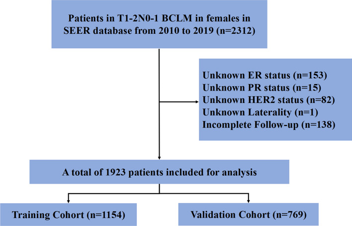
The SEER database, encompassing patients from 18 cancer registries and accounting for roughly 30% of the US population, served as the primary source of patient information for this study, which was obtained using the SEER*Stat software (version 8.4.0; National Cancer Institute, USA). Ethical approval and informed consent were not required for our study. Since the SEER database only collected information about organ metastases data from 2010, the demographic, tumor, treatment, and follow-up information of breast cancer patients from 2010 to 2019 was examined, and the inclusion criteria comprised: (1) female patients with primary T1-2N0-1 BCLM; (2) complete follow-up information. The exclusion criteria were displayed in Fig. 6.
Fig. 6.
The study flowchart of the selection process
Development and validation of the nomogram
Development
We randomly divided the patients into two groups for the development and internal validation of the nomogram, with 60% being assigned to the training cohort and 40% to the validation cohort. Univariate and multivariate Cox proportional hazards models were used to assess the independent risk factors associated with OS by calculating the hazard ratio and 95% CI in the training set. Variables with p < 0.05 in the univariate analysis were selected for the multivariate analysis to estimate the significance of each variable. Variables with statistical significance in the multivariate Cox regression analysis were selected as candidate variables for constructing the nomogram to predict survival. The R package ‘‘survival’’ was applied to calculate risk scores and ‘‘rms’’ was applied to construct the nomogram. The nomogram was then developed using a multivariate Cox regression model, with each variable individually listed and assigned a corresponding number of points based on its magnitude. The cumulative score for all variables is then aligned with a scale to predict OS at 1, 3, and 5 years.
Validation
The c-index and calibration curves were used to evaluate the performance of the nomogram and the AUC was used to test the accuracy in the training and validation cohorts. Additionally, a DCA was performed for both cohorts. We also constructed relevant mortality risk classification systems to distinguish patients with a high or low risk of mortality. Survival curves were plotted using the Kaplan–Meier method.
Statistical analysis
Age and tumor size were presented as median and percentile and other variables were presented as frequency and percentage. Age was compared using Mann–Whitney U test and other variables were compared using Pearson’s chi-square test. Statistical analyses were performed using the RStudio version 4.2.1. All tests of statistical significance were 2-sided, and a P-value < 0.05 was considered statistically significant.
Acknowledgements
The authors acknowledge the public access to the SEER database.
Author contributions
KW and LS contributed equally to this study and shared the first authorship. ZT and YC designed the study. KW extracted and analyzed the data. KW and LS interpreted the evidence and wrote the manuscript. KW and LS and YC revised the article. All authors contributed to the article and approved the submitted version.
Funding
This work was supported by a key research and development project of Zhejiang province (2021C03048), the key project of the Natural Science Foundation of Zhejiang Province (LZ20H160002).
Availability of data and materials
All data in this study are available from the corresponding author on reasonable request.
Declarations
Ethics approval and consent to participate
The data of this study was obtained from the SEER database. The patients’ data was public and anonymous, so this study did not require ethical approval and informed consent.
Competing interests
The authors declare no competing interests.
Footnotes
Publisher's Note
Springer Nature remains neutral with regard to jurisdictional claims in published maps and institutional affiliations.
Contributor Information
Yiding Chen, Email: ydchen@zju.edu.cn.
Zhe Tang, Email: 8xi@zju.edu.cn.
References
- 1.Sung H, Ferlay J, Siegel RL, Laversanne M, Soerjomataram I, Jemal A, et al. Global cancer statistics 2020: GLOBOCAN estimates of incidence and mortality worldwide for 36 cancers in 185 countries. CA Cancer J Clin. 2021;71:209–49. 10.3322/caac.21660 [DOI] [PubMed] [Google Scholar]
- 2.Giordano SH. Breast cancer in men. N Engl J Med. 2018;378:2311–20. 10.1056/NEJMra1707939 [DOI] [PubMed] [Google Scholar]
- 3.Burstein HJ, Curigliano G, Thürlimann B, Weber WP, Poortmans P, Regan MM, et al. Customizing local and systemic therapies for women with early breast cancer: the St Gallen international consensus guidelines for treatment of early breast cancer. Ann Oncol. 2021;2021(32):1216–35. 10.1016/j.annonc.2021.06.023 [DOI] [PMC free article] [PubMed] [Google Scholar]
- 4.Lau KH, Tan AM, Shi Y. New and emerging targeted therapies for advanced breast cancer. Int J Mol Sci. 2022;23:2288. 10.3390/ijms23042288 [DOI] [PMC free article] [PubMed] [Google Scholar]
- 5.Liang Y, Zhang H, Song X, Yang Q. Metastatic heterogeneity of breast cancer: molecular mechanism and potential therapeutic targets. Semin Cancer Biol. 2020;60:14–27. 10.1016/j.semcancer.2019.08.012 [DOI] [PubMed] [Google Scholar]
- 6.Ibragimova MK, Tsyganov MM, Kravtsova EA, Tsydenova IA, Litviakov NV. Organ-specificity of breast cancer metastasis. Int J Mol Sci. 2023;24:15625. 10.3390/ijms242115625 [DOI] [PMC free article] [PubMed] [Google Scholar]
- 7.Merloni F, Palleschi M, Gianni C, Casadei C, Curcio A, Romeo A, et al. Locoregional treatment of de novo stage IV breast cancer in the era of modern oncology. Front Oncol. 2023;13:1083297. 10.3389/fonc.2023.1083297 [DOI] [PMC free article] [PubMed] [Google Scholar]
- 8.National Cancer Institute. AJCC Cancer Staging Manual 8th Edition. Definitions Qeios; 2020. https://www.qeios.com/read/definition/28609
- 9.Sparano JA, Crager MR, Tang G, Gray RJ, Stemmer SM, Shak S. Development and validation of a tool integrating the 21-gene recurrence score and clinical-pathological features to individualize prognosis and prediction of chemotherapy benefit in early breast cancer. J Clin Oncol. 2021;39:557–64. 10.1200/JCO.20.03007 [DOI] [PMC free article] [PubMed] [Google Scholar]
- 10.Hansen J, Auprich M, Ahyai SA, de la Taille A, van Poppel H, Marberger M, et al. Initial prostate biopsy: development and internal validation of a biopsy-specific nomogram based on the prostate cancer antigen 3 assay. Eur Urol. 2013;63:201–9. 10.1016/j.eururo.2012.07.030 [DOI] [PubMed] [Google Scholar]
- 11.Harbeck N, Penault-Llorca F, Cortes J, Gnant M, Houssami N, Poortmans P, et al. Breast cancer. Nat Rev Dis Primer. 2019;5:66. 10.1038/s41572-019-0111-2 [DOI] [PubMed] [Google Scholar]
- 12.Tao Z, Shi A, Lu C, Song T, Zhang Z, Zhao J. Breast cancer: epidemiology and etiology. Cell Biochem Biophys. 2015;72:333–8. 10.1007/s12013-014-0459-6 [DOI] [PubMed] [Google Scholar]
- 13.Kim BK, Choi JE, Youn HJ, Park HS, Kim D, Oh SJ, et al. Clinicopathological features and prognosis associated with breast cancer laterality: a nationwide study from the Korean Breast Cancer Society. Ann Surg Treat Res. 2022;103:119–28. 10.4174/astr.2022.103.3.119 [DOI] [PMC free article] [PubMed] [Google Scholar]
- 14.Schonberg MA, Marcantonio ER, Ngo L, Li D, Silliman RA, McCarthy EP. Causes of death and relative survival of older women after a breast cancer diagnosis. J Clin Oncol. 2011;29:1570–7. 10.1200/JCO.2010.33.0472 [DOI] [PMC free article] [PubMed] [Google Scholar]
- 15.Smith BD, Jiang J, McLaughlin SS, Hurria A, Smith GL, Giordano SH, et al. Improvement in breast cancer outcomes over time: are older women missing out? J Clin Oncol. 2011;29:4647–53. 10.1200/JCO.2011.35.8408 [DOI] [PubMed] [Google Scholar]
- 16.Walsh SM, Zabor EC, Stempel M, Morrow M, Gemignani ML. Does race predict survival for women with invasive breast cancer? Cancer. 2019;125:3139–46. 10.1002/cncr.32296 [DOI] [PMC free article] [PubMed] [Google Scholar]
- 17.Gehlert S, Hudson D, Sacks T. A critical theoretical approach to cancer disparities: breast cancer and the social determinants of health. Front Public Health. 2021;9: 674736. 10.3389/fpubh.2021.674736 [DOI] [PMC free article] [PubMed] [Google Scholar]
- 18.Aizer AA, Chen M-H, McCarthy EP, Mendu ML, Koo S, Wilhite TJ, et al. Marital status and survival in patients with cancer. J Clin Oncol Off J Am Soc Clin Oncol. 2013;31:3869–76. 10.1200/JCO.2013.49.6489 [DOI] [PMC free article] [PubMed] [Google Scholar]
- 19.Zhai Z, Zhang F, Zheng Y, Zhou L, Tian T, Lin S, et al. Effects of marital status on breast cancer survival by age, race, and hormone receptor status: a population-based study. Cancer Med. 2019;8:4906–17. 10.1002/cam4.2352 [DOI] [PMC free article] [PubMed] [Google Scholar]
- 20.Kennecke H, Yerushalmi R, Woods R, Cheang MCU, Voduc D, Speers CH, et al. Metastatic behavior of breast cancer subtypes. J Clin Oncol Off J Am Soc Clin Oncol. 2010;28:3271–7. 10.1200/JCO.2009.25.9820 [DOI] [PubMed] [Google Scholar]
- 21.Duan XF, Dong NN, Zhang T, Li Q. The prognostic analysis of clinical breast cancer subtypes among patients with liver metastases from breast cancer. Int J Clin Oncol. 2013;18:26–32. 10.1007/s10147-011-0336-x [DOI] [PubMed] [Google Scholar]
- 22.Cardoso F, Harbeck N, Fallowfield L, Kyriakides S, Senkus E. Locally recurrent or metastatic breast cancer: ESMO clinical practice guidelines for diagnosis, treatment and follow-up. Ann Oncol. 2012;23:11–9. 10.1093/annonc/mds232 [DOI] [PubMed] [Google Scholar]
- 23.Soran A, Ozmen V, Ozbas S, Karanlik H, Muslumanoglu M, Igci A, et al. Randomized trial comparing resection of primary tumor with no surgery in stage IV breast cancer at presentation: protocol MF07-01. Ann Surg Oncol. 2018;25:3141–9. 10.1245/s10434-018-6494-6 [DOI] [PubMed] [Google Scholar]
- 24.Soran A, Ozmen V, Ozbas S, Karanlik H, Muslumanoglu M, Igci A, et al. Primary surgery with systemic therapy in patients with de novo stage IV breast cancer: 10-year follow-up; protocol MF07-01 randomized clinical trial. J Am Coll Surg. 2021;233:742. 10.1016/j.jamcollsurg.2021.08.686 [DOI] [PubMed] [Google Scholar]
- 25.Fitzal F, Bjelic-Radisic V, Knauer M, Steger G, Hubalek M, Balic M, et al. Impact of breast surgery in primary metastasized breast cancer: outcomes of the prospective randomized phase III ABCSG-28 POSYTIVE trial. Ann Surg. 2019;269:1163. 10.1097/SLA.0000000000002771 [DOI] [PubMed] [Google Scholar]
- 26.Khan SA, Zhao F, Goldstein LJ, Cella D, Basik M, Golshan M, et al. Early local therapy for the primary site in De Novo stage IV breast cancer: results of a randomized clinical trial (EA2108). J Clin Oncol Off J Am Soc Clin Oncol. 2022;40:978–87. 10.1200/JCO.21.02006 [DOI] [PMC free article] [PubMed] [Google Scholar]
- 27.Harris E, Barry M. Meta-analysis to determine if surgical resection of the primary tumour in the setting of stage IV breast cancer impacts on survival. Ann Surg Oncol. 2013;20:2828–34. 10.1245/s10434-013-2998-2 [DOI] [PubMed] [Google Scholar]
- 28.Rashaan ZM, Bastiaannet E, Portielje JEA, van de Water W, van der Velde S, Ernst MF, et al. 2012. Surgery in metastatic breast cancer Patients with a favorable profile seem to have the most benefit from surgery. Eur J Surg Oncol. 38 56 [DOI] [PubMed]
- 29.Xiong Y, Shi X, Hu Q, Wu X, Long E, Bian Y. A nomogram for predicting survival in patients with breast cancer liver metastasis: a population-based study. Front Oncol. 2021;11: 600768. 10.3389/fonc.2021.600768 [DOI] [PMC free article] [PubMed] [Google Scholar]
- 30.EBCTCG (Early Breast Cancer Trialists’ Collaborative Group), McGale P, Taylor C, Correa C, Cutter D, Duane F, et al. Effect of radiotherapy after mastectomy and axillary surgery on 10-year recurrence and 20-year breast cancer mortality: meta-analysis of individual patient data for 8135 women in 22 randomised trials. Lancet Lond Engl. 2014;383:2127–35. 10.1016/S0140-6736(14)60488-8 [DOI] [PMC free article] [PubMed] [Google Scholar]
- 31.Regierer AC, Wolters R, Ufen M-P, Weigel A, Novopashenny I, Köhne CH, et al. An internally and externally validated prognostic score for metastatic breast cancer: analysis of 2269 patients. Ann Oncol. 2014;25:633–8. 10.1093/annonc/mdt539 [DOI] [PMC free article] [PubMed] [Google Scholar]
- 32.Gerratana L, Fanotto V, Bonotto M, Bolzonello S, Minisini AM, Fasola G, et al. Pattern of metastasis and outcome in patients with breast cancer. Clin Exp Metastasis. 2015;32:125–33. 10.1007/s10585-015-9697-2 [DOI] [PubMed] [Google Scholar]
- 33.Ji L, Cheng L, Zhu X, Gao Y, Fan L, Wang Z. Risk and prognostic factors of breast cancer with liver metastases. BMC Cancer. 2021;21:238. 10.1186/s12885-021-07968-5 [DOI] [PMC free article] [PubMed] [Google Scholar]
Associated Data
This section collects any data citations, data availability statements, or supplementary materials included in this article.
Data Availability Statement
All data in this study are available from the corresponding author on reasonable request.


