Abstract
Background
Thyroid storm (TS), a life-threatening condition that can damage multiple organs, has limited therapeutic options. Hypercytokinemia is a suggested background, but the pathological condition is unclear and there are no appropriate animal models. We aimed to develop a TS mouse model by administration of triiodothyronine and lipopolysaccharide, and then to examine the effects of ghrelin on this model.
Methods
We evaluated the use of serum IL-6 levels as a representative marker of hypercytokinemia in patients with TS. To establish the mouse model, preliminary experiments were conducted to determine the non-lethal doses of triiodothyronine and lipopolysaccharide when administered individually. As a TS model, C57BL/6 mice were administered with triiodothyronine 1.0 mg/kg (subcutaneously, once daily for seven consecutive days) and lipopolysaccharide 0.5 mg/kg (intraperitoneally, on day 7) to develop a lethal model with approximately 30% survival on day 8. We assessed the survival ratio, mouse sepsis scores and blood biomarkers (IL-6, metanephrine, alanine aminotransferase) and evaluated the effects of ghrelin 300 µg/kg on these parameters in TS model.
Results
Serum IL-6 was increased in patients with TS compared with those with Graves’ disease as the diseased control (18.2 vs. 2.85 pg/mL, P < .05, n = 4 each). The dosage for the murine TS model was triiodothyronine 1.0 mg/kg and lipopolysaccharide 0.5 mg/kg. The TS model group had increased mouse sepsis score, serum IL-6, metanephrine and alanine aminotransferase. In this model, the ghrelin improved the survival rate to 66.7% (P < .01, vs. 0% [saline-treated group]) as well as the mouse sepsis score, and it decreased the serum IL-6 and metanephrine.
Conclusion
We established an animal model of TS that exhibits pathophysiological states similar to human TS with induction of serum IL-6 and other biomarkers by administration of T3 and LPS. The results suggest the potential effectiveness of ghrelin for TS in humans.
Supplementary Information
The online version contains supplementary material available at 10.1186/s12902-024-01680-8.
Keywords: Cytokine, IL-6, Metanephrine, Animal model, Thyrotoxicosis, Graves’ disease
Background
Thyroid storm (TS) is a potentially fatal medical emergency which can affect multiple organs. It is caused by a combination of thyrotoxicosis and severe physical stresses. Graves’ disease is the most common underlying thyroid disease, and infection is the most common triggering factor [1]. Excessive thyroid hormone action can result in liver damage, tachycardia and heart failure due to increased sympathetic activity, and the fatality rate of patients with TS is as high as approximately 10%. Hypercytokinemia has been postulated as a pathogenesis of TS [2, 3]. The key cytokine implicated in hypercytokinemia is IL-6 [4]. The elevated serum IL-6 in patients with thyrotoxicosis has been previously reported [5, 6].
Administration of lipopolysaccharide (LPS) is often used as a sepsis model because it elevates endotoxin, and there have been reports of elevations in various inflammatory cytokines, including IL-6 [7–10]. This model may prove valuable for simulating infection, one of the most prevalent triggers for TS.
Ghrelin has been shown to exert various physiological effects including anti-inflammatory effects, sympathomimetic inhibition effects, and growth hormone secretion-promoting effects [11]. The anti-inflammatory effect of ghrelin has already been observed in a sepsis model [12]. Based on these findings, we hypothesized that the anti-inflammatory properties of ghrelin might be able to effectively ameliorate TS.
In the present study, we initially compared serum IL-6 levels between patients with TS and those with Graves’ disease without TS. Subsequently, we established a lethal model simulating TS by inducing “thyrotoxicosis” by exogenous administration of thyroid hormones, and by inducing “sepsis” by intraperitoneal administration of LPS. Finally, we used this mouse model to test the efficacy of ghrelin on TS.
Materials and methods
Subjects for evaluating serum IL-6 levels
Peripheral blood samples were collected from four untreated patients with TS who visited the Wakayama Medical University Hospital between May 2018 and January 2020, and we measured serum IL-6 (SRL, Tokyo, Japan). Diagnosis of TS was made according to the diagnostic criteria of the Japan Thyroid Association [1]. Serum IL-6 was similarly measured in four patients with untreated Graves’ disease without TS and this was compared with that in the TS group. Additionally, clinical data including TSH, free T3 (FT3), free T4 (FT4), aminotransferase (ALT), and C-reactive protein (CRP) were collected and subjected to comparative analysis of the two groups.
We obtained ethical approval for use of an opt-out consent approach for this study, and none of the subjects declined participation. The study protocol was approved by the Wakayama Medical University Institutional Ethical Review Board (No.3714) and was conducted in accordance with the principles of the Declaration of Helsinki.
Mice
Seven-week-old male C57BL/6 mice were procured from Japan SLC (Hamamatsu, Japan). They were housed in a room with controlled temperature, humidity and light intensity (12 h light/12 hours dark cycle, lights on at 08:00) and had free access to water and standard mouse food. After a one-week acclimation period, these mice were used in the experiments. Mice were euthanised by cervical dislocation under isoflurane inhalation anaesthesia and blood samples were taken from the inferior vena cava, as well as liver and heart samples. Animal care and all experimental procedures were performed in accordance with AVMA Guidelines for the Euthanasia of Animals 2020 [13] and the Wakayama Medical University Guidelines for Animal Experiments, with the approval of the Institutional Animal Care and Use Committee (No. 819).
Murine sepsis score
The murine sepsis score (MSS) was used to assess the severity of sepsis and humane endpoints. MSS is calculated by consideration of items including appearance, level of consciousness, activity, response to stimulus, eyes, respiration rate, and respiration quality, and each of these variables is given a score between 0 and 4. Mice with an MSS score of ≥ 21 reportedly have a 100% mortality rate within 24 h [14], so we euthanized mice with an MSS score of ≥ 21.
Creation of a thyrotoxicosis mouse model
Triiodothyronine (T3; Sigma Aldrich, Missouri, USA) was subcutaneously injected into 8-week-old male C57BL/6 mice for consecutive 7 days, and blood samples were collected on day 8 to measure serum FT3. The control group received a vehicle (0.02 N NaOH). The T3 dose was set at 1.0 mg/kg or 3.0 mg/kg with reference to a previous report that used a dose of 1.0 mg/kg [15], aiming to achieve a blood concentration 2–5 times higher than that observed in the control group for the serum FT3 levels.
Creation of a TS mouse model
LPS (serotype O55:B5; Sigma Aldrich, St. Louis, MO) was administered intraperitoneally to the thyrotoxicosis mouse model on day 7. The control group received a vehicle (saline). The LPS dose was determined to be 0.2 mg/kg or 0.5 mg/kg with reference to previous reports that used a dose of 0, 0.1, 1.0, or 10 mg/kg [10], aiming to achieve a survival rate of approximately 30% at 24 h after administration. Additionally, MSS was assessed in surviving subjects at 4-hour intervals up to 24 h following administration of LPS. Blood samples were collected 4 h after administration of LPS in the mouse model of TS and they were analyzed for metanephrine to assess sympathetic nerve activity [16], ALT to assess liver damage, and IL-6 to evaluate hypercytokinemia. TS model mice were sacrificed by cervical dislocation, and the heart and liver were taken with scissors and fixed in formalin neutral buffer solution and embedded in paraffin. Sections were stained with hematoxylin and eosin (HE). Each of these processes was carried out as an independent experiment.
Protocol for evaluating the Effect of ghrelin in a TS mouse model
Ghrelin (PEPTIDE INSTITUTE, INC., Osaka, Japan) was subcutaneously injected into the TS model mice on day 7 immediately after LPS injection. The dose was determined at 300 µg/kg with reference to a previous report [10]. The control group received a vehicle (saline). We evaluated the effect of ghrelin on MSS, the survival rate, and serum biomarkers.
Biochemical measurement
FT3 levels were measured by enzyme immunoassay method (TOSOH, Tokyo, Japan). Plasma metanephrine (ImmuSmol, Bordeaux, France, Catalog #BA E-8100, RRID: AB_2940855) and serum IL-6 (Enzo Life Science, Farmingdale, NY, Catalog #ADI-900-045, RRID: AB_2940854) were measured using specific ELISA kits according to the manufacturer’s manual. Serum ALT was measured using the EnzyChrom™ Alanine Transaminase Assay Kit (BioAssay Systems, Hayward, CA).
Statistical analysis
Two groups were compared with Student’s t tests or Mann-Whitney U tests. Three or more groups were compared with one-way analysis of variance (ANOVA) and Bonferroni post hoc tests. Log-rank tests were used to compare survival. Statistical analyses were performed using JMP software, version 14 (SAS Institute Inc, Cary, NC). P < .05 was considered to be statistically significant.
Results
Serum IL-6 levels in patients with TS
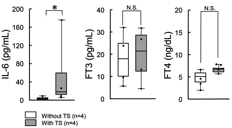
The triggers for TS were infection in three cases and intestinal necrosis in one case (Table 1). Among patients with TS, C-reactive protein was clearly elevated into two cases, and only slightly elevated in the other two. The medians and interquartile ranges of IL-6 levels in the four untreated patients with Graves’ disease without TS were 2.85 [1.98, 4.00] pg/mL, similar to previously reported levels (Fig. 1) [6]. In contrast, IL-6 levels were significantly elevated in the patients with TS, 18.2 [13.0, 60.1] pg/mL (P < .05), although their thyroid hormone levels were comparable with those of the four patients with Graves’ disease without TS (Table 1; Fig. 1).
Table 1.
Clinical characteristics of patients with Graves’ disease with/without thyroid storm
| Gender | Age (y) |
JTA criteria | BWPS | IL-6 (pg/mL) | TSH (µIU/mL) | FT3 (pg/mL) | FT4 (ng/dL) | ALT (IU/L) | CRP (mg/dL) | Triggers | Outcome | |
|---|---|---|---|---|---|---|---|---|---|---|---|---|
|
With Thyroid Storm |
female | 80 | Definite | 60 | 175.0 | < 0.005 | 15.27 | 7.14 | 98 | 10.02 | Infection | Survival |
| female | 45 | Definite | 45 | 13.5 | < 0.005 | 31.37 | > 7.77 | 20 | 14.43 | Infection | Survival | |
| female | 55 | Definite | 45 | 11.4 | < 0.005 | 27.98 | > 7.77 | 22 | 0.19 | Infection | Survival | |
| female | 75 | Definite | 85 | 22.8 | < 0.005 | 6.37 | 6.69 | 20 | 0.65 | Intestinal Necrosis | Survival | |
|
Without Thyroid Storm |
female | 62 | ― | ― | 5.2 | < 0.005 | 11.46 | 4.39 | 35 | 0.06 | ― | ― |
| female | 33 | ― | ― | 1.6 | < 0.005 | 8.25 | 3.21 | 23 | 0.04 | ― | ― | |
| female | 39 | ― | ― | 3.6 | < 0.005 | > 32.55 | > 7.77 | 40 | 0.04 | ― | ― | |
| female | 41 | ― | ― | 2.1 | < 0.005 | 24.62 | 5.78 | 49 | < 0.02 | ― | ― | |
| P value | N/A | 0.11 | N/A | N/A | < 0.05 | N/A | 0.50 | 0.07 | 0.44 | 0.17 | N/A | N/A |
Fig. 1.
Elevated Serum IL-6 Levels in Patients with Thyroid Storm Compared to Untreated Graves’ Disease. Serum IL-6 levels were measured in four patients with TS and compared with those of four patients with untreated Graves’ disease. The patients with Graves’ disease had IL-6 level of 2.85 [1.98, 4.00] pg/mL, whereas patients with TS exhibited a significantly elevated IL-6 level, 18.2 [13.0, 60.1] pg/mL. There were no significant differences in FT3 and FT4 levels. Data are presented as median [interquartile range (IQR)]. *P < .05. TS: thyroid storm. IL-6: interleukin-6. FT3: free T3. FT4: free T4
Creation of a thyrotoxicosis mouse model
In the T3 1.0 mg/kg group (n = 8), serum FT3 levels were approximately three times higher (15.3 ± 4.5 vs. 4.5 ± 0.7 pg/ml, P < .05) than those in the control group (n = 8). In the T3 3.0 mg/kg group (n = 5), they were approximately five times higher (23.0 ± 5.1 vs. 4.5 ± 0.7 pg/ml, P < .05) among survivors. Two out of five mice in the 3.0 mg/kg group died, while none of the eight mice in the 1.0 mg/kg group died. Based on these findings, we determined that the appropriate T3 dose for the TS mouse model is 1.0 mg/kg, which is not lethal when administered alone.
Creation of a TS mouse model
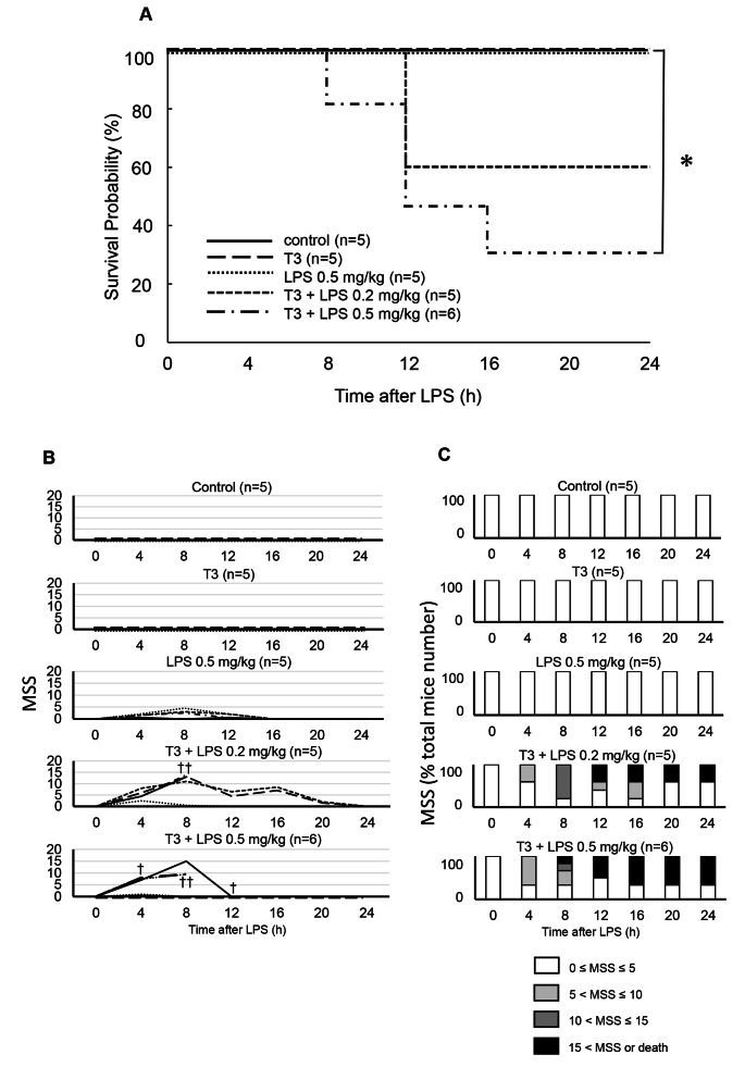
Survival was 100% in the control group (n = 5), T3 1.0 mg/kg alone group (n = 5), and LPS 0.5 mg/kg alone group (n = 5) (Fig. 2A). In contrast, survival at 24 h post-dose was 60% in the T3 + LPS 0.2 mg/kg group (n = 5) and 33% in the T3 + LPS 0.5 mg/kg group (n = 6) (Fig. 2A). After 24 h of administration, MSS was not increased in the control group or in the T3 alone group; it showed only a mild increase of < 5 points in the LPS 0.5 mg/kg alone group (Fig. 2B and C). However, in the T3 + LPS 0.2 mg/kg and T3 + LPS 0.5 mg/kg groups, mice showed an increase of ≥ 15 points in MSS (Fig. 2B and C). The parameter with the largest increase in these groups was the level of consciousness. Based on these results, we further analyzed the T3 + LPS 0.5 mg/kg group as a TS mouse model. There were no obvious changes in heart and liver pathology findings between the T3 alone group and the TS model group (Supplementary Fig. 1).
Fig. 2.
Survival and mouse sepsis score in thyrotoxicosis models. LPS 0.2 mg/kg or 0.5 mg/kg was administered intraperitoneally to mice models of thyrotoxicosis on day 7, and survival and MSS were assessed up to 24 h after administration. Survival was 100% in the control (n = 5), T3 alone (n = 5) and LPS 0.5 mg/kg alone (n = 5), but 60% in the T3 + LPS 0.2 mg/kg group (n = 5) and 33% in the T3 + LPS 0.5 mg/kg group (n = 6) (A). * P = .05. Twenty-four hours after administration, MSS did not increase in the control group or in the T3 alone group, but a mild increase of < 5 points was observed in the LPS 0.5 mg/kg alone group. Additionally, several mice in the T3 + LPS 0.2 mg/kg and T3 + LPS 0.5 mg/kg groups showed an increase of > 15 points in their MSS (B, C). Three mice in the T3 + LPS 0.2 mg/kg group and four mice in the T3 + LPS 0.5 mg/kg group died at 4–12 h (shown as †). B, C are cumulative data as time progresses. MSS: mouse sepsis score. LPS: lipopolysaccharide. T3: triiodothyronine
Measurement of blood biomarkers in a TS mouse model
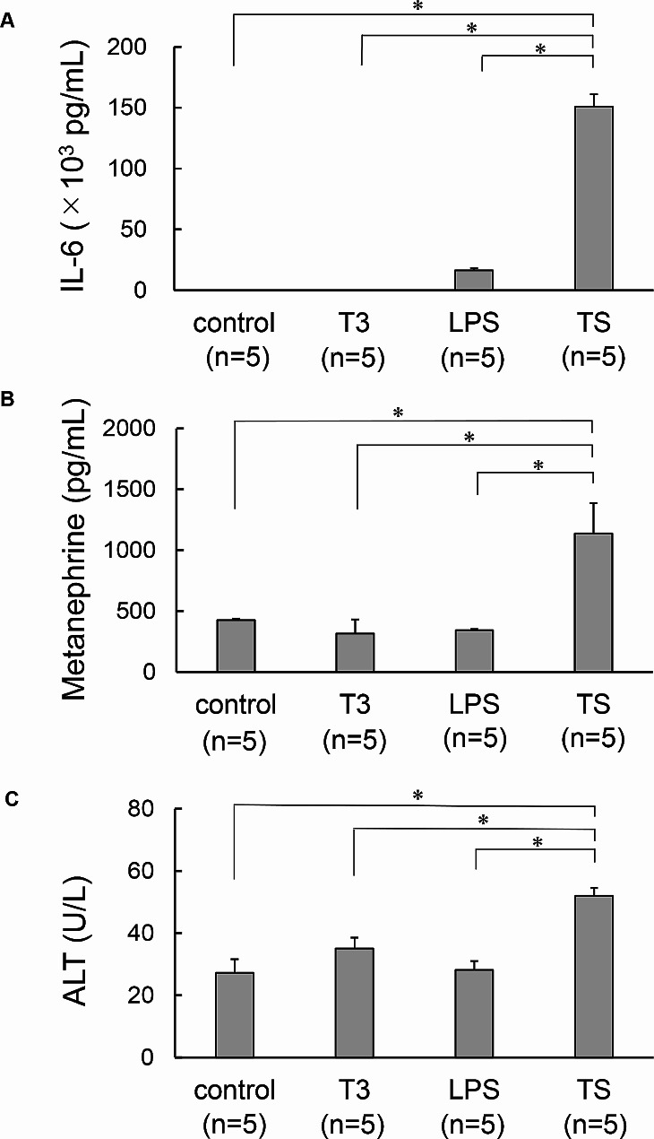
In the TS model group there were significant increases compared with the T3 alone and LPS alone groups in serum IL-6 (control group, T3 alone group, LPS alone group, TS model group; 47.4 ± 3.8, 47.7 ± 7.5, 16415 ± 1800, 150846 ± 10204 pg/ml, respectively [Fig. 3A]), metanephrine (425 ± 11.5, 315 ± 116, 343 ± 9.6, 1137 ± 249 pg/ml, respectively [Fig. 3B]) and ALT (27 ± 4.4, 35 ± 3.5, 28 ± 2.9, 52 ± 2.6 U/L, respectively [Fig. 3C]).
Fig. 3.
Elevated serum IL-6, metanephrine, and ALT levels in thyroid storm mouse model. Blood samples were taken 4 h after administration of LPS in the TS model mice. IL-6, metanephrine, and ALT were measured. Compared with the T3 alone and LPS alone groups, the TS model group had significant increases in serum IL-6 (*P < .05, A), metanephrine (*P < .05, B), and ALT (*P < .05, C). Data are shown as mean ± SE. TS: thyroid storm. LPS: lipopolysaccharide. T3: triiodothyronine. IL-6: interleukin-6. ALT: alanine aminotransferase
Effect of ghrelin in a TS mouse model
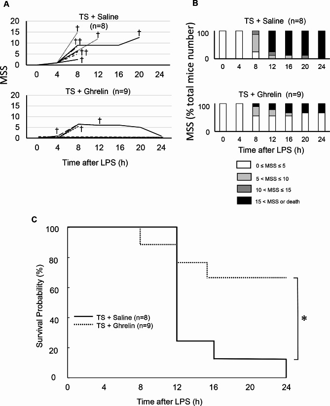
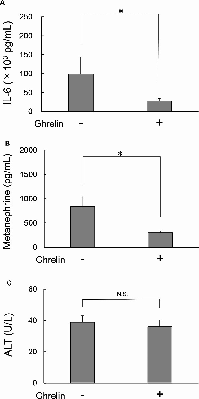
Compared with the saline-treated TS model group (n = 8), the ghrelin-treated group (n = 9) showed significantly lower MSS (P < .05, Fig. 4A and B) and improved survival to 66.7% (vs. 0% [vehicle group], P < .01) (Fig. 4C). Regarding blood biomarkers, serum IL-6 (TS model group vs. ghrelin-treated group: 99208 ± 45266 vs. 27923 ± 6842 pg/ml, P < .05) and metanephrines (838 ± 220 vs. 297 ± 39.8 pg/ml, P < .05) were significantly lower in the ghrelin-treated group. ALT was not significantly different (39 ± 4.1 vs. 36 ± 4.4 U/L, P = .66) between the groups (Fig. 5A, B and C).
Fig. 4.
Effect of ghrelin on survival and sepsis scores in thyroid storm mouse model. Ghrelin (300 µg/kg) was injected subcutaneously on day 7 in the TS model mice, and we analyzed the changes in MSS and survival. Compared with the saline-treated TS model group (n = 8), the ghrelin group (n = 9) showed a significant reduction in MSS (P < .05, A, B) and improved survival to 66.7% (vs. 0% [saline-treated], * P < .05, C). Eight mice in the TS model group and three mice in the saline-treated TS model group died at 4–20 h (shown as †). A and B are cumulative data as time progresses. TS: thyroid storm. MSS : mouse sepsis score. LPS : lipopolysaccharide
Fig. 5.
Effect of ghrelin on blood biomarkers in thyroid storm mouse model. Ghrelin (300 µg/kg, n = 10) or saline (n = 10) was injected subcutaneously on day 7 in a mouse model of TS and we analyzed the changes in blood biomarkers. Blood samples were taken 4 h after administration of LPS. In the ghrelin-treated group, serum IL-6 and metanephrines were significantly reduced, while ALT showed no significant difference (A, B, and C). Data are shown as mean ± SE. *P < .05. TS: thyroid storm. LPS: lipopolysaccharide. IL-6: interleukin-6. ALT: alanine aminotransferase
Discussion
Hypercytokinemia has been postulated as a pathogenesis of TS, but there are no previous reports in which there was measurement of cytokines in actual patients. The present study confirms that serum IL-6 is markedly elevated in patients with TS compared with in patients with Graves’ disease.
To elucidate the pathogenesis of TS with development of therapeutic agents, the establishment of animal models has been anticipated. It is difficult to show stable thyrotoxicosis in animal models of Graves’ disease mediated by anti-thyrotropin (TSH) receptor antibodies. However, the development of TS does not require the involvement of immunological mechanisms; thyrotoxicosis induced by administration of exogenous thyroid hormone can be the underlying condition for TS. Nationwide surveys of TS in Japan also included cases of TS in which the underlying thyroid disease was not Graves’ disease, but actually destructive thyroiditis [1].
In the animal experiments, we successfully induced pathophysiological changes similar to those observed in human TS by externally administering thyroid hormone and LPS to mice. To reproduce the pathogenesis of TS, in which thyrotoxicosis is combined with infection, we administered T3 on a daily basis beforehand (once per day for seven consecutive days), followed by concurrent LPS administration (once, on day 7). This approach not only significantly increased IL-6, metanephrines and ALT in blood biomarkers, it also increased MSS and mortality. In our animal experiments, plasma metanephrine levels did not increase in the T3-only treatment group, similar to previous reports on thyrotoxicosis [17]. While plasma metanephrine were indeed elevated in the TS model, there are no existing reports measuring metanephrine in TS patients. TS may induce liver damage due to circulatory and/or metabolic disturbances in the liver [18]. Additionally, beta-blockers have been shown to be effective against cardiovascular symptoms of Graves’ disease and TS, so sympathomimetic effects could be involved in these conditions [20]. No obvious changes in heart and liver pathology were observed, possibly because of the short time of 4 h after LPS administration. The elevated levels of metanephrines and ALT observed in our model were thought to resemble the pathologies seen in human TS. The marked elevation of IL-6 in our model is also consistent with the aforementioned results observed in patients with TS, which could support the reproduction of hypercytokinemia. Our model is therefore considered to highly resemble human TS, characterized by liver injury, heightened sympathetic activation, and hypercytokinemia.
In this model, neither T3 nor LPS alone caused an increase in MSS or mortality, but their combination resulted in a significant increase. The synergistic effect of thyroid hormone excess and infection was consistent with the condition observed in human TS. In patients with thyrotoxicosis, macrophages, osteoblasts, and other cells are known to release IL-6 in response to T3 in patients with thyrotoxicosis [5, 6]. The release of IL-6 is therefore assumed to be more enhanced when thyrotoxicosis is complicated by infection. Furthermore, IL-6 itself has been recognized as an exacerbating factor in sepsis [21]. Hypercytokinemia can therefore cause multi-organ failure and death in TS.
The TS model in the present study was used to investigate whether ghrelin is useful in the treatment of TS. Ghrelin treatment was shown to significantly reduce IL-6 and metanephrine levels and to improve survival and MSS. The anti-inflammatory and sympathetic nerve activity suppressing effects of ghrelin are suggested by these findings to be highly beneficial for the treatment of TS [11]. ALT did not show significant changes within the initial 4 h after ghrelin administration, but this lack of significant change can be attributed to the long half-life of approximately 25 h [22] .
Ghrelin is known to bind to GHS-R1a expressed on monocytes and macrophages, directly inhibiting the production of IL-6 and IL-1β [23]. In addition, ghrelin binding to GHS-R1a expressed on the nerve endings of the vagal afferent tract activates the vagus nerve and indirectly suppresses NF-κB signaling in macrophages [24]. Ghrelin has been demonstrated to have positive effects on survival, the inhibition of inflammatory cytokine production, and the reduction of acute kidney and lung injuries in sepsis models [12, 25]. Clinical trials of ghrelin administration in humans have reported anti-inflammatory effects and improved exercise tolerance and food intake in patients with chronic respiratory failure [26]. Ghrelin has also been shown to reduce blood noradrenaline levels, to maintain left ventricular contractility, and to improve muscle strength and exercise tolerance in patients with chronic heart failure [27]. Moreover, the ghrelin receptor agonist anamorelin has been recently approved for use in Japan to manage cancer-related cachexia [28]. As clinical research progresses, indications for administration of ghrelin are expected to expand to include sepsis, chronic respiratory failure, and chronic heart failure. Our study suggests that in the future, ghrelin could be clinically applicable in the treatment of TS.
This study has some limitations. Firstly, due to the rarity of TS, it was challenging to collect a large sample of patients, and this resulted in a small sample of patients in this study that could be evaluated for serum IL-6 levels. However, the difference in serum IL-6 levels between patients with and without TS was statistically significant. Secondly, TS can be triggered by a variety of factors. Further investigation is required to determine whether our animal model is also applicable to TS caused by other triggers. Thirdly, although the dosage and timing of ghrelin administration used in the present study were based on the results and findings of previous studies [10], further studies might be required to find the optimal dosage and timing. Finally, there has been no elucidation of molecular mechanisms such as the interaction between ghrelin and cytokine signaling. Further challenging research is therefore necessary to clarify these questions.
Conclusions
We successfully created an animal model of TS by administering a combination of thyroid hormones and LPS to mice. There were pathophysiological changes similar to those in human TS. This model proved to be appropriate for studying TS and it is thought that it will significantly contribute to the understanding of its pathogenesis. The administration of ghrelin in this model suggests its potential effectiveness in treating TS and improving prognosis.
Electronic supplementary material
Below is the link to the electronic supplementary material.
Supplementary Material 1: Fig. 1. Pathological analysis of heart and liver tissues in thyroid storm mouse model. Heart and liver tissues were taken from the T3 alone group and the TS model group. There were no obvious changes in pathology before and after LPS administration. Scale bars in left panels and right panels represent 250 μm and 50 μm, respectively. T3: triiodothyronine. TS: thyroid storm. LPS: lipopolysaccharide.
Acknowledgements
We thank members of the First Department of Medicine, and the Animal Research Facility at Wakayama Medical University for cooperating in the research. We acknowledge proofreading and editing by Benjamin Phillis at the Clinical Study Support Center at Wakayama Medical University.
Author contributions
C.K., Y.F. and T.A. conceived and planned the experiments, and analyzed the data. C.K., Y.F. and A.D. carried out the experiments. T.A. and T.M. supervised and administered the project. C.K. wrote the article with support from K.T., S.M., H.I., H.A., H.F., M.N. and T.M. All authors discussed the results and commented on the article.
Funding
This study was supported by funds from the Ministry of Education, Science, Culture, Sports and Technology of Japan, and the Ministry of Health, Labor and Welfare of Japan.
Data availability
All data supporting the findings of this study are available within the paper and its Supplementary Information.
Declarations
Ethics approval and consent to participate
We obtained ethical approval for use of an opt-out method for this study, and none of the subjects declined participation. Therefore, informed consent was not required. This study protocol was approved by the Wakayama Medical University Institutional Ethical Review Board (No.3714). Animal care and all experimental procedures were performed in accordance with the Wakayama Medical University Guidelines for Animal Experiments, with the approval of the Institutional Animal Care and Use Committee (No. 819).
Consent for publication
Not applicable.
Competing interests
The authors declare no competing interests.
Footnotes
Publisher’s Note
Springer Nature remains neutral with regard to jurisdictional claims in published maps and institutional affiliations.
References
- 1.Akamizu T, Satoh T, Isozaki O, et al. Diagnostic criteria, clinical features, and incidence of thyroid storm based on nationwide surveys. Thyroid. 2012;22:661–79. 10.1089/thy.2011.0334. 10.1089/thy.2011.0334 [DOI] [PMC free article] [PubMed] [Google Scholar]
- 2.Pokhrel B, Aiman W, Bhusal K, Thyroid S. StatPearls [Internet];2022 Oct 6. https://www.ncbi.nlm.nih.gov/books/NBK448095/?report=printable [Last accessed: 1/31/2024].
- 3.Spaulding SW. A large nationwide survey of patients hospitalized with thyroid storm has been used to develop a different approach to characterizing the disease. Clin Thyroidol. 2012;24(6):5–7. [Google Scholar]
- 4.Tisoncik JR, Korth MJ, Simmons CP, et al. Into the eye of the cytokine storm. Microbiol Mol Biol Rev. 2012;76(1):16–32. 10.1128/MMBR.05015-11. 10.1128/MMBR.05015-11 [DOI] [PMC free article] [PubMed] [Google Scholar]
- 5.Siddiqi A, Monson JP, Wood DF, et al. Serum cytokines in thyrotoxicosis. J Clin Endocrinol Metab. 1999;84(2):435–9. 10.1210/jcem.84.2.5436. 10.1210/jcem.84.2.5436 [DOI] [PubMed] [Google Scholar]
- 6.Salvi M, Pedrazzoni M, Girasole G, et al. Serum concentrations of proinflammatory cytokines in Graves’ disease: effect of treatment, thyroid function, ophthalmopathy and cigarette smoking. Eur J Endocrinol. 2000;143:197–202. 10.1530/eje.0.1430197. 10.1530/eje.0.1430197 [DOI] [PubMed] [Google Scholar]
- 7.Zhou L, Liu Z, Wang Z, et al. Astragalus polysaccharides exerts immunomodulatory effects via TLR4-mediated MyD88-dependent signaling pathway in vitro and in vivo. Sci Rep. 2017;7:44822. 10.1038/srep44822. 10.1038/srep44822 [DOI] [PMC free article] [PubMed] [Google Scholar]
- 8.Huang H, Liu T, Rose JL, et al. Sensitivity of mice to lipopolysaccharide is increased by a high saturated fat and cholesterol diet. J Inflamm (Lond). 2007;4:22. 10.1186/1476-9255-4-22. 10.1186/1476-9255-4-22 [DOI] [PMC free article] [PubMed] [Google Scholar]
- 9.Yang J, Wise L, Fukuchi KI. TLR4 cross-talk with NLRP3 inflammasome and Complement Signaling pathways in Alzheimer’s Disease. Front Immunol. 2020;11:724. 10.3389/fimmu.2020.00724. 10.3389/fimmu.2020.00724 [DOI] [PMC free article] [PubMed] [Google Scholar]
- 10.Hataya Y, Akamizu T, Hosoda H, et al. Alterations of plasma ghrelin levels in rats with lipopolysaccharide-induced wasting syndrome and effects of ghrelin treatment on the syndrome. Endocrinology. 2003;144:5365–71. 10.1210/en.2003-0427. 10.1210/en.2003-0427 [DOI] [PubMed] [Google Scholar]
- 11.Van der Lely AJ, Tschöp M, Heiman ML, et al. Biological, physiological, pathophysiological, and pharmacological aspects of ghrelin. Endocr Rev. 2004;25(3):426–57. 10.1210/er.2002-0029. 10.1210/er.2002-0029 [DOI] [PubMed] [Google Scholar]
- 12.Chorny A, Anderson P, Gonzalez-Rey E, et al. Ghrelin protects against experimental sepsis by inhibiting high-mobility group box 1 release and by killing bacteria. J Immunol. 2008;180:8369–77. 10.4049/jimmunol.180.12.8369. 10.4049/jimmunol.180.12.8369 [DOI] [PubMed] [Google Scholar]
- 13.Edition AVMA, Guidelines for the Euthanasia of Animals. : 2020 Edition. https://www.avma.org/sites/default/files/2020-02/Guidelines-on-Euthanasia-2020.pdf [Last accessed: 5/7/2024].
- 14.Shrum B, Anantha RV, Xu SX, et al. A robust scoring system to evaluate sepsis severity in an animal model. BMC Res Notes. 2014;7:233. 10.1186/1756-0500-7-233. 10.1186/1756-0500-7-233 [DOI] [PMC free article] [PubMed] [Google Scholar]
- 15.Chabowski A, Zendzian-Piotrowska M, Mikłosz A, et al. Fiber specific changes in sphingolipid metabolism in skeletal muscles of hyperthyroid rats. Lipids. 2013;48:697–704. 10.1007/s11745-013-3769-3. 10.1007/s11745-013-3769-3 [DOI] [PMC free article] [PubMed] [Google Scholar]
- 16.Eisenhofer G, Friberg P, Pacak K, et al. Plasma metadrenalines: do they provide useful information about sympatho-adrenal function and catecholamine metabolism? Clin Sci. 1995;88:533–42. 10.1042/cs0880533. 10.1042/cs0880533 [DOI] [PubMed] [Google Scholar]
- 17.Ratge D, Hansel-Bessey S, et al. Altered plasma catecholamines and numbers of alpha- and beta-adrenergic receptors in platelets and leucocytes in hyperthyroid patients normalized under antithyroid treatment. Acta Endocrinol. 1985;110(1):75–82. 10.1530/acta.0.1100075. 10.1530/acta.0.1100075 [DOI] [PubMed] [Google Scholar]
- 18.Satoh T, Isozaki O, Suzuki A, et al. 2016 guidelines for the management of thyroid storm from the Japan Thyroid Association and Japan Endocrine Society (First Edition). Endocr J. 2016;63(12):1025–64. 10.1507/endocrj.EJ16-0336. 10.1507/endocrj.EJ16-0336 [DOI] [PubMed] [Google Scholar]
- 19.Burch HB, Wartofsky L. Life-threatening thyrotoxicosis. Thyroid storm. Endocrinol Metab Clin North Am. 1993;22:263–77. 10.1016/S0889-8529(18)30165-8 [DOI] [PubMed] [Google Scholar]
- 20.Silva JE, Bianco SDC. Thyroid-adrenergic interactions: physiological and clinical implications. Thyroid 2008; Feb 18(2):157–65; 10.1089/thy.2007.0252 [DOI] [PubMed]
- 21.Honda S, Sato K, Totsuka N, et al. Marginal zone B cells exacerbate endotoxic shock via interleukin-6 secretion induced by Fca/mR-coupled TLR4 signalling. Nat Commun. 2016;7:11498. 10.1038/ncomms11498. 10.1038/ncomms11498 [DOI] [PMC free article] [PubMed] [Google Scholar]
- 22.Smith AK, Ropella GEP, McGill MR, et al. Contrasting model mechanisms of alanine aminotransferase (ALT) release from damaged and necrotic hepatocytes as an example of general biomarker mechanisms. PLoS Comput Biol. 2020;16(6):e1007622. 10.1371/journal.pcbi.1007622. 10.1371/journal.pcbi.1007622 [DOI] [PMC free article] [PubMed] [Google Scholar]
- 23.Dixit VD, Schaffer EM, Pyle RS, et al. Ghrelin inhibits leptin- and activation-induced proinflammatory cytokine expression by human monocytes and T cells. J Clin Invest. 2004;114(1):57–66. 10.1172/JCI21134. 10.1172/JCI21134 [DOI] [PMC free article] [PubMed] [Google Scholar]
- 24.Wu R, Dong W, Cui X, et al. Ghrelin down-regulates proinflammatory cytokines in sepsis through activation of the vagus nerve. Ann Surg. 2007;245(3):480–6. 10.1097/01.sla.0000251614.42290.ed. 10.1097/01.sla.0000251614.42290.ed [DOI] [PMC free article] [PubMed] [Google Scholar]
- 25.Chang L, Zhao J, Yang J, et al. Therapeutic effects of ghrelin on endotoxic shock in rats. Eur J Pharmacol. 2003;473(2–3):171–6. 10.1016/s0014-2999(03)01972-1. 10.1016/s0014-2999(03)01972-1 [DOI] [PubMed] [Google Scholar]
- 26.Matsumoto N, Miki K, Tsubouchi H, et al. Ghrelin administration for chronic respiratory failure: a randomized dose-comparison trial. Lung. 2015;193:239–47. 10.1007/s00408-015-9685-y. 10.1007/s00408-015-9685-y [DOI] [PubMed] [Google Scholar]
- 27.Nagaya N, Moriya J, Yasumura Y, et al. Effects of ghrelin administration on left ventricular function, exercise capacity, and muscle wasting in patients with chronic heart failure. Circulation. 2004;110:3674–9. 10.1161/01.CIR.0000149746.62908.BB. 10.1161/01.CIR.0000149746.62908.BB [DOI] [PubMed] [Google Scholar]
- 28.Nishie K, Sato S, Hanaoka M. Anamorelin for cancer cachexia. Drugs Today (Barc). Mar; 2022;58(3):97–104. 10.1358/dot.2022.58.3.3381585. 10.1358/dot.2022.58.3.3381585 [DOI] [PubMed] [Google Scholar]
Associated Data
This section collects any data citations, data availability statements, or supplementary materials included in this article.
Supplementary Materials
Supplementary Material 1: Fig. 1. Pathological analysis of heart and liver tissues in thyroid storm mouse model. Heart and liver tissues were taken from the T3 alone group and the TS model group. There were no obvious changes in pathology before and after LPS administration. Scale bars in left panels and right panels represent 250 μm and 50 μm, respectively. T3: triiodothyronine. TS: thyroid storm. LPS: lipopolysaccharide.
Data Availability Statement
All data supporting the findings of this study are available within the paper and its Supplementary Information.


