ABSTRACT
Ten cadaveric heads fixed and injected were dissected in the operative position. An enlarged subfrontal approach was adopted. The clival bone was drilled as much as possible under direct microscopic vision. Dissection in blind angles was avoided until the clival dura was exposed. The rigid 4–mm endoscope (angled 0 degrees and 30 degrees) was secured in a holder so the surgical cavity could be inspected. The residual bone was drilled under endoscopic visualization. The amount of bone removed was measured and compared with that removed under microscopic view. Blind angles in both microscopic and endoscopic views were recorded. The additional area of clival bone removed under endoscopic visualization compared with microscopic visualization was 467 mm2 (range, 176 to 753 mm2; standard deviation, 208.8 mm2).The amount of additional bone removed under endoscopy was inversely and significantly related to the minimal distance between the vertical segment of the two cavernous carotid arteries (p = 0.04). The endoscope is of great value in the removal of clival bone through the extended subfrontal approach. Its use improves the visualization of angles that are blind under the microscope.
Keywords: Anatomy, clivus, endoscopy, microsurgery, skull base
The extended subfrontal approach to the clivus1 was introduced as a modification of the approach to the skull base described by Derome in 1972.2 The basic concept of the modification was to enlarge the cranial bone opening by adding a bilateral fronto–orbital osteotomy combined with an ethmoidectomy to expose the sphenoclival area. As a result, less retraction on the frontal lobes is required and a wider exposure of the clivus is possible. However, some blind angles remain and removal of the lateral bone may be difficult in the deep and narrow operative field. This report describes a new technique to remove the clival bone under endoscopic visualization through a modified transcranial subfrontal orbitoethmoidal approach.
MATERIALS AND METHODS
Ten adult cadaveric heads (5 males and 5 females) of unknown age with no pathology of the skull and brain were injected and preserved in a solution of formalin and glutaraldehyde. During dissection the heads were fixated in a three–point clamp and placed in the same position used during surgery.
A bicoronal skin incision was begun at the level of the zygomatic arch less than 1 cm in front of one tragus and extended to the other side, crossing the vertex 4 cm behind the hairline. The skin flap was elevated from the skull to the supraorbital rim bilaterally and 1 cm below the frontonasal suture. The supraorbital vessels and nerves were released from their foramina and preserved. The periorbita was dissected from the roof of both orbits at least 3 cm posteriorly to the orbital rim and medially to the anterior ethmoidal artery.
A bilateral frontal craniotomy was performed just above the supraorbital ridge. The dura of the frontal lobes was elevated from the orbital roof and crista galli. The dural sleeves of the olfactory grooves were cut bilaterally, and the crista galli was removed. The dura was elevated from the planum sphenoidale.
A bilateral fronto–orbitonasal bone flap was extended from the frontonasal junction inferiorly and the border of the frontal craniotomy superiorly. The first cut in the orbital roof was made bilaterally in an anteroposterior direction and then from lateral to medial while the frontal dura and periorbita were protected with retractor blades. A horizontal cut posterior to the cribriform plate joined the two orbital cuts. Another horizontal osteotomy through the nasal and ethmoidal bones at the level of the ethmoidal foramina extended posteriorly to the anterior ethmoidal foramina arteries. This cut was connected with the orbital osteotomies on the orbital roofs. When necessary, the fronto–orbitoethmoidal bone flap was removed with the aid of a chisel and mallet.
A self–retaining retractor was placed under the frontal lobes in the midline, and the operating microscope was brought into the surgical field. The middle and posterior ethmoid cells were removed to enlarge the exposure. The optic nerves were decompressed superiorly, laterally, and medially at the optic canal. The planum sphenoidale and the anterior wall of the sphenoid were resected, and the mucosa and bony septa were removed. The sella, cavernous sinuses, and carotid arteries were unroofed with a curved handpiece air drill (Midas Rex Company, Fort Worth, TX).
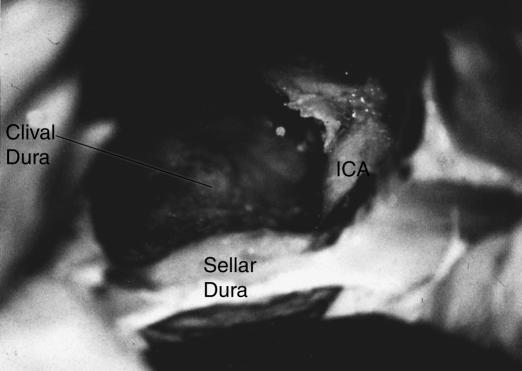
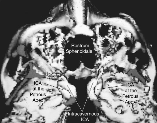
The anterior and posterior vertical segments of the intracavernous portions of the internal carotid arteries (ICAs) were exposed. The arteries were followed retrogradely to the point where they entered the cavernous sinus, allowing part of the petrous apex to be resected (Fig. 1).
Figure 1.
Microscopic view of the specimen after microsurgical drilling.
Bone resection was continued as much as possible under the sella toward the posterior clinoid processes and then proceeded caudally toward the foramen magnum and occipital condyles. Bone removal was extended as much as possible by changing the position of the microscope to expose the clival dura from the foramen magnum up to the bone of the posterior clinoid processes. Drilling ceased when no more bone could be removed safely under an unobstructed microscopic view (i.e., the contact point between burr and bone was clearly visible during drilling). Three authors confirmed this condition by examining each specimen separately. The area of clivus exposed under microscopic visualization (“M” area) was measured using a millimetric paper.
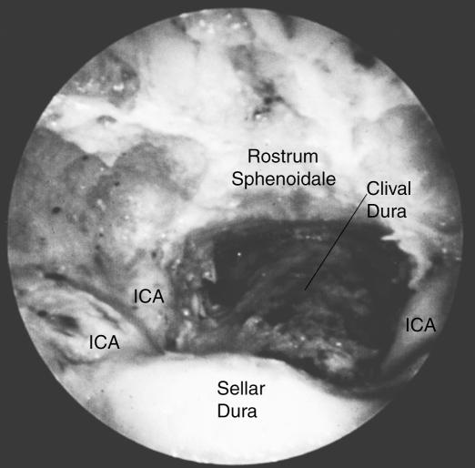
A 0–degree– and a 30–degree–angled, rigid, 4–mm diameter endoscope (Karl Storz Endoscopy America Company, Culver City, CA) connected to a video recorder was introduced and secured by an electropneumatic holder (Mitaka Kohki Company, Toyko, Japan). A 0–degree lens was introduced first and a 30–degree endoscope was used as a second step. Drilling was then continued under endoscopic visualization (Fig. 2). The amount of bone removed from the clivus was determined by measuring the area of additional bone removal obtained under the endoscopic visualization (“E” area).
Figure 2.
Endoscopic view of the specimen after endoscopic drilling.
A CT scan was made after both the microscopic and endoscopic drilling. The areas of bone removal were compared with particular attention to the amount of bone still present in the posterior clinoid, petrous apex, and condylar areas.
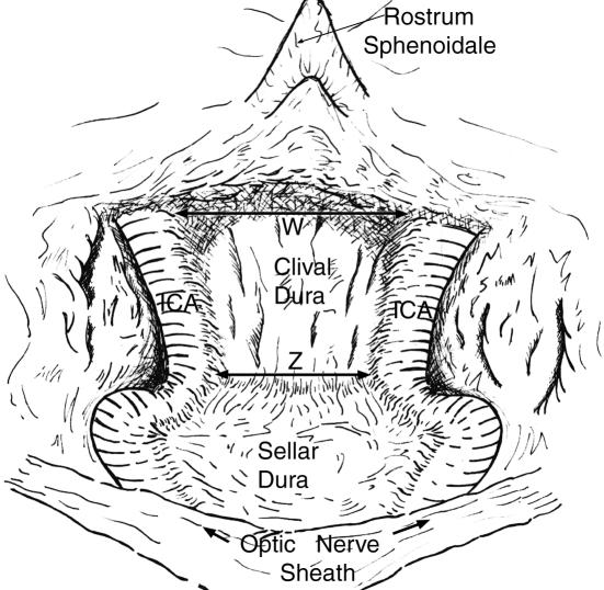
The measurements before and after endoscopic drilling were compared and the difference, range, and standard deviation were calculated. The percentage of the increased area after endoscopy from “M” into “E” was defined as “Δperc.” The location of the blind angles was recorded under both the microscope and endoscope. To evaluate the safety of drilling, particular attention was paid to the subsellar, petrous apex, and condylar areas. Under endoscopic view the distance between ICAs at the petrous apex (distance “W”) and the minimal distance between the vertical posterior segments of the cavernous ICA (distance “Z”) were measured (Fig. 3).
Figure 3.
The distance between the two carotid arteries was measured at two points, “W” and “Z” ( see text for explanation).
The following variables considered in the statistical analysis were performed with SAS software: (1) the areas of bone removal after microscopic (M) and endoscopic (E) drilling; (2) the difference between (M) and (E) expressed as a percentage (Δperc); (3) the distances between the carotid arteries at the petrous apex (W) and at the vertical segment (Z). The variables were distributed normally, as demonstrated by the Shapiro–Wilk statistic. Therefore, parametric analyses were performed. Pearson's correlation coefficient and linear regression models were used to evaluate the relationship among all variables considered.
RESULTS
The area of clival bone removed under microscopic visualization ranged from 416 to 616 mm2 (mean, 515.9 mm2; SD, 63.12 mm2). The area of bone removed under endoscopic visualization ranged from 680 to 1353 mm2 (mean, 983.5 mm2; SD, 217.2 mm2). The difference in the amount of bone removed under endoscopic visualization ranged from 176 to 753 mm2 (mean, 467.6mm2; SD, 208.8 mm2). The percentage of the amount of additional bone removed under endoscopy compared with under microscopy ranged from 34.9 to 174 % (mean, 92.2 %; SD, 42.2 %). The mean distance between the two ICAs at the petrous apex and the mean minimal distance in the vertical segment was 19.7 mm (SD ± 2.3 mm; range, 16 to 24 mm) and 15.3 mm (SD, 3.4 mm; range, 10 to 21 mm), respectively.
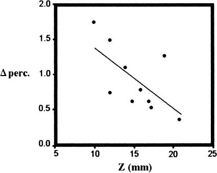
A significant correlation between “Δperc” and “Z” (r = 0.65, p = 0.04) was found. To further define the relationship of “Δperc” with “Z” and “W”, linear regression models were calculated. “Δperc” was inversely correlated with “Z” (p = 0.04, r2 = 0.42, b = –0.09 where “b” is the angular coefficient). No relationship was found between “Δperc” and “W” (p = 0.39) (Fig. 4).
Figure 4.
Linear regression model showing the relationship between “Δperc” and “Z” (b = –0.09, r2 = 0.42). Model equation: Δperc = 2.2402 + b · Z.
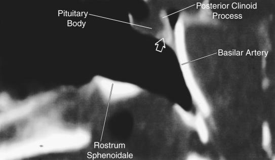
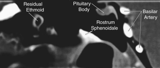
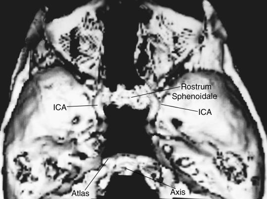
The blind angles under microscopic view were located (1) at the dorsum sellae and the area immediately under the sella, (2) posterior and lateral to the vertical segment of the carotid artery, and (3) above and lateral to the condyles. Under endoscopic visualization all these angles were associated with a residual amount of bone left after the microscopic drilling which could be safely removed under the endoscope. Under microscopic visualization the dorsum sellae was never totally removed (Fig. 5), whereas under endoscopy the dorsum sellae was removed completely in 8 cases (Figs. 6 and 7) and subtotally in 2.
Figure 5.
Sagittal CT reconstruction after the microscopic approach. Residual bone is evident at the dorsum sellae (arrow).
Figure 6A.
Sagittal CT reconstruction after the endoscopic approach in the same specimen. The dorsum sellae has been removed completely.
Figure 7.
3D–CT reconstruction of a specimen with a wide space between the ICAs after the endoscopic approach.
DISCUSSION
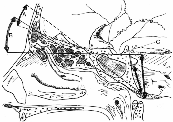
The extended subfrontal approach to the clivus was proposed as an improvement on the transbasal approach described by Derome.1, 2 The basic idea was to combine this approach with an orbitonasal osteotomy. The aim of the modification was to reduce traction on the frontal lobes and to improve vision in a deep and narrow operative field such as the clival area (Fig. 8). The indications for this approach include infiltrative tumors that involve the clival bone (e.g., chordomas and chondrosarcomas).3
Figure 8.
The extended subfrontal approach. The orbital osteotomy (B) increases the exposure of the clivus compared with the standard frontal craniotomy (A) while brain retraction is minimized. The blind area (C) can be reached with the help of the endoscope (see Fig. 6).
Compared with transfacial routes, the extended subfrontal approach has three advantages. First, it allows closure of the clival dura by reflecting a pedicled pericranial flap packed by fatty tissue in the operative field. Second, it allows decompression of both optic nerves, which is frequently necessary due to a tumoral extension. Finally, for extensive tumors, it can be combined with a frontotemporo–orbitozygomatic or a subtemporo–infratemporo–transzygomatic approach.
Complete resection of these tumors requires removing bone beyond the radiological border of a lesion.1, 4, 5 Although T2–weighted magnetic resonance imaging shows demarcated margins, knowing where and when to stop bone resection during surgery is difficult. Therefore, it is extremely important to have a direct view of the macroscopic appearance of the clival bone around the lesion.5
Despite the improvement offered by the extended subfrontal approach, microscopic visualization is still limited. Furthermore, the angle of vision under the microscope must be changed according to the clival area the surgeon wants to reach.6 Consequently, vision in the deepest area of the field and the space available to introduce instruments are reduced. Moreover, a tumoral extension lateral to the petrous ICA is difficult to reach under microscopic vision.7
Because of the intensity of their light and high optical quality, only rigid lens endoscopes fixed in the desired position by a simple mechanical holder have been shown to be safe and effective during surgery.8, 9 Safe drilling requires continuous direct vision of the drill burr. Therefore, the position of the endoscope must be changed frequently. However, the multiple maneuvers required to move the endoscope can slow the procedure considerably. The mechanical holder adopted in our study was easy to use because the electropneumatic control system allowed delicate and rapid movements. As a result, positioning the endoscope in the operative field was precise and quick. Endoscopy requires little space to work in deep corridors; compared with drilling under microscopic visualization, less retraction on the frontal lobes is needed. This advantage helps to avoid damage to the frontal lobes during surgery.
Both the 0– and 30–degree–angled lens endoscopes should be used in two steps. This strategy makes it easier to understand the anatomy and to maintain orientation. Our data indicate that the endoscope improves vision in a deep, reversed, cone–shaped operative field and allows safe removal of bone in areas that are blind under microscopic vision. As shown by the dissections and confirmed by CT, the blind angles under microscopy are located under and behind the sella, lateral and posterior to the intrapetrous ICAs, at the foramen lacerum segment, and superoposteriorly to the occipital condyles. Endoscopy visualizes these angles and allows the surgeon to work under direct vision, making bone removal safe and accurate.
Our data suggest that the improvement in vision offered by endoscopy is more evident in narrow corridors because the distance between the ICAs at the vertical segment (“Z” distance) is minimal. In fact, the percentage of additional bone removed under endoscopy was inversely related to that distance, as the statistical analysis on this small number of anatomical specimens suggested. To confirm the significance of this finding, a larger number of specimens should be dissected. In most of our specimens, the dorsum sellae, typically considered to be a blind spot unresectable through the transbasal approach1, 2 under microscopic vision, was removed totally under endoscopy.
In our experience, the flat two–dimensional vision provided by the endoscope does not represent a problem during dissection. Although the sharpness of the pictures seen on the monitor is still inferior to direct microscopic vision, it is sufficient to perform the dissection safely.
An endoscopic approach to the clivus in the attempt to remove a clival tumor was reported early in the literature. However, it was conducted through an endonasal trans–sphenoidal route.10 The idea of using the extended subfrontal approach to reach the clivus through an endoscopic technique has never been reported. We removed an extended portion of the viscerocranium to reduce traction on the brain and to improve visualization of deeply located areas. Endoscopy improved visualization to a remarkable level. This study confirms the value of the endoscope for visualizing blind angles compared with the microscope. Endoscopic visualization improved the amount of bone that could be removed, thereby enhancing the safety and radical removal of some clival tumors.
Figure 6B.
3D–CT reconstruction after the endoscopic approach in the same specimen. The dorsum sellae has been removed completely even when the distance between the ICAs is minimal.
REFERENCES
- Sekhar LN, Nanda A, Sen CN, Snyderman CN, Janecka IP. The extended frontal approach to tumors of the anterior, middle and posterior skull base. J Neurosurg. 1992;76:198–206. doi: 10.3171/jns.1992.76.2.0198. [DOI] [PubMed] [Google Scholar]
- Derome P, Akerman M, Anquez L. Les tumeurs spheno–ethmoidales. Possibilites d'exerese et de reparation chirurgicales. Neurochirurgie (suppl) 1972;18:1–164. [PubMed] [Google Scholar]
- Gay E, Sekhar LN, Rubinstein E, et al. Chordomas and chondrosarcomas of the cranial base: results and follow–up of 60 patients. Neurosurgery. 1995;36:887–897. doi: 10.1227/00006123-199505000-00001. [DOI] [PubMed] [Google Scholar]
- Brown AP, Spetzler RF. [comment]. Neurosurgery. 1995;36:896. doi: 10.1227/00006123-199502000-00032. [DOI] [PubMed] [Google Scholar]
- Sen CN, Sekhar LN, Schramm VL, Janecka IP. Chordoma and chondrosarcoma of the cranial base: an 8–year experience. Neurosurgery. 1989;25:931–941. doi: 10.1097/00006123-198912000-00013. [DOI] [PubMed] [Google Scholar]
- Sekhar LN, Raso JL, Tzortzidis F. Extended frontal transbasal approach: anatomy. New York: Thieme Verlag. 1999:76–81. In: Sekhar LN, De Oliveira E, eds. Cranial Microsurgery, Approaches and Techniques. [Google Scholar]
- Derome P, Visot A, Monteil JP, Maestro JL. Management of cranial chordomas. New York: Futura. 1987:607–622. In: Sekhar LN, Schramm VL, eds. Tumors of the Cranial Base. Diagnosis and Treatment. [Google Scholar]
- Perneczky A, Fries G. Endoscope–assisted brain surgery: part 1-evolution, basic concept and current technique. Neurosurgery. 1998;42:219–224. doi: 10.1097/00006123-199802000-00001. [DOI] [PubMed] [Google Scholar]
- Jho HD, Carrau RL, Ko Y, Daly MA. Endoscopic pituitary surgery: an early experience. Surg Neurol. 1997;47:213–223. doi: 10.1016/s0090-3019(96)00452-1. [DOI] [PubMed] [Google Scholar]
- Jho HD, Carrau RL, McLaughlin MR, Somaza SC. Endoscopic transsphenoidal resection of a large chordoma in the posterior fossa. Acta Neurochir (Wien) 1997;139:343–348. [PubMed] [Google Scholar]


