Abstract
Background
The current early warning model for organ damage in critically ill patients has certain limitations. Based on the pathological mechanism, the establishment of an early warning system for organ damage in critically ill children using cytokines profile has not been explored. The aim of this study is to explore the predicting value of cytokines in critically ill patients.
Methods
There were 200 critically pediatric patients and 49 general patients between August 22, 2018 and April 28, 2023 from Children’s Hospital of Soochow University enrolled in this study. The clinical information was retrospectively collected and analyzed. The cytokine profiles of these patients were detected by flow cytometry. Receiver operating characteristic (ROC) curves were plotted to determine the association between the cytokines and organ injury.
Results
There were no statistically significant differences in gender, age and underlying disease between critically ill patients and general patients. The interleukin (IL)-6 (P<0.001), IL-10 (P<0.001), IL-17A (P=0.001), tumor necrosis factor-α (TNF-α) (P=0.02) and interferon-γ (IFN-γ) (P=0.02) level in the critically patients were significantly higher than those in the general patients. The results showed that the incidence of acute gastrointestinal injury (AGI) and acute kidney injury (AKI) in critically ill patients was 39% and 23.5%, respectively. Moreover, there were 4% and 3.5% patients with the occurrence of cardiac arrest and acute live injury. The IFN-γ level was increased in these patients with acute liver injury compared to those without these organ injuries, but reduced in the patients with AGI compared to those without. The patients with AKI showed a significant increase in IL-10 in contrast to those without. The IL-2, IL-4, IL-6, IL-10 and IL-17A were higher in patients with acute liver failure (ALF), but TNF-α was reduced, compared to those without. The IL-2, IL-4, IL-6 and IL-10 were significantly increased in the patients with cardiac arrest compared to those without. When IL-10 was higher than 279.45 pg/mL, the sensitivity and specificity for predicting cardiac arrest were 0.875 and 0.927, respectively. While the sensitivity and specificity of IL-6 (more than 1,425.6 pg/mL) were 0.625 and 0.844, respectively. However, no synergistic effect of IL-6 and IL-10 was observed for predicting cardiac arrest. Additionally, the IL-17A (more than 21.6 pg/mL) was a good predictor for the incidence of ALF (sensitivity =0.714, specificity =0.876).
Conclusions
The cytokines profile was different between critically ill patients with organ injury and those without organ injury. The IL-6 and IL-10 levels were good predictors for cardiac arrest in critically ill patients. Additionally, higher IL-17A predicted the incidence of ALF of the critically ill patients.
Keywords: Multiple organ dysfunction syndrome (MODS), cytokines, critically ill patients, cardiac arrest
Highlight box.
Key findings
• The cytokines profile was different between critically ill patients with organ injury and those without organ injury. The interleukin (IL)-6 and IL-10 levels were good predictors for cardiac arrest in critically ill patients. Additionally, higher IL-17A predicted the incidence of acute liver failure of the critically ill patients.
What is known and what is new?
• Cytokine has been reported as an ideal factor for predicting the severity and prognosis of certain diseases. However, no ones have explored the predictive value for the multiple organ dysfunction syndrome (MODS) in critically ill pediatric patients.
• The present study presented that IL-6 and IL-10 can be used to predict the occurrence of cardiac arrest in critically ill patients.
What is the implication, and what should change now?
• It is necessary to routinely monitor the cytokines profile for critically ill patients, which is of significance for evaluating the occurrence of MODS.
Introduction
Multiple organ dysfunction syndrome (MODS) is a common complication in critically ill patients and one of the main causes of death in patients at the intensive care unit (1). Early warning of the occurrence of MODS can enable patients to receive effective intervention at an early stage, thereby improving patient outcomes (2,3). A series of studies and model have been explored, but all of them have certain limitations (4,5). In order to better find the indicators of early warning model for the occurrence of MODS, we aim to explore the pathological mechanism.
As we all know, the occurrence of the MODS is a complex pathophysiological response, involving the release and interaction of a variety of inflammatory cytokines, and these ultimately lead to vascular endothelial injury, microcirculation disorder, tissue hypoxia and cell damage. Since the inflammatory response is an important part of the mechanism of MODS development, monitoring inflammatory mediators and controlling the inflammatory response are essential to ameliorate the MODS. Therefore, based on the data of previous studies, we designed a feasible cytokine panel for routine detection in critically ill patients, and then used these indicators to predict the occurrence of organ damage, which was performed by logistic regression model. To the best of our knowledge, no one has assessed the incidence of organ injury in critically ill patients based on cytokines profile. Cytokine has been widely used in clinical practice such as distinguishing disease severity (6,7), determining pathogen type (8-10) and the long-term prognosis of the disease (11,12). Thus, we believe that this method is feasible and reliable. We present this article in accordance with the TRIPOD reporting checklist (available at https://tp.amegroups.com/article/view/10.21037/tp-24-95/rc).
Methods
Study population and data collection
There were 200 critically pediatric patients between August 22, 2018 to April 28, 2023 from Children’s Hospital of Soochow University enrolled in this study. Sex- and age-matched approximate-general children (49 cases) were recruited from other department of our center as a control group. This was a respectively cohort study. The primary endpoint was the incidence of organ injury within 7 days post admission. The information including sex, age, underlying diseases and organ injury indicators was collected and analyzed. All of the patients underwent cytokines profile test. The inclusion criteria were (I) age between 1 month to 18 years old; (II) critically ill patients meet criteria for severe disease; (III) the length of hospital stay is more than 7 days and the clinical information was complete. The exclusion criteria were: (I) patients who declined blood test; (II) use of immunomodulatory drugs prior to hospitalization; (III) definite inflammatory disease prior to admission. The criteria for organ injury were based on international guidelines and/or the criteria from other studies (13-24). The study was conducted in accordance with the Declaration of Helsinki (as revised in 2013). The study was approved by institutional ethics board of Children’s Hospital of Soochow University (No. 2023CS034) and informed consent was taken from all the patients or their parents/legal guardians.
Cytokines assay
According to the previous researches, the cytokine panel including interleukin (IL)-2, IL-4, IL-6, IL-10, IL-17A, tumor necrosis factor-α (TNF-α) and interferon-γ (IFN-γ) was designed and measured. Blood samples from patients were collected on admission. The patient’s peripheral blood was collected using ethylenediaminetetraacetic acid (EDTA) anticoagulant tubes. After centrifuge, the serum was extracted for the cytokines test through flow cytometry. The flow cytometry procedure was performed according to the kit instructions, which were purchased from Jiangxi Saiji Biotechnology Co., Ltd. (Nanchang, China). All tests utilized blinded specimen as quality control. The intra-assay and inter-assay coefficients of variation were <5 and <10%, respectively. When the cytokine level was below the threshold value but undetectable, we use the threshold value to represent their titers.
Statistical analysis
The cytokine levels were log-transformed, and then the differences in cytokine levels between the control and the critically ill group were compared using the t-test. Multivariate logistics regression analysis was conducted to identify independent factors (cytokines) within 7 days post admission that were associated with the incidence of MODS. To determine the association between these factors and the occurrence of MODS, 95% confidence intervals (CIs) were estimated with adjustment for independent risk factors. Following to previous reports, variables with a P value <0.1 were included in the receiver operating characteristic (ROC) analysis. The area under the curve (AUC) was calculated to assess the prognostic value of the predicting factor. SPSS 27.0.1 software was used for this analysis. P<0.05 was determined as statistically significant.
Results
Clinical information of the subjects
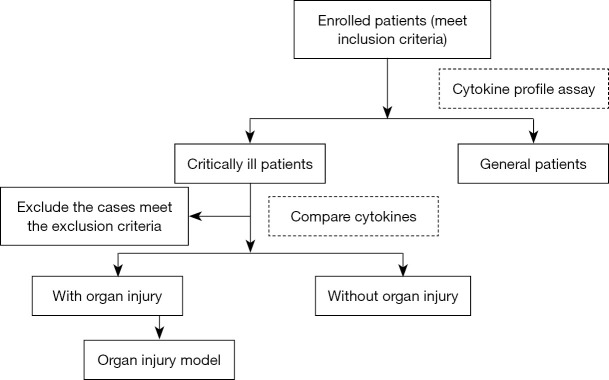
The flow chart of this study is depicted in Figure 1. A total of 249 children were enrolled in the study, including 200 critically ill children and 49 general patients from the same period. The information was collected within 7 days post-cytokine profiling. Fifty-nine percent of the critically ill children were male, and a similar proportion of males in the general patient group, and there was no statistically significant difference in gender between the two groups. The median age of critically ill children was 6.17 years, while the median age of the general patients was 6.38 years, with no significant difference in age between the two groups. Underlying diseases included initial leukemia, relapsed leukemia, post-chimeric antigen receptor (CAR) T cell/chemo therapy, post-bone marrow transplantation, sepsis, severe pneumonia, severe encephalitis, myocarditis, arthritis and hemophagocytic syndrome, with no difference in underlying diseases between the two groups (Table 1).
Figure 1.
The flow chart of this study.
Table 1. The clinical information of the patients in this study.
| Variables | Critically ill patients (n=200) | General patients (n=49) | P |
|---|---|---|---|
| Age (median, years) | 6.17 | 6.38 | 0.56 |
| Male, n (%) | 118 (59.0) | 29 (59.2) | 0.56 |
| Underlying disease, n (%) | 0.06 | ||
| Initial leukemia | 9 (4.5) | 9 (18.4) | |
| Relapsed leukemia | 11 (5.5) | 3 (6.1) | |
| Post-CAR T cell/chemo therapy | 44 (22.0) | 12 (24.5) | |
| Post-bone marrow transplantation | 7 (3.5) | 2 (4.1) | |
| Sepsis | 47 (23.5) | 11 (22.4) | |
| Severe pneumonia | 1 (0.5) | 1 (2.0) | |
| Severe encephalitis | 39 (19.5) | 4 (8.2) | |
| Myocarditis | 4 (2.0) | 1 (2.0) | |
| Arthritis and hemophagocytic syndrome | 37 (18.5) | 6 (12.2) | |
| Solid tumors | 1 (0.5) | 0 |
CAR, chimeric antigen receptor.
Cytokine level between these two groups
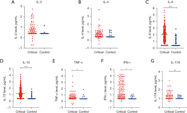
The serum cytokines profile (IL-2, IL-4, IL-6, IL-10, TNF-α, IFN-γ and IL-17A) of these enrolled patients were performed. Flow cytometry was utilized to determine cytokine levels in this study. If certain cytokines were detectable but below the measurable threshold, the threshold value was used to indicate the cytokine level. The minimum threshold for all other cytokines was 2.5 pg/mL, except for IL-17A, which was 10 pg/mL. For the critically ill patients, the median levels of IL-2, IL-4, IL-6, IL-10, TNF-α, IFN-γ, IL-17A were 2.5, 2.5, 104.25, 15.35, 2.5, 2.55 and 10 pg/mL, respectively. For the general patients, the median levels of IL-2, IL-4, IL-6, TNF-α, IFN-γ, IL-17A were 2.5, 2.5, 2.5, 2.5, 2.5 and 10 pg/mL, respectively. Furthermore, the cytokine levels were compared between these two groups. The levels of IL-6 (P<0.001), IL-10 (P<0.001), TNF-α (P=0.02), IL17A (P=0.001) and IFN-γ (P=0.02) in the critically patients were significantly higher than those in the general patients (Figure 2).
Figure 2.
The comparation of cytokines between critically ill patients and general patients. (A) IL-2, (B) IL-4, (C) IL-6, (D) IL-10, (E) TNF-α, (F) IFN-γ, (G) IL-17A were compared in these two groups, respectively. Note: general patients (control), critically ill patients (critical). The cytokine levels were log-transformed, and then the differences of cytokines between the control and the critically ill group were compared using the t-test. *, P<0.05; **, P<0.01; ***, P<0.001. IL, interleukin; TNF-α, tumor necrosis factor-α; IFN-γ, interferon-γ.
The incidences of organ dysfunction in the critically ill patients and cytokine distribution
Firstly, the incidence of organ dysfunction was explored in the critically ill patients. The results showed that the incidence of acute gastrointestinal injury (AGI) was 39% (78/200). The incidence of severe acute respiratory distress syndrome was 1.5% (3/200). The incidences of acute liver injury (ALI) and acute liver failure (ALF) were 56% (112/200) and 3.5% (7/200), respectively. The incidences of acute myocardial injury (AMI) and cardiac arrest were 40% (80/200) and 4% (8/200), respectively. The incidence of acute kidney injury (AKI) was 23.5% (47/200). For the neurological system, there was 36.5% (73/200) patients with neurological abnormalities. Due to the large number of patients with hematologic malignancies in our center, coagulation was also evaluated. The results showed that the incidence of coagulation abnormalities and mucocutaneous hemorrhage were 60.5% and 22%, respectively. For the therapy, we observed that 60 (30%) and 64 (32%) should receive the continuous renal replacement therapy (CRRT) and vasodilator drugs, respectively.
The relationship between cytokines and organ injury was further analyzed in critically patients. The data revealed differences in cytokine levels between patients with organ injury and those without. The median of IFN-γ level was 2.5 pg/mL in patients with AGI, which was lower than in those without AGI (2.5 vs. 4.4 pg/mL, P=0.01). A significant increase in IL-10 was observed in the patients with acute liver injury compared to those without (18.45 vs. 10.30 pg/mL, P=0.01). A similar trend was observed in patients with ALF compared to those without (33 vs. 14.3 pg/mL, P=0.006). Additionally, IL-10, IL-2, IL-4, IL-6 and IL-17A levels were significantly higher in patients with ALF than in those without, while TNF-α levels were lower in patients with ALF. Patients with AKI showed an increase in IL-10 compared to those without (21.6 vs. 12.2 pg/mL, P=0.01). There were 40% critically ill patients occurred with acute myocardial injury, the IL-6 was significantly higher in these patients compared to those without AMI (150.75 vs. 51.15 pg/mL, P=0.03). Levels of IL-6 and IL-10 were also elevated in patients with cardiac arrest compared to those without (148.5 vs. 83.65 pg/mL, P=0.001; 41.85 vs. 14.1 pg/mL, P<0.001). In contract to the patients without neurological abnormalities, IL-6 levels were increased in patients with neurological abnormalities (145.8 vs. 61.2 pg/mL, P=0.002). Significant differences in IL-10 and IL-6 were also found between patients with coagulation abnormalities and those without (16.1 vs. 13.9 pg/mL, P<0.001; 145.8 vs. 45 pg/mL, P=0.02) (Table 2).
Table 2. Comparison of cytokines of organ injury in critically ill patients.
| Organ injury | N (%) | IL-2 (pg/mL) | IL-4 (pg/mL) | IL-6 (pg/mL) | IL-10 (pg/mL) | TNF-α (pg/mL) | IFN-γ (pg/mL) | IL-17A (pg/mL) | |||||||||||||
|---|---|---|---|---|---|---|---|---|---|---|---|---|---|---|---|---|---|---|---|---|---|
| Value | P | Value | P | Value | P | Value | P | Value | P | Value | P | Value | P | ||||||||
| AGI | 0.45 | 0.82 | 0.17 | 0.99 | 0.41 | 0.01 | 0.32 | ||||||||||||||
| Yes | 78 (39.0) | 2.5 (2.5, 2.5) |
2.5 (2.5, 2.65) |
149.5 (13.45, 653.025) |
14.1 (4.75, 57.9) |
2.5 (2.5, 2.5) |
2.5 (2.5, 11.9) |
10 (10, 10) |
|||||||||||||
| No | 122 (61.0) | 2.5 (2.5, 2.5) |
2.5 (2.5, 2.575) |
68.25 (11.925, 732.375) |
15.35 (5.5, 84.45) |
2.5 (2.5, 2.5) |
4.4 (2.5, 81.225) |
10 (10, 10.45) |
|||||||||||||
| Severe ARDS | 0.51 | 0.41 | 0.09 | 0.85 | 0.51 | 0.36 | 0.63 | ||||||||||||||
| Yes | 3 (1.5) | 2.5 (2.5, 2.5) |
2.5 (2.5, 26.9) |
627.9 (315.2, 18,605.55) |
11 (6.75, 135.4) |
2.5 (2.5, 20.6) |
2.5 (2.5, 37.4) |
10 (10, 132.05) |
|||||||||||||
| No | 197 (98.5) | 2.5 (2.5, 16.25) |
2.5 (2.5, 2.6) |
86.5 (13.2, 686.6) |
15.1 (5.2, 70.5) |
2.5 (2.5, 2.5) |
2.5 (2.5, 55) |
10 (10, 10) |
|||||||||||||
| ALI | 0.54 | 0.74 | 0.76 | 0.01 | 0.49 | 0.03 | 0.74 | ||||||||||||||
| Yes | 112 (56.0) | 2.5 (2.5, 2.5) |
2.5 (2.5, 2.5) |
154.15 (22.25, 804.75) |
18.45 (6.7, 76.275) |
2.5 (2.5, 2.5) |
3.3 (2.5, 60.025) |
10 (10, 10) |
|||||||||||||
| No | 88 (44.0) | 2.5 (2.5, 2.5) |
2.5 (2.5, 2.825) |
45.75 (9.25, 318.525) |
10.3 (3.925, 61.125) |
2.5 (2.5, 2.5) |
2.5 (2.5, 51.55) |
10 (10, 10.15) |
|||||||||||||
| ALF | 0.005 | 0.01 | 0.02 | 0.006 | 0.04 | 0.31 | 0.02 | ||||||||||||||
| Yes | 7 (3.5) | 2.5 (2.5, 2.5) |
2.5 (2.5, 7.8) |
530.9 (98.75, 700.4) |
33 (12.05, 93.65) |
2.5 (2.5, 4.55) |
2.5 (2.5, 87.55) |
22 (10, 23) |
|||||||||||||
| No | 193 (96.5) | 2.5 (2.5, 2.5) |
2.5 (2.5, 2.5) |
80.8 (11.6, 627.9) |
14.3 (5.2, 70.5) |
2.5 (2.5, 2.5) |
2.5 (2.5, 55) |
10 (10, 10) |
|||||||||||||
| AKI | 0.97 | 0.61 | 0.18 | 0.01 | 0.50 | 0.71 | 0.69 | ||||||||||||||
| Yes | 47 (23.5) | 2.5 (2.5, 2.7) |
2.5 (2.5, 3.35) |
151.5 (13.25, 633.25) |
21.6 (9.15, 103.35) |
2.5 (2.5, 2.9) |
4.3 (2.5, 71.45) |
10 (10, 10) |
|||||||||||||
| No | 153 (76.5) | 2.5 (2.5, 2.5) |
2.5 (2.5, 2.5) |
73.6 (11.6, 686.6) |
12.2 (4.4, 60.3) |
2.5 (2.5, 2.5) |
2.5 (2.5, 71.45) |
10 (10, 10) |
|||||||||||||
| AMI | 0.64 | 0.29 | 0.03 | 0.15 | 0.53 | 0.61 | 0.97 | ||||||||||||||
| Yes | 80 (40.0) | 2.5 (2.5, 2.5) |
2.5 (2.5, 2.625) |
150.75 (18.5, 791.775) |
17.2 (6.175, 69.075) |
2.5 (2.5, 2.5) |
2.5 (2.5, 20.75) |
10 (10, 10) |
|||||||||||||
| No | 120 (60.0) | 2.5 (2.5, 2.5) |
2.5 (2.5, 2.525) |
51.15 (11.475, 530.4) |
12.2 (5.075, 79.55) |
2.5 (2.5, 2.5) |
2.8 (2.5, 73.125) |
10 (10, 10.3) |
|||||||||||||
| Neurological abnormalities | 0.051 | 0.22 | 0.002 | 0.78 | 0.16 | 0.69 | 0.40 | ||||||||||||||
| Yes | 73 (36.5) | 2.5 (2.5, 2.5) |
2.5 (2.5, 2.5) |
145.8 (11.4, 773.6) |
11.8 (4.9, 47.7) |
2.5 (2.5, 2.5) |
2.5 (2.5, 29.6) |
10 (10, 10) |
|||||||||||||
| No | 127 (63.5) | 2.5 (2.5, 2.5) |
2.5 (2.5, 2.75) |
61.2 (13.6, 514.4) |
17.7 (5.5, 83.5) |
2.5 (2.5, 2.5) |
3.2 (2.5, 64.4) |
10 (10, 10) |
|||||||||||||
| Coagulation abnormalities | 0.03 | 0.22 | 0.02 | <0.001 | 0.15 | 0.38 | 0.17 | ||||||||||||||
| Yes | 121 (60.5) | 2.5 (2.5, 2.5) |
2.5 (2.5, 2.5) |
145.8 (18.2, 773.6) |
16.1 (5.3, 76) |
2.5 (2.5, 2.5) |
3.2 (2.5, 81) |
10 (10, 10) |
|||||||||||||
| No | 79 (39.5) | 2.5 (2.5, 2.5) |
2.5 (2.5, 2.8) |
45 (10.3, 479.85) |
13.9 (5.15, 61.45) |
2.5 (2.5, 2.5) |
2.5 (2.5, 17.6) |
10 (10, 10) |
|||||||||||||
| Cardiac arrest | 0.01 | 0.02 | 0.001 | <0.001 | 0.15 | 0.56 | 0.11 | ||||||||||||||
| Yes | 8 (4.0) | 2.5 (2.5, 3.55) |
2.65 (2.5, 5.3) |
148.5 (57.775, 502.775) |
41.85 (13.45, 325.4) |
2.5 (2.5, 2.5) |
2.6 (2.5, 571.1) |
10 (10, 14.3) |
|||||||||||||
| No | 192 (96.0) | 2.5 (2.5, 2.5) |
2.5 (2.5, 2.5) |
83.65 (12.8, 727.45) |
14.1 (5.2, 69.075) |
2.5 (2.5, 2.5) |
2.5 (2.5, 51.55) |
10 (10, 10) |
|||||||||||||
Data are presented as median (interquartile range). IL, interleukin; TNF-α, tumor necrosis factor-α; IFN-γ, interferon-γ; AGI, acute gastrointestinal injury; ARDS, acute respiratory distress syndrome; ALI, acute liver injury; ALF, acute liver failure; AKI, acute kidney injury; AMI, acute myocardial injury.
Predicting value of cytokines for the organ injury in critically ill patients
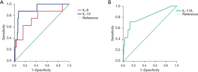
The cytokines with statistical significance were further analyzed for their ability to predict organ injury. ROC curves were plotted to assess the prediction value of the selected cytokines for the organ injury. Significant differences in IL-10 between critically ill patients and general patients were indicated. The AUC for IL-10 (more than 279.45 pg/mL) in predicting cardiac arrest was the largest (AUC =0.913, sensitivity =0.875, specificity =0.927), followed by the IL-6 (more than 1,425.6 pg/mL) for cardiac arrest (AUC =0.754, sensitivity =0.625, specificity =0.844). However, no synergistic effect of combining IL-6 with IL-10 was observed. Other cytokines were also evaluated. Furthermore, the results indicted the IL-17A (more than 21.6 pg/mL) was a good predictor for ALF (AUC =0.763, sensitivity =0.714, specificity =0.876) (Figure 3). Previous studies have reported a linear relationship between IL-2 and IL-10 in predicting organ injury, but we did not observe such a relationship in our results. Similar results were found for IL-10 and IL-6 (Figure S1).
Figure 3.
The predicting value of cytokines were assessed. (A) The predicting value of IL-6 and IL-10 for the incidence of cardiac arrest; (B) The predicting value of IL-17A for acute liver failure. IL, interleukin.
Discussion
MODS is a common complication for the critically ill patients. Thus, healthcare workers have been striving to predict the occurrence of MODS so that patients can receive effective intervention as early as possible (1,25). Several biomarkers have displayed promising performance. For example, transcriptomic profiles have been found to be altered in children with MODS (9,26). However, the approach is not clinically feasible. Red cell distribution width has shown satisfactory predictive ability, but it is limited to predicting characteristics specific to certain age groups and types of diseases, thus limiting its utility (27). Oxygen debt has been identified as a convenient and rapid predictor during the coronavirus disease 2019 (COVID-19) pandemic, but its forecasting effectiveness is poor, similar to the neutrophil index (28,29). Immunologically relevant biomarkers have been a focus in recent years (25,30,31), and cytokines have been identified as ideal factors for predicting the severity and prognosis of various diseases (4,6-8,10-13,32-38). Furthermore, the advantages of cytokine evaluation have been demonstrated, including simple collection, long prediction time and excellent predictive performance (11,28). This supports our hypothesis that cytokine levels can be utilized to predict organ injury.
Our results showed that AKI and ALI happened in the critically ill patients with the occurrence of 23.5% and 56%, in line with previous studies (39,40). The cytokines profile was different between critically ill patients and general patients. Higher levels cytokines in critically ill patients suggest a more active immune system compared to general patients. Specifically, levels of IL-6, IL-10, IL-17A, TNF-α and IFN-γ levels were elevated in the critically ill patients compared to general patients, which further confirms the conclusions that cytokines are associated with the development of disease severity (6,32,33,41). What is more, the patients with acute liver injury showed higher concentration with various cytokines, including the IL-2, IL-4, IL-6, IL-10, TNF-α and IL-17A, which meets the notion that these inflammatory mediators are correlated with the liver failure described more recently (42). There is study reported that IL-10 participated in the development of AKI (43), which is consistent with our results showing elevated IL-10 in the patients with acute kidney.
After observing these significant differences, we wondered if these factors could be used to predict organ injury. By modeling, we found the unique value of IL-6 and IL-10 in predicting the occurrence of cardiac arrest, and IL-17A serves as a good predictor for the incidence of ALF in our study. IL-10, according to existing researches, is a cytokine that suppresses the immune response (44-48). Increasing serum IL-10 level in most cases is accompanied by a strong inflammatory response, implying severed tissue damage. IL-10 prevents macrophage activity and induces fibrosis of inflammatory tissues, which may explain the reason why it could predict cardiac arrest. Similarly, IL-17A is a member of the IL-10 family, the similar function may explain its performance in predicting ALF. IL-6 is usually regarded as pro-inflammatory factors (49,50), inducing anti-inflammatory factors like IL-10 and cortisol section through negative feedback regulation to suppress the inflammatory, which could prevent the occurrence of cytokines release syndrome. But previous research has shown that increasing IL-10 levels are relevant to the worse outcome of some diseases. We suppose that the increasing level of IL-6 and IL-10 may indicate rapidly worsening of some diseases and causing the cardiac arrest. However, the performance of its prognosis value requires further investigation. We also attempted to combine IL-6 and IL-10 for prognosis, but the combined indicators showed poor performance compared to IL-10 alone. A previous study reported a linear regression between IL-6 and IL-10 (32). In a meta-analysis, the combination of IL-6 and IL-10 accurately predicted the severity of a specific disease, with the sensitivity and specificity of 83.3% and 100% respectively (33). However, our results are inconsistent with theirs, possibly due the small sample size.
This study has some limitations. Firstly, there is an inevitable selection bias in the design of the retrospective study, and the potential influence of unmeasured variables and unknown confounding factors that cannot be ruled out in this study. Secondly, the sample size was small and there was a lack of subgroup analysis of specific diseases, which may lead to the lack of certain generalization. Thirdly, if the level of IL-2, IL-4, IFN-γ and IL-17A were less than the thresholds, the thresholds were set as the value of some patients. This may introduce bias in the cytokine values, highlighting the need for more accurate quantification to further evaluate the significance of these cytokines in future studies. Moreover, we plan to conduct a multicenter and prospective study in this area, to further validate our conclusions. Additionally, molecular biology and genomics methods can be used to study the association between cytokines and organ injury, aiming to uncover the specific mechanism by which cytokines contribute to the development of MODS in children.
Conclusions
The cytokines profile was different between critically ill patients with organ injury and those without organ injury. The IL-6 and IL-10 levels were higher in the patient with cardiac arrest compared to those without, and they serve as predictors of cardiac arrest in critically ill patients. Moreover, elevated IL-17A levels indicate ALF in critically ill patients.
Supplementary
The article’s supplementary files as
Acknowledgments
Funding: Design of the study and collection, analysis and interpretation of data was supported by funds from Suzhou Science and Technology Development Project (No. SKY2022178) and Gusu Health Talent Training Project (Nos. GSWS2019050 and GSWS2020044). Writing the manuscript was supported by the Natural Science Foundation of Jiangsu Province (Nos. BK20210097 and BK20211077) and National Science Foundation of China (No. 82300182).
Ethical Statement: The authors are accountable for all aspects of the work in ensuring that questions related to the accuracy or integrity of any part of the work are appropriately investigated and resolved. The study was conducted in accordance with the Declaration of Helsinki (as revised in 2013). The study was approved by institutional ethics board of Children’s Hospital of Soochow University (No. 2023CS034) and informed consent was taken from all the patients or their parents/legal guardians.
Footnotes
Reporting Checklist: The authors have completed the TRIPOD reporting checklist. Available at https://tp.amegroups.com/article/view/10.21037/tp-24-95/rc
Conflicts of Interest: All authors have completed the ICMJE uniform disclosure form (available at https://tp.amegroups.com/article/view/10.21037/tp-24-95/coif). The authors have no conflicts of interest to declare.
Data Sharing Statement
Available at https://tp.amegroups.com/article/view/10.21037/tp-24-95/dss
References
- 1.Typpo KV, Petersen NJ, Hallman DM, et al. Day 1 multiple organ dysfunction syndrome is associated with poor functional outcome and mortality in the pediatric intensive care unit. Pediatr Crit Care Med 2009;10:562-70. 10.1097/PCC.0b013e3181a64be1 [DOI] [PMC free article] [PubMed] [Google Scholar]
- 2.Supthut W, Nuding S, Wienke A, et al. Relationship between cardiac output, heart rate and microcirculation in patients with multiorgan dysfunction syndrome. Med Klin Intensivmed Notfmed 2023. doi:. 10.1007/s00063-023-01086-9 [DOI] [PubMed] [Google Scholar]
- 3.Copeland CC, Young A, Grogan T, et al. Preoperative risk stratification of critically ill patients. J Clin Anesth 2017;39:122-7. 10.1016/j.jclinane.2017.03.044 [DOI] [PubMed] [Google Scholar]
- 4.Elemam NM, Hammoudeh S, Salameh L, et al. Identifying Immunological and Clinical Predictors of COVID-19 Severity and Sequelae by Mathematical Modeling. Front Immunol 2022;13:865845. 10.3389/fimmu.2022.865845 [DOI] [PMC free article] [PubMed] [Google Scholar]
- 5.Liu X, DuMontier C, Hu P, et al. Clinically Interpretable Machine Learning Models for Early Prediction of Mortality in Older Patients with Multiple Organ Dysfunction Syndrome: An International Multicenter Retrospective Study. J Gerontol A Biol Sci Med Sci 2023;78:718-26. 10.1093/gerona/glac107 [DOI] [PMC free article] [PubMed] [Google Scholar]
- 6.Zhang J, Wang J, Gong Y, et al. Interleukin-6 and granulocyte colony-stimulating factor as predictors of the prognosis of influenza-associated pneumonia. BMC Infect Dis 2022;22:343. 10.1186/s12879-022-07321-6 [DOI] [PMC free article] [PubMed] [Google Scholar]
- 7.Shafiek HK, El Lateef HMA, Boraey NF, et al. Cytokine profile in Egyptian children and adolescents with COVID-19 pneumonia: A multicenter study. Pediatric Pulmonology 2021;56:3924-33. 10.1002/ppul.25679 [DOI] [PMC free article] [PubMed] [Google Scholar]
- 8.Angulo J, Martínez-Valdebenito C, Marco C, et al. Serum levels of interleukin-6 are linked to the severity of the disease caused by Andes Virus. PLoS Negl Trop Dis 2017;11:e0005757. 10.1371/journal.pntd.0005757 [DOI] [PMC free article] [PubMed] [Google Scholar]
- 9.Pandey N, Jain A, Garg RK, et al. Serum levels of IL-8, IFNγ, IL-10, and TGF β and their gene expression levels in severe and non-severe cases of dengue virus infection. Arch Virol 2015;160:1463-75. 10.1007/s00705-015-2410-6 [DOI] [PubMed] [Google Scholar]
- 10.Zhang Y, Mei S, Zhou Y, et al. Cytokines as the good predictors of refractory Mycoplasma pneumoniae pneumonia in school-aged children. Sci Rep 2016;6:37037. 10.1038/srep37037 [DOI] [PMC free article] [PubMed] [Google Scholar]
- 11.Li JB, Xie MR, Duan ML, et al. Over-expression of programmed death-ligand 1 and programmed death-1 on antigen-presenting cells as a predictor of organ dysfunction and mortality during early sepsis: a prospective cohort study. World J Emerg Med 2023;14:179-85. 10.5847/wjem.j.1920-8642.2023.041 [DOI] [PMC free article] [PubMed] [Google Scholar]
- 12.Alonso-Domínguez J, Gallego-Rodríguez M, Martínez-Barros I, et al. High Levels of IL-1β, TNF-α and MIP-1α One Month after the Onset of the Acute SARS-CoV-2 Infection, Predictors of Post COVID-19 in Hospitalized Patients. Microorganisms 2023;11:2396. 10.3390/microorganisms11102396 [DOI] [PMC free article] [PubMed] [Google Scholar]
- 13.Liu S, Wang C, Guo J, et al. Serum Cytokines Predict the Severity of Coronary Artery Disease Without Acute Myocardial Infarction. Front Cardiovasc Med 2022;9:896810. 10.3389/fcvm.2022.896810 [DOI] [PMC free article] [PubMed] [Google Scholar]
- 14.McDonagh TA, Metra M, Adamo M, et al. 2021 ESC Guidelines for the diagnosis and treatment of acute and chronic heart failure. Eur Heart J 2021;42:3599-726. 10.1093/eurheartj/ehab368 [DOI] [PubMed] [Google Scholar]
- 15.Senior JR. Alanine aminotransferase: a clinical and regulatory tool for detecting liver injury-past, present, and future. Clin Pharmacol Ther 2012;92:332-9. 10.1038/clpt.2012.108 [DOI] [PubMed] [Google Scholar]
- 16.Lameire N, Van Biesen W, Vanholder R. Acute renal failure. Lancet 2005;365:417-30. 10.1016/S0140-6736(05)17831-3 [DOI] [PubMed] [Google Scholar]
- 17.Pickkers P, Darmon M, Hoste E, et al. Acute kidney injury in the critically ill: an updated review on pathophysiology and management. Intensive Care Med 2021;47:835-50. 10.1007/s00134-021-06454-7 [DOI] [PMC free article] [PubMed] [Google Scholar]
- 18.Chinese Abdominal Intensive Care Association . Asia society for emergency and critical care medicine. Expert consensus on enteral nutrition for gastrointestinal dysfunction in critically ill patients. Chin J Dig Surg 2021;20:1123-36. [Google Scholar]
- 19.Expert consensus group on the diagnosis and treatment of acute hemorrhagic coagulation dysfunction, Shao M, Xue M, et al. Expert consensus on diagnosis and treatment of acute hemorrhagic coagulation dysfunction. Chin J Emerg Med 2020;29:780-7.
- 20.Standl T, Annecke T, Cascorbi I, et al. The Nomenclature, Definition and Distinction of Types of Shock. Dtsch Arztebl Int 2018;115:757-68. 10.3238/arztebl.2018.0757 [DOI] [PMC free article] [PubMed] [Google Scholar]
- 21.Scheffer IE, Berkovic S, Capovilla G, et al. ILAE classification of the epilepsies: Position paper of the ILAE Commission for Classification and Terminology. Epilepsia 2017;58:512-21. 10.1111/epi.13709 [DOI] [PMC free article] [PubMed] [Google Scholar]
- 22.Chronic Obstructive Pulmonary Disease Group of Chinese Thoracic Society. Chronic Obstructive Pulmonary Disease Committee of Chinese Association of Chest Physician . Zhonghua Jie He He Hu Xi Za Zhi 2021;44:170-205. 10.3760/cma.j.cn112147-20210109-00031 [DOI] [PubMed] [Google Scholar]
- 23.Bergeron N, Dubois MJ, Dumont M, et al. Intensive Care Delirium Screening Checklist: evaluation of a new screening tool. Intensive Care Med 2001;27:859-64. 10.1007/s001340100909 [DOI] [PubMed] [Google Scholar]
- 24.Teasdale G, Jennett B. Assessment of coma and impaired consciousness. A practical scale. Lancet 1974;2:81-4. 10.1016/s0140-6736(74)91639-0 [DOI] [PubMed] [Google Scholar]
- 25.Gourd NM, Nikitas N. Multiple Organ Dysfunction Syndrome. J Intensive Care Med 2020;35:1564-75. 10.1177/0885066619871452 [DOI] [PubMed] [Google Scholar]
- 26.Novak T, Crawford JC, Hahn G, et al. Transcriptomic profiles of multiple organ dysfunction syndrome phenotypes in pediatric critical influenza. Front Immunol 2023;14:1220028. 10.3389/fimmu.2023.1220028 [DOI] [PMC free article] [PubMed] [Google Scholar]
- 27.Duchnowski P, Hryniewiecki T, Kuśmierczyk M, et al. Red cell distribution width as a predictor of multiple organ dysfunction syndrome in patients undergoing heart valve surgery. Biol Open 2018;7:bio036251. 10.1242/bio.036251 [DOI] [PMC free article] [PubMed] [Google Scholar]
- 28.Kong T, Park YS, Lee HS, et al. The delta neutrophil index predicts development of multiple organ dysfunction syndrome and 30-day mortality in trauma patients admitted to an intensive care unit: a retrospective analysis. Sci Rep 2018;8:17515. 10.1038/s41598-018-35796-4 [DOI] [PMC free article] [PubMed] [Google Scholar]
- 29.Perez-Garzon M, Poveda-Henao C, Bastidas-Goyes A, et al. Oxygen Debt as Predictor of Mortality and Multiple Organ Dysfunction Syndrome in Severe COVID-19 Patients: A Retrospective Study. J Intensive Care Med 2024;39:358-67. 10.1177/08850666231208433 [DOI] [PubMed] [Google Scholar]
- 30.Munro N. Immunology and Immunotherapy in Critical Care: An Overview. AACN Adv Crit Care 2019;30:113-25. 10.4037/aacnacc2019415 [DOI] [PubMed] [Google Scholar]
- 31.Hall MW, Greathouse KC, Thakkar RK, et al. Immunoparalysis in Pediatric Critical Care. Pediatr Clin North Am 2017;64:1089-102. 10.1016/j.pcl.2017.06.008 [DOI] [PMC free article] [PubMed] [Google Scholar]
- 32.Zhang Y, Li B, Ning B. Evaluating IL-6 and IL-10 as rapid diagnostic tools for Gram-negative bacteria and as disease severity predictors in pediatric sepsis patients in the intensive care unit. Front Immunol 2022;13:1043968. 10.3389/fimmu.2022.1043968 [DOI] [PMC free article] [PubMed] [Google Scholar]
- 33.Dhar SK, K V, Damodar S, et al. IL-6 and IL-10 as predictors of disease severity in COVID-19 patients: results from meta-analysis and regression. Heliyon 2021;7:e06155. 10.1016/j.heliyon.2021.e06155 [DOI] [PMC free article] [PubMed] [Google Scholar]
- 34.Zhang Y, Zhou Y, Li S, et al. The Clinical Characteristics and Predictors of Refractory Mycoplasma pneumoniae Pneumonia in Children. PLoS One 2016;11:e0156465. 10.1371/journal.pone.0156465 [DOI] [PMC free article] [PubMed] [Google Scholar]
- 35.Hong Y, Lin X, Zhang C, et al. Initial indicators for the prognosis of Acinetobacter Baumannii bacteremia in children. BMC Infect Dis 2023;23:640. 10.1186/s12879-023-08639-5 [DOI] [PMC free article] [PubMed] [Google Scholar]
- 36.Han H, Ma Q, Li C, et al. Profiling serum cytokines in COVID-19 patients reveals IL-6 and IL-10 are disease severity predictors. Emerg Microbes Infect 2020;9:1123-30. 10.1080/22221751.2020.1770129 [DOI] [PMC free article] [PubMed] [Google Scholar]
- 37.Seethaler A, Stenner M, McNally A, et al. IL-8 and IFN-γ as Preoperative Predictors of the Outcome of Tonsillectomy. Ear Nose Throat J 2021;100:822S-827S. 10.1177/0145561320910682 [DOI] [PubMed] [Google Scholar]
- 38.Yang X, Zeng J, Yu X, et al. PCT, IL-6, and IL-10 facilitate early diagnosis and pathogen classifications in bloodstream infection. Ann Clin Microbiol Antimicrob 2023;22:103. 10.1186/s12941-023-00653-4 [DOI] [PMC free article] [PubMed] [Google Scholar]
- 39.Kaddourah A, Basu RK, Bagshaw SM, et al. Epidemiology of Acute Kidney Injury in Critically Ill Children and Young Adults. N Engl J Med 2017;376:11-20. 10.1056/NEJMoa1611391 [DOI] [PMC free article] [PubMed] [Google Scholar]
- 40.Jetton JG, Boohaker LJ, Sethi SK, et al. Incidence and outcomes of neonatal acute kidney injury (AWAKEN): a multicentre, multinational, observational cohort study. Lancet Child Adolesc Health 2017;1:184-94. 10.1016/S2352-4642(17)30069-X [DOI] [PMC free article] [PubMed] [Google Scholar]
- 41.Jarczak D, Nierhaus A. Cytokine Storm-Definition, Causes, and Implications. Int J Mol Sci 2022;23:11740. 10.3390/ijms231911740 [DOI] [PMC free article] [PubMed] [Google Scholar]
- 42.Casulleras M, Zhang IW, López-Vicario C, et al. Leukocytes, Systemic Inflammation and Immunopathology in Acute-on-Chronic Liver Failure. Cells 2020;9:2632. 10.3390/cells9122632 [DOI] [PMC free article] [PubMed] [Google Scholar]
- 43.Mu H, Zheng Q, Hao L. IL-10 -1082 A/G polymorphism is related with the risk and clinical characteristics of acute kidney injury: a case-control study. BMC Nephrol 2021;22:212. 10.1186/s12882-021-02410-1 [DOI] [PMC free article] [PubMed] [Google Scholar]
- 44.Hombach AA, Heiders J, Foppe M, et al. OX40 costimulation by a chimeric antigen receptor abrogates CD28 and IL-2 induced IL-10 secretion by redirected CD4(+) T cells. Oncoimmunology 2012;1:458-66. 10.4161/onci.19855 [DOI] [PMC free article] [PubMed] [Google Scholar]
- 45.Couper KN, Blount DG, Riley EM. IL-10: the master regulator of immunity to infection. J Immunol 2008;180:5771-7. 10.4049/jimmunol.180.9.5771 [DOI] [PubMed] [Google Scholar]
- 46.Fiorentino DF, Zlotnik A, Vieira P, et al. IL-10 acts on the antigen-presenting cell to inhibit cytokine production by Th1 cells. J Immunol 1991;146:3444-51. [PubMed] [Google Scholar]
- 47.Del Prete G, De Carli M, Almerigogna F, et al. Human IL-10 is produced by both type 1 helper (Th1) and type 2 helper (Th2) T cell clones and inhibits their antigen-specific proliferation and cytokine production. J Immunol 1993;150:353-60. [PubMed] [Google Scholar]
- 48.Deng B, Wehling-Henricks M, Villalta SA, et al. IL-10 triggers changes in macrophage phenotype that promote muscle growth and regeneration. J Immunol 2012;189:3669-80. 10.4049/jimmunol.1103180 [DOI] [PMC free article] [PubMed] [Google Scholar]
- 49.Steensberg A, Fischer CP, Keller C, et al. IL-6 enhances plasma IL-1ra, IL-10, and cortisol in humans. Am J Physiol Endocrinol Metab 2003;285:E433-7. 10.1152/ajpendo.00074.2003 [DOI] [PubMed] [Google Scholar]
- 50.Tanaka T, Narazaki M, Kishimoto T. Immunotherapeutic implications of IL-6 blockade for cytokine storm. Immunotherapy 2016;8:959-70. 10.2217/imt-2016-0020 [DOI] [PubMed] [Google Scholar]


