Abstract
Background
Cardiopulmonary bypass (CPB) can trigger a systemic inflammatory response during the perioperative period, which may lead to the consumption of the contact system and the production of neutrophil extracellular traps (NETs). This study attempted to determine whether the formation of NETs and contact activation are a vivid occurrence during CPB and whether they are related to post-operative atrial fibrillation (AF) and survival.
Methods
A prospective observational study was conducted in 97 patients who underwent aortic valve and/or aorta replacement surgery with CPB. Circulating markers of NETs [histone-DNA complex, cell-free double stranded DNA (dsDNA), neutrophil elastase] and the contact system [prekallikrein, high molecular weight kininogen (HMWK), activated factor XII (FXIIa)] were measured at four-time points: before surgery (T0), immediately after surgery (T1), 1 day after surgery (T2), and 3 days after surgery (T3).
Results
Elevated levels of circulating NETs markers were observed across post-CPB time. Significantly elevated levels of histone-DNA complex and cell-free dsDNA measured T3 were detected in patients with post-operative AF compared to those without. In logistic regression analysis, levels of histone-DNA complex and cell-free dsDNA measured at T3 were significant markers of risk for occurrence of AF. The levels of cell-free dsDNA measured T2 were significantly higher in non-survivors than in survivors. The level of cell-free dsDNA showed significant prognostic value.
Conclusions
NETs markers may be useful for the assessment of risk for post-operative AF and mortality. Conduct of additional research regarding the role of NETs as clinical markers and as a therapeutic target in CPB is anticipated.
Keywords: Cardiopulmonary bypass (CPB), neutrophil extracellular traps (NETs), atrial fibrillation (AF), contact system
Highlight box.
Key findings
• Elevated neutrophil extracellular traps (NETs) markers measured after surgery showed association with post-operative atrial fibrillation (AF).
What is known and what is new?
• NETs-induced proinflammatory processes contribute to development of post-operative complications after cardiac surgery including AF.
• In this study, there were increases in the levels of circulating NETs markers after surgery with cardiopulmonary bypass (CPB). Notably, when comparing patients with post-operative AF to those without, significantly elevated levels of histone-DNA complex and cell-free double stranded DNA were detected after surgery.
What is the implication, and what should change now?
• Patients with elevated levels of NETs markers at three days after CPB surgery have a high risk for occurrence of post-operative AF.
• Markers of NETs may be useful for assessment of risk for post-operative AF and mortality.
Introduction
Cardiopulmonary bypass (CPB) can trigger a systemic inflammatory response during the peri-operative period (1). The inflammatory response can be caused by multiple factors including blood contact with an extracorporeal circuit, ischemia-reperfusion injury, and neutrophil activation (1,2). The activation of neutrophils, elicited by various stimuli (3) leads to the formation of neutrophil extracellular traps (NETs) consisting of histones, DNA, neutrophil elastase, and myeloperoxidase, etc. (4). Among these constituents, DNA provides a negatively charged surface, thus promoting the intrinsic coagulation system through activation of coagulation factor XII (FXII) (5). FXII assumes a pivotal role as a protease in the initiation of the contact system, which drives both the kallikrein-kinin system and the intrinsic coagulation system (6). The contact system includes coagulation factor XII and XI, prekallikrein, and high molecular weight kininogen (HMWK).
Findings from recent studies have indicated that NETs-induced proinflammatory processes might play a contributory role in the development of post-operative complications after cardiac surgery including atrial fibrillation (AF) (7,8). AF has been reported to occur in 30% to 50% of patients after cardiac surgery, which contributes to the potential for poor outcomes of cardiac surgery (9). The post-operative AF has been reported to be associated with local and systemic inflammations (10). Inflammation can lead to increased atrial conduction heterogeneity after cardiac surgery, which can lead to the development of AF (11,12). Interleukin-6 (IL-6), C-reactive protein (CRP), and mitochondrial damage associated molecular patterns levels have been proposed as predictors of post-operative AF (13-16). The use of predictive markers can enable accurate identification of patients at risk of post-operative AF, which can facilitate preventive management and reduce the potential for adverse outcomes associated with AF.
Because activation of neutrophils can occur during CPB, leading to the formation of NETs, the levels of NETs can increase during the perioperative period of CPB and may influence the occurrence of complications such as AF. This study attempted to determine whether the formation of NETs and the activation of the contact system is a vivid occurrence during CPB and whether they are related to post-operative AF and survival. Therefore, the levels of circulating markers of NETs [including histone-DNA complex, cell-free double stranded DNA (dsDNA), neutrophil elastase] and the levels of contact system markers [comprising prekallikrein, HMWK, activated factor XII (FXIIa)] were measured in 97 patients who had undergone CPB. We present this article in accordance with the STROBE reporting checklist (available at https://jtd.amegroups.com/article/view/10.21037/jtd-24-295/rc).
Methods
Study population and data collection
This was a prospective observational study in a single institution. A total of 97 patients who underwent elective surgery for aortic valve and/or aortic replacement with the use of CPB at Seoul National University Hospital spanning from May 2021 to May 2022 were enrolled in the study. The types of cardiac surgery performed included as follows: aortic valve replacement only (AVR group, n=46), aortic valve and ascending aorta replacement (AVR + aorta group, n=28), and aorta replacement only (aorta group, n=23). Blood samples were collected at four distinct time points throughout the perioperative period, before surgery (T0), immediately after surgery (T1), 1 day after surgery (T2), and 3 days after surgery (T3). These samples were collected to measure the circulating NETs markers and the contact system markers. A review of medical charts was performed to obtain detailed clinical and demographic information including age, sex, past medical history such as AF and chronic kidney disease, results of laboratory tests, dates of admission and discharge, date and cause of death, date and type of surgery, CPB time, body temperature during CPB, and occurrence of AF. The study was conducted in accordance with the Declaration of Helsinki (as revised in 2013). The study was approved by the Institutional Review Board of Seoul National University Hospital (IRB No. 2104-231-1217). Written informed consent was obtained from all participants.
Patients with documented AF in their medical charts or electrocardiogram records were classified as individuals who had experienced post-operative AF. One patient with pre-existing AF was classified as not having post-operative AF. Acute kidney injury (AKI) was defined according to the acute kidney injury network (AKIN) classification system. Five patients who had chronic kidney disease mentioned in a discharge summary and/or renal consultation note were excluded from analysis on AKI.
Measurement of the circulating markers
Samples of peripheral blood were collected in sodium citrate tubes, which were then transferred to a conventional laboratory. Upon their arrival, the samples were centrifuged at a force of 1,550 ×g for 15 minutes, following which they were stored at − 80 ℃. Measurement of the histone-DNA complex levels was performed using a cell death detection ELISA kit (Roche Diagnostics, Indiana, USA). For the determination of cell-free dsDNA levels, Quant-iT PicoGreen dsDNA reagent (Molecular Probes, Eugene, Oregon, USA) and a Fluoroskan Ascent microplate fluorometer (Thermo Fisher Scientific Inc., Waltham, Massachusetts, USA) were performed. The levels of neutrophil elastase were measured using a human neutrophil elastase platinum ELISA kit (eBioscience, Vienna, Austria). Measurement of the levels of prekallikrein and HMWK antigens was performed using respective ELISA kits (Cloud-Clone Co., Houston, TX, USA). Lastly, the Measurement of activated factor XII activity was performed using a Factor XIIa test kit (CoaChrom Diagnostica, Maria Enzersdorf, Austria). Reference ranges for all circulating markers were established through analysis of samples obtained from healthy volunteers (n=45, Table S1).
Statistical analysis
Comparisons of continuous data were performed using the Mann-Whitney U test and the Kruskal-Wallis test. Comparison of categorical variables was performed using the chi-square test. The type I error rate of the Mann-Whitney U test and the chi-square test was controlled using the Bonferroni procedure for multiple comparisons. Analysis of correlations was performed using Spearman’s correlation coefficient. The determination of optimal cutoff values for the circulating markers for AF at 3 days after surgery and 1 day after surgery were determined in the analysis using the receiver operating characteristic (ROC) curve analysis. For risk assessment of circulating markers for the occurrence of AF, logistic regression analysis adjusted for age and sex was performed, where the levels of circulating markers detected at 3 days after surgery were dichotomized based on the optimal cutoff values. Assessment of all-cause mortality was performed using Kaplan-Meier survival analysis with an optimal cutoff value at 1 day after surgery. Intragroup comparisons of the serially assessed data were performed using a paired t-test. Intergroup comparisons of the serially assessed data were performed using linear mixed models, which included group, time, and group-by-time as fixed effects. MedCalc Software version 20.215 (MedCalc Software, Ostend, Belgium) was used for the determination of optimal cutoff values from analysis using ROC curves and other statistical analyses were performed using IBM SPSS Statistics version 27.0 (IBM Corp., Armonk, NY, USA). P<0.05 was considered statistically significant. GraphPad Prism version 9.3.0 (GraphPad Software, San Diego, CA, USA) was used for visualization of data.
Results
Patient characteristics
The median age of the patients was 70 years [interquartile range (IQR), 63–77 years] (Table 1). The median age in the AVR + aorta group was younger than that in the Aorta group, and the difference was statistically significant. Among the patients, there were 51 male and 46 female patients. The median CPB time was shorter in the AVR group compared with the AVR + aorta groups and the Aorta groups, and the CPB temperature was higher in the AVR group compared with the AVR + aorta groups and the Aorta group. Furthermore, when comparing the CPB temperature between the AVR + aorta group and the Aorta group, the CPB temperature was higher in the AVR + aorta group. Out of 97 patients, 29 (29.9%) patients developed post-operative AF. The median time of onset of post-operative AF was 3 days (IQR, 2–4 days). Out of 92 patients without chronic kidney diseases, 42 (45.7%) patients developed post-operative AKI. One patient in the AVR group died 28 days after surgery and two patients in the Aorta group died on days 9 and 10 after surgery, as a consequence of septic shock. The proportion of hypertension exhibited a lower prevalence within the AVR + aorta group as opposed to the Aorta group. No discernible distinction in the proportion of diabetes and chronic kidney disease was observed among groups.
Table 1. Clinical and demographic characteristics of patients.
| Characteristics | Total (n=97) | Cardiac surgery | ||
|---|---|---|---|---|
| AVR (n=46) | AVR + aorta (n=28) | Aorta (n=23) | ||
| Age (years) | 70 [63–77] | 71 [65–78] | 66 [59–72] | 73† [69–77] |
| Gender (M/F) | 51/46 | 22/24 | 16/12 | 13/10 |
| Cardiopulmonary bypass | ||||
| Time (min) | 136 [105–168] | 103 [86–125] | 150* [137–179] | 162* [134–193] |
| Temperature (℃) | 29.5 [27.4–31.5] | 31.4 [30.7–32.1] | 28.1* [27.0–29.0] | 27.0*† [25.4–28.1] |
| Complication | ||||
| Atrial fibrillation | 29 (29.9) | 13 (28.3) | 8 (28.6) | 8 (34.8) |
| Acute kidney injurya | 42 (45.7) | 17 (40.5) | 13 (46.4) | 12 (54.5) |
| Death | 3 (3.1) | 1 (2.2) | 0 (0.0) | 2 (8.7) |
| Comorbidity | ||||
| Hypertension | 65 (67.0) | 30 (65.2) | 14 (50.0) | 21† (91.3) |
| Diabetes | 20 (20.6) | 11 (23.9) | 4 (14.3) | 5 (21.7) |
| Chronic kidney disease | 5 (5.2) | 4 (8.7) | 0 (0.0) | 1 (4.3) |
Data are shown as median [interquartile range] or n (%). a, values are based on 92 patients without chronic kidney diseases (42 patients in the aortic valve replacement group, 28 patients in the aortic valve replacement and ascending aorta surgery groups, and 22 patients in the aorta surgery group); *, P<0.05 vs. AVR group; †, P<0.05 vs. AVR + aorta group. M, male; F, female; AVR, aortic valve replacement; AVR + aorta, aortic valve replacement and ascending aorta surgery; Aorta, aorta replacement surgery.
Changes of circulating markers in regard to surgery type
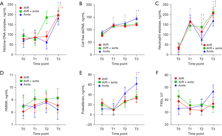
Perioperative changes of circulating markers based on three types of surgery are shown in Figure 1. Statistically significant increases in the levels of histone-DNA complex were detected T3 compared with T0 in all three types of surgery (Figure 1A). Significant increases in the levels of cell-free dsDNA and neutrophil elastase were observed T1 and these elevations persisted in an augmented state until T3 compared with the baseline at T0 in all three types, as illustrated in Figure 1B,1C. Regarding contact system markers, a decrease in the level of prekallikrein was observed at T1 for all three types of surgery, although statistical significance was not reached in the Aorta group. Significantly elevated levels of prekallikrein were detected in the Aorta group at T3 compared with T0 (Figure 1E). No significant changes in HMWK and FXIIa were observed across time (Figure 1D,1F).
Figure 1.
Perioperative changes in markers of neutrophil extracellular traps and the contact system based on cardiac surgery type across four time points (T0, before surgery; T1, immediately after surgery; T2, 1 day after surgery; T3, 3 days after surgery). (A) Histone-DNA complex, (B) cell-free dsDNA, (C) neutrophil elastase, (D) HMWK, (E) prekallikrein, (F) FXIIa. *P<0.05 vs. T0, †P<0.05 vs. T1, ‡P<0.05 vs. T2 in paired t-test. Red line and sign, aortic valve replacement (AVR, n=46); green line and sign, aortic valve and ascending aorta replacement (AVR + aorta, n=28); blue line and sign, aorta replacement (Aorta, n=23). AVR, aortic valve replacement; AVR + aorta, aortic valve replacement and ascending aorta surgery; Aorta, aorta replacement surgery; dsDNA, double stranded DNA; HMWK, high molecular weight kininogen; FXIIa, activated factor XII.
At baseline time of T0, the percentage of total patients who showed increased circulating levels of the NETs and contact system compared with reference range were shown (Table S1). The histone-DNA complex, cell free dsDNA, neutrophil elastase, HMWK, prekallikrein and FXIIa at T0 were increased in 5.15%, 19.59%, 0%, 46.39%, 32.99% and 30.93%, respectively.
Changes of circulating markers in regard to the occurrence of AF
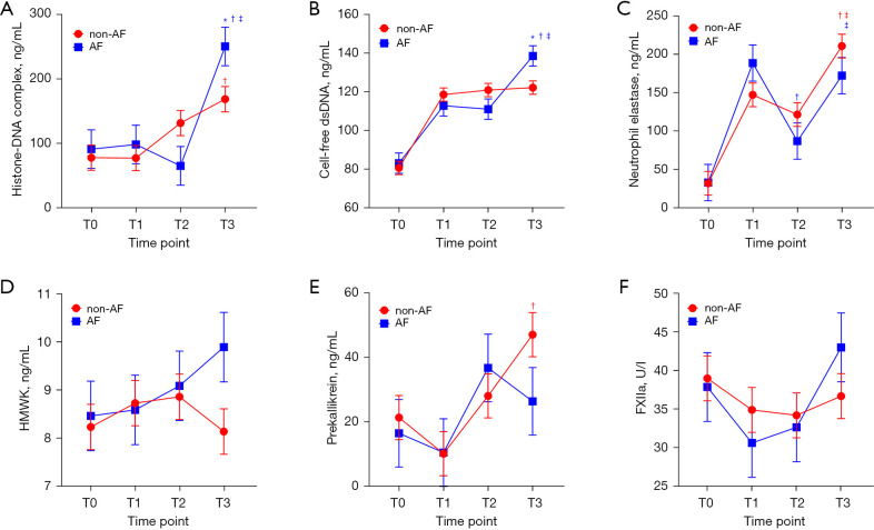
Perioperative changes of circulating markers between patients with AF (n=29) and those without (n=68) are shown in Figure 2. Notably, histone-DNA complex and cell-free dsDNA showed a sudden increase in patients with AF at T3, compared with T2, while these markers showed no significant increase in patients without AF at T3, compared with T2 (Figure 2A,2B). Neutrophil elastase showed an increasing trend at T3 compared with 1 day after surgery (T2) in both patients with and without AF (Figure 2C). HMWK showed an increasing trend after surgery in patients with AF, but without statistical significance (Figure 2D). The levels of prekallikrein showed a significant increase at T3 compared with T1 in patients without AF (Figure 2E). No specific trend was observed for FXIIa levels (Figure 2F).
Figure 2.
Perioperative changes in markers of neutrophil extracellular traps and the contact system based on AF across four-time points (T0, before surgery; T1, immediately after surgery; T2, 1 day after surgery; T3, 3 days after surgery). (A) Histone-DNA complex, (B) cell-free dsDNA, (C) neutrophil elastase, (D) HMWK, (E) prekallikrein, (F) FXIIa. *P<0.05 vs. non-AF at linear mixed model; †P<0.05 vs. T1, ‡P<0.05 vs. T2 in paired t-test. Red line and sign, patients without post-operative AF (n=68); blue line and sign, patients with post-operative AF (n=29). AF, atrial fibrillation; dsDNA, double stranded DNA; HMWK, high molecular weight kininogen; FXIIa, activated factor XII.
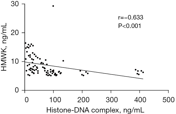
At the T1 time point, the levels of histone-DNA complex showed a reverse correlation with HMWK (r=−0.633, P<0.001) (Figure 3). No significant correlation was observed between the levels of histone-DNA complex and levels of prekallikrein and FXIIa (data not shown). No statistically significant perioperative changes in circulating markers were observed between patients with AKI (n=42) and those without (n=50) (data not shown).
Figure 3.

Correlation of histone-DNA complex with HMWK. Spearman’s correlation coefficients for pairwise comparisons of histone-DNA complex and HMWK at T1 (immediately after surgery). HMWK, high molecular weight kininogen.
Risk assessment for circulating markers for the occurrence of AF
The high level of histone-DNA complex (>400 ng/mL) at T3 showed a significantly high odds ratio for risk associated with the occurrence of post-operative AF, as delineated in Table 2. Results from the calculation of delta-changes (∆) as values at T3 minus values at T2 indicated that a high level of ∆ histone-DNA complexT3-T2 (>263 ng/mL) was a significant marker for the occurrence of AF. Moreover, the presence of high levels of cell-free dsDNA (>138 ng/mL) and ∆ cell-free dsDNAT3-T2 (>8.86 ng/mL) were significant markers for the occurrence of AF. Neutrophil elastase and three contact system markers did not show statistical significance at T3.
Table 2. Predictive circulating markers at the T3 time point (3 days after surgery) for post-operative occurrence of atrial fibrillation.
| Markers | Odds ratio | 95% CI | P value |
|---|---|---|---|
| NETs markers | |||
| Histone-DNA complex (>400 vs. ≤400 ng/mL) | 14.532 | 1.560–135.413 | 0.02 |
| ∆ Histone-DNA complexT3-T2 (>263 vs. ≤263 ng/mL) | 4.761 | 1.213–18.691 | 0.03 |
| Cell-free dsDNA (>138 vs. ≤138 ng/mL) | 2.896 | 1.076–7.797 | 0.04 |
| ∆ Cell-free dsDNAT3-T2 (>8.86 vs. ≤8.86 ng/mL) | 5.117 | 1.910–13.709 | 0.001 |
| Neutrophil elastase (>426 vs. ≤426 ng/mL) | 0.494 | 0.094–2.592 | 0.40 |
| Contact system markers | |||
| Prekallikrein (>7.57 vs. ≤7.57 ng/mL) | 0.607 | 0.171–2.157 | 0.44 |
| HMWK (>12.6 vs. ≤12.6 ng/mL) | 3.124 | 0.912–10.697 | 0.07 |
| FXIIa (>63.9 vs. ≤63.9 U/I) | 2.714 | 0.789–9.338 | 0.11 |
Age- and sex-adjusted multivariate logistic regression was performed for the assessment of atrial fibrillation risk. ∆ Histone-DNA complexT3-T2 was calculated according to changes in histone-DNA complex levels between T2 (1 day after surgery) and T3 (3 days after surgery). ∆ Cell-free dsDNAT3-T2 was calculated according to changes in cell-free dsDNA levels between T2 and T3. NETs, neutrophil extracellular traps; dsDNA, double stranded DNA; HMWK, high molecular weight kininogen; FXIIa, activated factor XII; CI, confidence interval.
Prognostic value of cell-free dsDNA
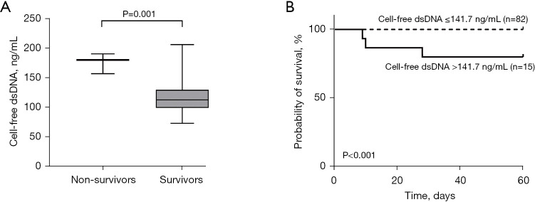
Significantly higher levels of cell-free dsDNA were detected at T2 in non-survivors (n=3) compared to survivors (n=94), as illustrated in Figure 4A. Conversely, no significant differences in other markers at T2 and all markers at other time points were observed between non-survivors and survivors (data not shown). In Kaplan-Meier analysis, patients were divided into two distinct groups according to the cutoff value of cell-free dsDNA at T2 determined from ROC analysis. A significantly lower survival rate was observed for patients with high levels of cell-free dsDNA (>141.7 ng/mL) (Figure 4B).
Figure 4.
Prognostic value of cell-free dsDNA at T2 (1 day after surgery). (A) Differences in cell-free dsDNA levels at T2 between non-survivors (n=3) and survivors (n=94). (B) Kaplan-Meier survival analysis stratified for cell-free dsDNA at T2. dsDNA, double stranded DNA.
Discussion
The findings elucidated by this study demonstrated that the levels of circulating markers of NETs were elevated across post-CPB time, suggesting that an inflammatory response that varied in intensity was triggered by CPB (17). In particular, significantly elevated levels of histone-DNA complex and cell-free dsDNA were detected 3 days after surgery in patients with post-operative AF compared to those without. NETs could induce injury to the mitochondria of cardiomyocytes (18). Large numbers of free radicals capable of oxidizing many intracellular targets, including sodium channels, are released as a result of dysfunctional mitochondria (19). These changes can directly alter the excitability of cardiomyocytes. In addition, myeloperoxidase (MPO) is released to endothelial cells by activated neutrophils via a direct CD11b/CD18-integrin (20). MPO promotes fibrosis, thereby increasing susceptibility to AF (21). Accordingly, elevated formation of NETs is considered to influence the occurrence of AF.
Unlike the two NETs markers, the level of neutrophil elastase was not significantly elevated in patients with AF. Release of neutrophil elastase can occur in a non-specific inflammatory response (22). Its levels are likely to be increased even in patients without AF due to the inflammatory potential associated with surgery. In addition, the release of neutrophil elastase occurs during the initial process of NETs formation, and the release of NETs finally occurs via a downstream signaling cascade for chromatin de-condensation (23-25). Therefore, it is believed that AF is associated more with the final formation of NETs than with the early stage of inflammation.
In the results of logistic regression analysis, levels of histone-DNA complex and cell-free dsDNA measured at 3 days after surgery were significant markers of risk for the occurrence of AF. Although the association of several inflammatory markers, including post-operative neutrophil/lymphocyte ratio (26), IL-6 (13), and mitochondrial damage-associated molecular patterns (16) with post-operative AF has been reported, to the best of our knowledge, research on circulating markers of NETs has not been previously reported. The findings of this study demonstrated for the first time the significance of NETs markers as factors in the assessment of the risk for occurrence of AF after CPB surgery.
Circulating markers were measured at four-time points in this study, however, high levels of histone-DNA complex and cell-free dsDNA were significant markers for the risk of AF only at 3 days after surgery. Measurement of NETs markers post-operatively, rather than pre-operatively, will likely provide information regarding the development of post-operative AF. Given that post-operative AF is typically reported with a peak incidence between days 2 and 4 after surgery (27) and that the results of our study also showed a median onset of 3 days, the observed elevation of NETs markers at 3 days after surgery could be a consequence of post-operative AF, in addition to being a causal factor. In the future, testing at more detailed time points may be required for clarification of the causal relationship between NETs and AF.
According to our results, increased levels of prekallikrein were observed at 3 days after surgery compared with levels measured before surgery in patients who underwent aorta replacement surgery (aorta group), but not in other surgery. This could be explained by the significantly lower body temperature detected during CPB in the Aorta group compared with other surgery. The lower the temperature during CPB, the less prekallikrein is degraded (28), thus it is possible that increased levels of prekallikrein detected after surgery in the Aorta group were caused by low temperature.
A negative correlation was observed between levels of histone-DNA complex and HMWK immediately after surgery. NETs provide a negatively charged surface, therefore, increased formation of NETs could induce activation of the contact system through the negatively charged surface. During activation of the contact system, FXII is autocleaved into FXIIa, which causes the conversion of prekallikrein into kallikrein, and then kallikrein cleaves HMWK into bradykinin (29). Therefore, activation of the contact system proceeds with the consumption of prekallikrein and HMWK (30,31). The level of circulating HMWK is likely to show a decrease in patients with a high level of NETs formation. Although prekallikrein and FXIIa did not show a correlation with histone-DNA complex, their levels showed a substantial decrease at immediately after surgery compared to the initial assessment, suggesting its consumption due to activation of the contact system regardless of the extent of NETs formation.
Duration of CPB (32), high mobility group box 1 (33), and neutrophil-lymphocyte ratio (34) have been reported as risk factors for post-operative morbidity and mortality. Our findings demonstrated that the level of cell-free dsDNA measured on 1 day after surgery was of significant prognostic value. Inflammation triggered by CPB leads to the release of large numbers of NETs. Released NETs could cause occlusion of capillaries, impairment of microcirculation, tissue damage, and exacerbate inflammation. In addition, NETs or their components can cause damage to the endothelium, leading to the development of diffuse thrombosis, which can result in disseminated intravascular coagulation and acute organ injury, both of which can increase mortality (35).
However, our study is accompanied by several limitations. First, data were collected from a single center using a non-blinded design. Conduct of a multicenter study using a blinded design will be required for clinical application of NETs markers in CPB. Second, our study focused on aortic valve and/or aorta replacement surgery, thus caution should be used when attempting to extrapolate our results to other cardiac surgeries. Third, only three patients died after surgery. This small number of patients might not be adequate to determine the prognostic values. Conduct of future studies including large numbers of patients will be required. Finally, all patients in this study underwent cardiac surgery with CPB. Patients undergoing off-pump cardiac surgery as the control group would be required in order to clarify the mechanism of NETs formation in the context of CPB.
Conclusions
In summary, an increase in the level of NETs was observed after surgery in patients who underwent CPB. The high levels of histone-DNA complex and the high levels of cell-free dsDNA measured at 3 days after surgery were significant markers showing association with post-operative AF. Furthermore, the levels of cell-free dsDNA measured at 1 day after surgery showed significant prognostic value in patients who underwent CPB. Our findings suggest that markers of NETs may be useful for the assessment of risk for post-operative AF and mortality. The conduct of additional studies regarding the role of NETs as clinical markers and as a therapeutic target in CPB is anticipated.
Supplementary
The article’s supplementary files as
Acknowledgments
Funding: This research was supported by a grant of Korean Cell-Based Artificial Blood Project funded by the Korean government (The Ministry of Science and ICT, The Ministry of Trade, Industry and Energy, the Ministry of Health & Welfare, the Ministry of Food and Drug Safety) (grant number: HX23C1718).
Ethical Statement: The authors are accountable for all aspects of the work in ensuring that questions related to the accuracy or integrity of any part of the work are appropriately investigated and resolved. The study was approved by the Institutional Review Board of Seoul National University Hospital (IRB No. 2104-231-1217). Written informed consent was obtained from all participants. The study was conducted in accordance with the Declaration of Helsinki (as revised in 2013).
Footnotes
Reporting Checklist: The authors have completed the STROBE reporting checklist. Available at https://jtd.amegroups.com/article/view/10.21037/jtd-24-295/rc
Conflicts of Interest: All authors have completed the ICMJE uniform disclosure form (available at https://jtd.amegroups.com/article/view/10.21037/jtd-24-295/coif). The authors have no conflicts of interest to declare.
Data Sharing Statement
Available at https://jtd.amegroups.com/article/view/10.21037/jtd-24-295/dss
References
- 1.Warren OJ, Smith AJ, Alexiou C, et al. The inflammatory response to cardiopulmonary bypass: part 1--mechanisms of pathogenesis. J Cardiothorac Vasc Anesth 2009;23:223-31. 10.1053/j.jvca.2008.08.007 [DOI] [PubMed] [Google Scholar]
- 2.Paparella D, Yau TM, Young E. Cardiopulmonary bypass induced inflammation: pathophysiology and treatment. An update. Eur J Cardiothorac Surg 2002;21:232-44. 10.1016/S1010-7940(01)01099-5 [DOI] [PubMed] [Google Scholar]
- 3.Kaplan MJ, Radic M. Neutrophil extracellular traps: double-edged swords of innate immunity. J Immunol 2012;189:2689-95. 10.4049/jimmunol.1201719 [DOI] [PMC free article] [PubMed] [Google Scholar]
- 4.Vorobjeva NV, Chernyak BV. NETosis: Molecular Mechanisms, Role in Physiology and Pathology. Biochemistry (Mosc) 2020;85:1178-90. 10.1134/S0006297920100065 [DOI] [PMC free article] [PubMed] [Google Scholar]
- 5.Faymonville ME, Pincemail J, Duchateau J, et al. Myeloperoxidase and elastase as markers of leukocyte activation during cardiopulmonary bypass in humans. J Thorac Cardiovasc Surg 1991;102:309-17. 10.1016/S0022-5223(19)36564-X [DOI] [PubMed] [Google Scholar]
- 6.Weidmann H, Heikaus L, Long AT, et al. The plasma contact system, a protease cascade at the nexus of inflammation, coagulation and immunity. Biochim Biophys Acta Mol Cell Res 2017;1864:2118-27. 10.1016/j.bbamcr.2017.07.009 [DOI] [PubMed] [Google Scholar]
- 7.Ge L, Zhou X, Ji WJ, et al. Neutrophil extracellular traps in ischemia-reperfusion injury-induced myocardial no-reflow: therapeutic potential of DNase-based reperfusion strategy. Am J Physiol Heart Circ Physiol 2015;308:H500-9. 10.1152/ajpheart.00381.2014 [DOI] [PubMed] [Google Scholar]
- 8.Klopf J, Brostjan C, Eilenberg W, et al. Neutrophil Extracellular Traps and Their Implications in Cardiovascular and Inflammatory Disease. Int J Mol Sci 2021;22:559. 10.3390/ijms22020559 [DOI] [PMC free article] [PubMed] [Google Scholar]
- 9.Echahidi N, Pibarot P, O’Hara G, et al. Mechanisms, prevention, and treatment of atrial fibrillation after cardiac surgery. J Am Coll Cardiol 2008;51:793-801. 10.1016/j.jacc.2007.10.043 [DOI] [PubMed] [Google Scholar]
- 10.Maesen B, Nijs J, Maessen J, et al. Post-operative atrial fibrillation: a maze of mechanisms. Europace 2012;14:159-74. 10.1093/europace/eur208 [DOI] [PMC free article] [PubMed] [Google Scholar]
- 11.Ishii Y, Schuessler RB, Gaynor SL, et al. Inflammation of atrium after cardiac surgery is associated with inhomogeneity of atrial conduction and atrial fibrillation. Circulation 2005;111:2881-8. 10.1161/CIRCULATIONAHA.104.475194 [DOI] [PubMed] [Google Scholar]
- 12.Wang Z, Feng J, Nattel S. Idiopathic atrial fibrillation in dogs: electrophysiologic determinants and mechanisms of antiarrhythmic action of flecainide. J Am Coll Cardiol 1995;26:277-86. 10.1016/0735-1097(95)90845-F [DOI] [PubMed] [Google Scholar]
- 13.Gaudino M, Andreotti F, Zamparelli R, et al. The -174G/C interleukin-6 polymorphism influences postoperative interleukin-6 levels and postoperative atrial fibrillation. Is atrial fibrillation an inflammatory complication? Circulation 2003;108 Suppl 1:II195-9. 10.1161/01.cir.0000087441.48566.0d [DOI] [PubMed] [Google Scholar]
- 14.Olesen OJ, Vinding NE, Østergaard L, et al. C-reactive protein after coronary artery bypass graft surgery and its relationship with postoperative atrial fibrillation. Europace 2020;22:1182-8. 10.1093/europace/euaa088 [DOI] [PubMed] [Google Scholar]
- 15.Ishida K, Kimura F, Imamaki M, et al. Relation of inflammatory cytokines to atrial fibrillation after off-pump coronary artery bypass grafting. Eur J Cardiothorac Surg 2006;29:501-5. 10.1016/j.ejcts.2005.12.028 [DOI] [PubMed] [Google Scholar]
- 16.Sandler N, Kaczmarek E, Itagaki K, et al. Mitochondrial DAMPs Are Released During Cardiopulmonary Bypass Surgery and Are Associated With Postoperative Atrial Fibrillation. Heart Lung Circ 2018;27:122-9. 10.1016/j.hlc.2017.02.014 [DOI] [PubMed] [Google Scholar]
- 17.Beaubien-Souligny W, Neagoe PE, Gagnon D, et al. Increased Circulating Levels of Neutrophil Extracellular Traps During Cardiopulmonary Bypass. CJC Open 2020;2:39-48. 10.1016/j.cjco.2019.12.001 [DOI] [PMC free article] [PubMed] [Google Scholar]
- 18.He L, Liu R, Yue H, et al. Interaction between neutrophil extracellular traps and cardiomyocytes contributes to atrial fibrillation progression. Signal Transduct Target Ther 2023;8:279. 10.1038/s41392-023-01497-2 [DOI] [PMC free article] [PubMed] [Google Scholar]
- 19.Liu M, Liu H, Dudley SC, Jr. Reactive oxygen species originating from mitochondria regulate the cardiac sodium channel. Circ Res 2010;107:967-74. 10.1161/CIRCRESAHA.110.220673 [DOI] [PMC free article] [PubMed] [Google Scholar]
- 20.Jerke U, Rolle S, Purfürst B, et al. β2 integrin-mediated cell-cell contact transfers active myeloperoxidase from neutrophils to endothelial cells. J Biol Chem 2013;288:12910-9. 10.1074/jbc.M112.434613 [DOI] [PMC free article] [PubMed] [Google Scholar]
- 21.Rudolph V, Andrié RP, Rudolph TK, et al. Myeloperoxidase acts as a profibrotic mediator of atrial fibrillation. Nat Med 2010;16:470-4. 10.1038/nm.2124 [DOI] [PMC free article] [PubMed] [Google Scholar]
- 22.Pham CT. Neutrophil serine proteases: specific regulators of inflammation. Nat Rev Immunol 2006;6:541-50. 10.1038/nri1841 [DOI] [PubMed] [Google Scholar]
- 23.Papayannopoulos V, Metzler KD, Hakkim A, et al. Neutrophil elastase and myeloperoxidase regulate the formation of neutrophil extracellular traps. J Cell Biol 2010;191:677-91. 10.1083/jcb.201006052 [DOI] [PMC free article] [PubMed] [Google Scholar]
- 24.Hakkim A, Fuchs TA, Martinez NE, et al. Activation of the Raf-MEK-ERK pathway is required for neutrophil extracellular trap formation. Nat Chem Biol 2011;7:75-7. 10.1038/nchembio.496 [DOI] [PubMed] [Google Scholar]
- 25.Metzler KD, Goosmann C, Lubojemska A, et al. A myeloperoxidase-containing complex regulates neutrophil elastase release and actin dynamics during NETosis. Cell Rep 2014;8:883-96. 10.1016/j.celrep.2014.06.044 [DOI] [PMC free article] [PubMed] [Google Scholar]
- 26.Weedle RC, Da Costa M, Veerasingam D, et al. The use of neutrophil lymphocyte ratio to predict complications post cardiac surgery. Ann Transl Med 2019;7:778. 10.21037/atm.2019.11.17 [DOI] [PMC free article] [PubMed] [Google Scholar]
- 27.Dobrev D, Aguilar M, Heijman J, et al. Postoperative atrial fibrillation: mechanisms, manifestations and management. Nat Rev Cardiol 2019;16:417-36. 10.1038/s41569-019-0166-5 [DOI] [PubMed] [Google Scholar]
- 28.Engelman RM, Pleet AB, Rousou JA, et al. Influence of cardiopulmonary bypass perfusion temperature on neurologic and hematologic function after coronary artery bypass grafting. Ann Thorac Surg 1999;67:1547-55; discussion 1556. 10.1016/S0003-4975(99)00360-4 [DOI] [PubMed] [Google Scholar]
- 29.Kongsgaard UE, Smith-Erichsen N, Geiran O, et al. Different activation patterns in the plasma kallikrein-kinin and complement systems during coronary bypass surgery. Acta Anaesthesiol Scand 1989;33:343-7. 10.1111/j.1399-6576.1989.tb02921.x [DOI] [PubMed] [Google Scholar]
- 30.Campbell DJ, Dixon B, Kladis A, et al. Activation of the kallikrein-kinin system by cardiopulmonary bypass in humans. Am J Physiol Regul Integr Comp Physiol 2001;281:R1059-70. 10.1152/ajpregu.2001.281.4.R1059 [DOI] [PubMed] [Google Scholar]
- 31.Gallimore MJ, Jones DW, Winter M, et al. Changes in high molecular weight kininogen levels during and after cardiopulmonary bypass surgery measured using a chromogenic peptide substrate assay. Blood Coagul Fibrinolysis 2002;13:561-8. 10.1097/00001721-200209000-00012 [DOI] [PubMed] [Google Scholar]
- 32.Salis S, Mazzanti VV, Merli G, et al. Cardiopulmonary bypass duration is an independent predictor of morbidity and mortality after cardiac surgery. J Cardiothorac Vasc Anesth 2008;22:814-22. 10.1053/j.jvca.2008.08.004 [DOI] [PubMed] [Google Scholar]
- 33.Kim N, Lee S, Lee JR, et al. Prognostic role of serum high mobility group box 1 concentration in cardiac surgery. Sci Rep 2020;10:6293. 10.1038/s41598-020-63051-2 [DOI] [PMC free article] [PubMed] [Google Scholar]
- 34.Silberman S, Abu-Yunis U, Tauber R, et al. Neutrophil-Lymphocyte Ratio: Prognostic Impact in Heart Surgery. Early Outcomes and Late Survival. Ann Thorac Surg 2018;105:581-6. 10.1016/j.athoracsur.2017.07.033 [DOI] [PubMed] [Google Scholar]
- 35.Camicia G, Pozner R, de Larrañaga G. Neutrophil extracellular traps in sepsis. Shock 2014;42:286-94. 10.1097/SHK.0000000000000221 [DOI] [PubMed] [Google Scholar]


