Abstract
Background
Gastric cancer (GC) stands out as one of the most prevalent malignancies affecting the digestive system, characterized by a substantial incidence rate and mortality. Maternal embryonic leucine zipper kinase (MELK) has been implicated in the advancement of various cancer types and the modulation of the tumor microenvironment. This study aims to delve into the involvement of MELK in chemoresistance and the tumor microenvironment of GC.
Methods
The MELK expression was detected using quantitative real-time polymerase chain reaction (qRT-PCR), western blotting and immunohistochemistry. Lentiviral transfection was employed to establish stable cell lines with either overexpressed or silenced MELK. The impact of MELK on the chemoresistance of GC cells and the polarization of macrophages was investigated through in vitro and in vivo functional assays. Additionally, the correlation between MELK and the cytokines colony-stimulating factor 1 (CSF-1), as well as stromal macrophages, was analysed. The prognostic significance of MELK, CSF-1, and CD206 expression levels in clinical samples was further investigated.
Results
MELK was found to be highly expressed in chemoresistant GC cells and tissues. Furthermore, both in vitro and in vivo assays indicated that MELK overexpression conferred chemoresistance in GC cells. Additionally, MELK overexpression was observed to induce M2 macrophage polarization via the CSF-1/JAK2/STAT3 pathway, thereby contributing to chemoresistance within the tumor microenvironment. The expression of MELK in GC tissues from neoadjuvant chemotherapy patients correlated positively with CSF-1 and CD206. Moreover, patients with higher expression levels of MELK, CSF-1, or CD206 exhibited significantly shorter OS and DFS rates.
Conclusions
Our investigation underscores the critical role of MELK in promoting chemoresistance and inducing M2 macrophage polarization in GC. It proposes novel targets and methods for the treatment of GC, as well as prognostic factors for neoadjuvant chemotherapy.
Supplementary Information
The online version contains supplementary material available at 10.1186/s12935-024-03453-8.
Keywords: Gastric cancer, MELK, Chemoresistance, Tumor-associated macrophages, Polarization, Prognosis
Background
While combining surgery and perioperative chemotherapy has partially enhanced the therapeutic outcomes for gastric cancer (GC), it remains the fifth most prevalent malignancy and the fourth most common cause of cancer-related mortality worldwide [1]. 5-fluorouracil (5-Fu) is one of the most commonly prescribed antitumor drugs in perioperative chemotherapy for GC patients. However, chemoresistance stands out as a pivotal factor contributing to treatment failure and dismal prognoses among patients with GC [2]. Therefore, it is necessary to explore molecular targets of chemotherapy resistance in order to enhance the efficacy of treatment strategies.
Maternal embryonic leucine zipper kinase (MELK) is a serine/threonine kinase belonging to the AMP-activated protein kinase (AMP)/sucrose-non-fermenting kinase 1 (SNF1) family [3]. Studies have shown that MELK has vital function in multiple cellular processes, including cell cycle regulation, cell proliferation, cell apoptosis, mitotic progression, and RNA processing [4–6]. In addition, upregulation of MELK has been found in various human tumors, including breast cancer, glioblastomas, prostate cancer, colorectal cancer and GC [7–11]. It has been shown that higher MELK expression was closely related to poorer clinical outcomes, such as more advanced stages, vascular invasion and distant metastasis [3, 12]. Moreover, abnormal expression of MELK could confer therapy resistance in multiple types of cancers. For instance, MELK-dependent activation of FOXM1 regulates DNA repair and mediates resistance to doxorubicin in breast cancer [13, 14] In glioblastoma multiforme, JNK-driven MELK/c-JUN signaling maintains glioma stem cells in an immature state to facilitate therapy resistance in a p53-dependent manner [15]. Du et al. identified that MELK promoted cell migration and invasion through activating the FAK/paxillin signaling pathway and played a vital role in the development and occurrence of GC [16]. However, to date, the function of MELK in the development of chemoresistance of GC remains unexplored.
Tumor progression is influenced not only by the malignant biological characteristics of tumor cells, but also by the regulation and support provided by tumor microenvironment (TME) [17]. As one of the major cellular components of TME, tumor-associated macrophages (TAMs) exhibit various phenotypes and functional features, constituting the dominant immune cells involved in tumor growth, angiogenesis, metastasis and therapy resistance [18]. TAMs can be broadly separated into the classically activated (M1 phenotype) and the alternatively activated phenotype (M2 phenotype), depending on the stimulating signals from various cytokine or pathogen in the TME [19, 20]. As tumor progress, TAMs often shift towards the M2 phenotype, promoting tumor malignancy and contributing to poor prognosis in the majority of cases. In our previous study, we found that chemoresistant GC cells induced macrophages polarization towards M2 phenotype more effectively than parental GC cells. M2 macrophages, in turn, further promoted the chemoresistance of GC cells by activating the CXCL5/PI3K/AKT/mTOR signaling axis [21]. However, the mechanism underlying the M2 polarization of TAMs induced by chemoresistant GC cells remains unclear.
The present study illustrated that MELK expression was upregulated in chemoresistant GC cells and tissues. Both in vitro and in vivo experiments revealed that heightened MELK expression could increase resistance to 5-Fu in GC cells, whereas knocking down MELK could enhance sensitivity to 5-Fu. Then we found that MELK overexpression could induce macrophage polarization towards M2 phenotype through activating the colony-stimulating factor 1 (CSF-1)/Janus kinase 2 (JAK2)/signal transducer and activator of transcription 3 (STAT3) pathway, thereby contributing to chemoresistance within the TME. MELK expression in GC tissue following neoadjuvant chemotherapy correlated positively with CSF-1 and CD206. Patients with elevated expression levels of MELK, CSF-1, or CD206 exhibited significantly poorer prognoses. Our study delved into the interplay between MELK expression, GC chemoresistance and M2 polarization of TAMs. These findings suggest the potential utility of MELK and CSF-1 as predictive markers for chemotherapeutic effective and the prognosis, offering a novel avenue for precision therapy in patients with GC.
Materials and methods
Clinical samples
GC tissues were collected from 106 patients who underwent neoadjuvant chemotherapy with the 5-Fu-based regimen and radical gastrectomy between 2016 and 2018 at Peking Union Medical College Hospital. The clinical and demographic characteristics of patients were listed in Supplementary Table S1. Patients were divided into two groups based on the degree of pathological response, as evaluated according to the guidelines of the College of American Pathologists (CAP) [22]. CAP 0, CAP 1 and CAP 2 were defined as exhibiting pathological response (n = 63), whereas CAP 3 indicated no pathological response (n = 43). Additionally, fifteen normal gastric mucosal tissue samples were obtained via endoscopic biopsy. Tumor samples were collected with patients' written informed consent, and the study was reviewed and approved by the Institutional Review Board of Peking Union Medical College Hospital.
Immunohistochemistry staining
A total of 106 collected tumor specimens were fixed in 10% formaldehyde solution, embedded in paraffin, and then serially severed into slices (4 μm). After being deparaffinized and rehydrated by xylene and graded ethanol, antigen was retrieved using microwave heating with sodium citrate retrieval buffer (pH 6.0). The endogenous peroxidase was inactivated by treatment with 3% H2O2 for 10 min. Following treatment with blocking buffer to obstruct nonspecific bindings, the samples were incubated with primary antibodies: Anti-MELK (1:100, #11403-1-AP, Proteintech Group), Anti-CSF-1 (1:100, #ab233387, Abcam), and Anti-CD206 (1:100, #18704-1-AP, Proteintech Group) at 4 °C overnight. The next day, after PBS washing, horseradish peroxidase (HRP)-conjugated antibody anti-Rabbit IgG (1:100, #7074, Cell Signaling Technology) was added for 30 min’ incubation at room temperature. 3, 3ʹ-diaminobenzidine (DAB) regent was taken for brown staining subsequent to PBS washing, then all slices were re-dyed with hematoxylin staining solution, dehydrated, and sealed for microscopic examination. At least three slices were taken from each specimen and five independent fields were randomly chosen from each slice for microscopic examination. The immunoreactivity of MELK and CSF-1 was scored by multiplying the scores of percentage of positive cells (< 5% scores 0, 5–25% scores 1, 25–50% scores 2, 50–75% scores 3, and 75–100% scores 4) with the scores of staining intensity (no staining scores 0, weak staining scores 1, moderate staining scores 2, and strong staining scores 3). The overall scores of 0–3 were defined as low expression, while scores above 3 were considered high expression. For CD206 evaluation, the Olympus microscope was used to count the number of positive cells in five random 400-fold fields.
Cell lines and culture
Human gastric cancer cell lines MKN45 and HGC27, human mononuclear cells.
THP-1, and human gastric epithelial cell line GES-1 were obtained from the Cell Resource Center of Peking Union Medical College (Beijing, China). Parental gastric cancer cells were regarded as 5-Fu-sensitive (MKN45-S and HGC27-S) and 5-Fu-resistant cells (MKN45-R and HGC27-R) were generated as previously reported [21]. All cells were grown in RPMI-1640 medium (Gibco, Carlsbad, CA, USA) containing 10% fetal bovine serum (FBS) (Gibco) and 1% penicillin/streptomycin (Gibco) at 37 °C in a humidified incubator with a volume fraction of 5% CO2. For the generation of M0 macrophages, THP-1 cells were induced by 100 ng/ml phorbol 12-myristate 13-acetate (PMA) (Sigma-Aldrich, St. Louis, MO, USA) for 24 h.
Co-culture of cancer cells and macrophages
Transwell chambers (6-well plates, 0.4-μm pore size; Corning, NY, USA) were used for co-culture experiment. GC cells were seeded onto the membrane of upper chambers with 0.4-μm pore, while M0 macrophages were seeded onto the lower chambers of 6-well plates for 48 h co-culture. Macrophages were then harvested for subsequent experimental analysis.
Cell viability assays
GC Cell (MKN45-S LV-NC, MKN45-S LV-MELK, HGC27-S LV-NC, HGC27-S LV-MELK, MKN45-R sh-NC, MKN45-R sh-MELK, HGC27-R sh-NC, HGC27-R sh-MELK) viability was measured using Cell Counting Kit-8 (CCK-8; Dojindo, Kumamoto, Japan). Briefly, GC cells were planted in 96-well plates at a density of 5 × 10³ cells per well and cultured at 37 °C in a humidified incubator with 5% CO2 for 24 h. Then cells were incubated with culture medium containing varying concentrations of 5-Fu (0, 5, 10, 20, 40, 80, 120, 160, 200 μg/ml) for another 24 h. 10 μl of CCK-8 reagent was added to each well for 2 h incubation at 37 °C. The absorbance value at 450 nm was measured by microplate reader (Termo Scientific, Rockford, IL, USA).
Colony formation assay
GC cells (MKN45-S LV-NC, MKN45-S LV-MELK, HGC27-S LV-NC, HGC27-S LV-MELK, MKN45-R sh-NC, MKN45-R sh-MELK, HGC27-R sh-NC, HGC27-R sh-MELK) were seeded into 6-well plates at a density of 500 cells per well. After incubation for 24 h, the cells were treated with 5-Fu at a final concentration of 15 μg/ml, and the culture medium was replenished every 3–4 days. Following a two-week incubation period, visible colonies were rinsed with PBS twice and fixed with 4% paraformaldehyde for 15 min. 0.1% crystal violet solution was applied to staining the fixed colonies for 10 min. Colonies containing a minimum of 50 cells were later photographed and counted. The reason for using 15 μg/ml of 5-FU and the method for counting cell colonies were stated and uploaded as Supplementary Text S1.
Cell transfection
Lentiviral transfection was used to establish stable cell lines with overexpressed or silenced MELK. Lentiviral vectors were packaged with full-length MELK gene or shRNA targeting MELK by Genechem (Shanghai). MKN-45-S and HGC-27-S cells were transfected with overexpression lentivirus (LV-MELK) or its negative control (LV-NC), while MKN-45-R and HGC-27-R cells were transfected with shRNA targeting MELK lentivirus (sh-MELK) or its negative control (sh-NC). The full-length MELK gene sequence and three sequences of shRNA targeting MELK were listed in Supplementary Table S2 and S3. Multiplicity of infection (MOI) value was set at 10 based on our preliminary experiments. Cells were chosen for 2 weeks in a medium containing 2 μg/mL puromycin (Sangon Biotech, Shanghai, China), and the alive cells were defined as stable expression cells.
RNA extraction and quantitative real-time polymerase chain reaction (qRT-PCR)
Total RNA was isolated from the tissues and cells using TRIzol reagent (Invitrogen, Carlsbad, CA, USA) according to the manufacturer’s protocol. The RNA samples were reversed to cDNA using 1 μg of total RNA and 5 × PrimeScript RT reagent Kit (Takara, Dalian, China). Then qPCR was carried out by using TB Green Premix Ex Taq II (Takara, Dalian, China). The primer sequences of targeted genes are listed in Supplementary Table S4. The relative expression was calculated with GAPDH using 2−ΔΔCt method and experiment was conducted in triplicate.
Western blot analysis
Total protein was isolated out of cells and tissues using RIPA buffer (Termo Scientifc, Rockford, IL, USA) containing Halt Protease and Phosphatase inhibitor Cocktail (Termo Scientifc, Rockford, IL, USA). The lysates were sonicated followed by centrifugation at 12,000g for 15 min at 4 °C, and then the concentration of extracted protein was measured by BCA Protein Assay Kit (Beyotime, Shanghai, China). Approximately 30 µg of protein was transferred onto polyvinylidene fluoride (PVDF) membranes (Millipore, Billerica, MA, USA) using sodium dodecyl sulfate polyacrylamide gel electrophoresis (SDS-PAGE). Membranes were blocked with TBST solution incorporating 5% non-fat milk for 1 h at room temperature and then immunoblotted overnight at 4 °C with the primary antibodies against MELK (1:1000, #11403-1-AP, Proteintech Group), Jak2 (1:1000, #3230, Cell Signaling Technology), p-Jak2 (1:1000, #3771, Cell Signaling Technology), STAT3 (1:1000, #9139, Cell Signaling Technology), p-STAT3 (1:1000, #9145, Cell Signaling Technology), GAPDH (1:1000, #5174, Cell Signaling Technology). On the following day, the membranes were incubated with corresponding HRP-labeled secondary antibody (1:5000, #7074, Cell Signaling Technology) at room temperature for 1 h. The antibody-antigen complex was visualized using SuperSignal West Pico PLUS Chemiluminescent Substrate (Termo Scientific, Rockford, IL, USA) in a Kodak Image station (Tanon, China).
Enzyme-linked immunosorbent assay (ELISA)
The concentrations of CSF-1 in the cultured media of GC cells were measured by CSF-1 ELISA kits (Cell Signaling Technology, MA, USA) in accordance with the manufacturer’s instructions. The immunoreactive was evaluated by a microplate reader (Termo Scientific, Rockford, IL, USA), and the concentrations were estimated from the standard curve.
Apoptosis assay
Cell apoptosis was measured using Annexin-V-FITC Apoptosis Detection Kit (Dojindo, Kumamoto, Japan) in accordance with the manufacturer’s instructions. Cells were harvested and washed twice with ice-cold PBS and incubated in 100 μl binding buffer in the presence of 5 μl Annexin V-FITC and 5 μl propidium iodide for 15 min in dark conditions. Then apoptosis was quantified using BD Accuri C6 Plus flow cytometer (BD Biosciences, San Jose, CA, USA). The experiment was conducted in triplicate and data were analyzed using FlowJo software (Tree Star, Oregon, OR, USA).
Flow cytometry analysis of macrophages
Macrophages were washed twice using PBS and filtered through a 100 μm cell strainer for flow cytometry analysis. Then 1 × 10⁶ cells in 100 μl staining buffer were stained with FITC-CD11b (BioLegend, San Diego, CA, USA) and PE-CD206 antibodies (BioLegend, San Diego, CA, USA) in darkness for 15 min at 4 °C. Finally, the stained cells were analyzed by BD Accuri C6 Plus flow cytometer (BD Biosciences, San Jose, CA, USA). The process was conducted in triplicate and data were transferred and analyzed using FlowJo software (Tree Star, Oregon, OR, USA).
Mouse tumor xenograft and drug resistance assay in vivo
Male BALB/c nude mice (Sinogenetic Biotechnology, Beijing), aged 4–5 weeks, were randomly divided into four groups (five mice each group). Approximately 1 × 10⁷ cells were resuspended in 200 µL of PBS and then inoculated subcutaneously into the armpit of each mouse. 10 days after the inoculation, the tumor-bearing mice were administrated intraperitoneally with 5-Fu (30 mg/kg) every 3 days. Tumor lengths and widths were measured using calipers and tumor volumes were calculated according to the formula: length × width2/2. Mice were sacrificed at the end of 3 weeks after the administration. Tumors were carefully separated and weighed. The procedure abided by the National Institutes of Health guide for the care and use of Laboratory animals and approved by the Ethics Committee of Animal Experiments of Peking Union Medical College Hospital.
Statistical analysis
Data were shown as mean ± standard deviation (SD) of more than three separate experiments to ensure accuracy. Statistical comparisons between groups were performed by Student’s t-test or one-way analysis of variance (ANOVA). Correlations were assessed using Spearman rank-order correlation. Actuarial rates of survival were analyzed and compared using Kaplan–Meier methods and log-rank tests. All statistical analyses were performed using SPSS 22.0 (SPSS Inc., Chicago, IL, USA) in conjunction with GraphPad Prism 8 (GraphPad Prism Software, Inc., San Diego, CA, USA) and p-value < 0.05 indicated a statistically significant difference (∗p < 0.05, ∗∗p < 0.01, ∗∗∗p < 0.001).
Results
MELK was upregulated in chemoresistant GC cells and tissues
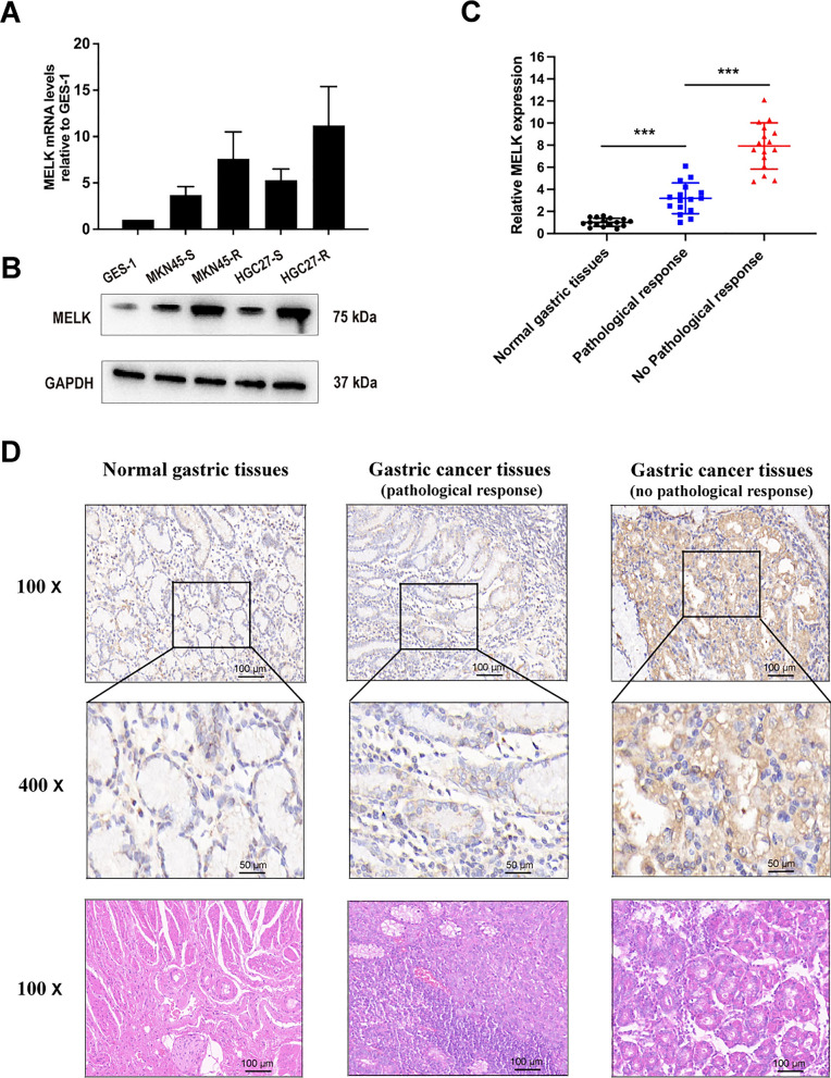
To investigate the potential role of MELK in GC progression, we initially assessed MELK expression in GES-1 cells, as well as the parental GC cells MKN45-S and HGC27-S, along with their chemoresistant counterparts MKN45-R and HGC27-R. Our findings revealed significantly higher levels of MELK expression in GC cells compared to GES-1 cells (Fig. 1A, B). In addition, the chemoresistant GC cells showed higher MELK expression than their parental cells (Fig. 1A, B). We further examined MELK expression in human gastric tissues, including samples from 15 individuals with normal gastric mucosa and samples from patients with or without response to neoadjuvant chemotherapy (n = 63 and n = 43, respectively). As depicted in Fig. 1C and D, MELK expression in tumor tissues was notably higher compared to that in normal gastric mucosal tissues. Particularly noteworthy was the significantly higher MELK expression observed in tumor tissues from patients who did not exhibit a pathological response to neoadjuvant chemotherapy, compared to those who did. These results indicated that MELK expression was upregulated in both GC cells and tumor tissues, particularly in chemoresistant contexts, highlighting its potential role in mediating resistance mechanisms.
Fig. 1.
MELK was highly expressed in chemoresistant GC cells and tissues. A, B MELK expression in gastric epithelial cells, parental and chemoresistant GC cells were evaluated by qRT-PCR and western blot assay. C, D MELK expression in normal gastric tissues and GC tissues was evaluated by qRT-PCR and IHC. Data were presented as mean ± SD of at least three independent experiments. ***p < 0.001
MELK could lead to 5-Fu-resistance in GC cells
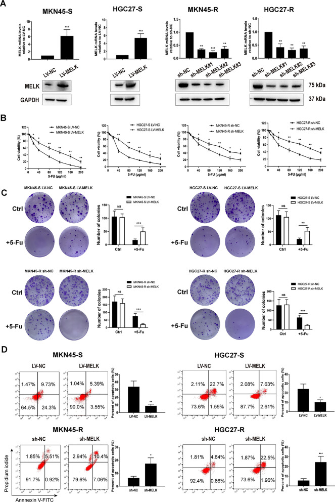
Stably transfected GC cells with MELK overexpression, MELK knockdown, and their corresponding negative control cells were established and then validated using qRT-PCR and western blotting. The results showed that the knockdown efficiency was higher using sh-MELK#2 compared with sh-MELK#1 and sh-MELK#3 (Fig. 2A), therefore, sh-MELK#2 was selected for further experiments. GC cells with MELK overexpression, MELK knockdown and their corresponding negative control cells were respectively incubated with gradient concentrations of 5-Fu (0, 5, 10, 20, 40, 80, 120, 160, 200 μg/ml) for 24 h. CCK-8 assay indicated that MELK overexpression significantly increased 5-Fu-resistance of parental GC cells, while MELK knockdown significantly reduced 5-Fu-resistance in chemoresistant GC cells (Fig. 2B). Similarly, MELK overexpression remarkably increased colony formation of parental GC cells in the presence of 15 μg/ml 5-Fu, and conversely MELK knockdown decreased colony formation of chemoresistant GC cells (Fig. 2C). Moreover, the apoptotic flow cytometry assay demonstrated that MELK overexpression inhibited apoptosis of parental GC cells induced by 5-Fu (Fig. 2D). In contrast, MELK suppression significantly increased apoptosis in chemoresistant GC cells.
Fig. 2.
Effect of MELK on 5-Fu-resistance of GC cells in vitro. A MELK expressions in GC cells with MELK overexpression or knockdown were detected and validated by qRT-PCR and western blot. B GC cells with MELK overexpression or knockdown were incubated with different concentrations of 5-Fu (0, 5, 10, 20, 40, 80, 120, 160, 200 μg/ml) for 24 h, and then cell viability was detected by CCK-8 assay. C GC cells treated with or without 5-Fu for two weeks, the numbers of colonies were counted. D Apoptotic flow cytometry assay was performed to verify that MELK inhibited 5-Fu-induced apoptosis. Data were presented as mean ± SD of three independent experiments. *p < 0.05, **p < 0.01, ***p < 0.001
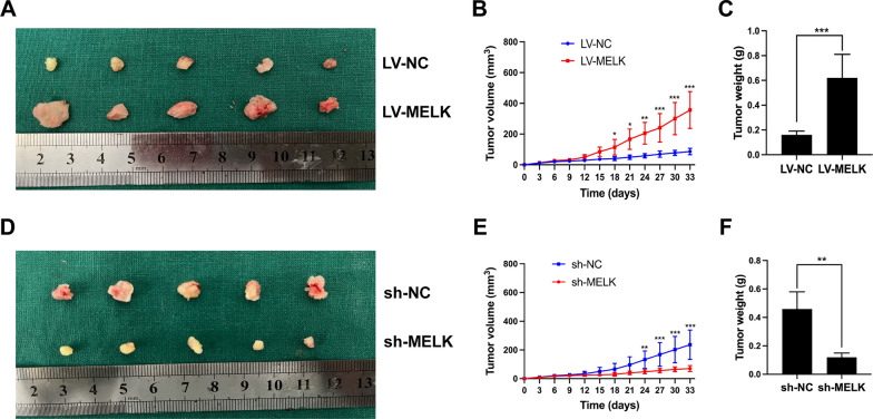
Since our in vitro study revealed that MELK promotes the chemoresistance of GC cells, we conducted further investigation to explore its role in vivo. MKN45-S cells overexpressing MELK, MKN45-R cells silencing MELK, and their corresponding negative control cells were subcutaneously implanted into the armpits of mice (n = 5) followed by the administration of 5-Fu. The results demonstrated that tumors originating from MKN45-S cells overexpressing MELK exhibited accelerated growth, resulting in larger tumor volume and weight compared to those in the control group (Fig. 3A–C). Conversely, MELK knockdown significantly attenuated the chemoresistance of MKN45-R cells, manifesting in slower tumor growth and smaller tumor volume and weight compared to those in the control group (Fig. 3D–F). Therefore, our finding suggest that MELK contributes to 5-Fu-resistance in GC cells.
Fig. 3.
Effect of MELK on 5-Fu-resistance of MKN45 cells in vivo. A–C The comparison of tumor volume and weight in nude mice inoculated with MKN45-S cells overexpressing MELK and negative control cells. D–F The comparison of tumor volume and weight in nude mice inoculated with MKN45-R cells silencing MELK and negative control cells. *p < 0.05, **p < 0.01, ***p < 0.001
MELK could induce M2 polarization of macrophages
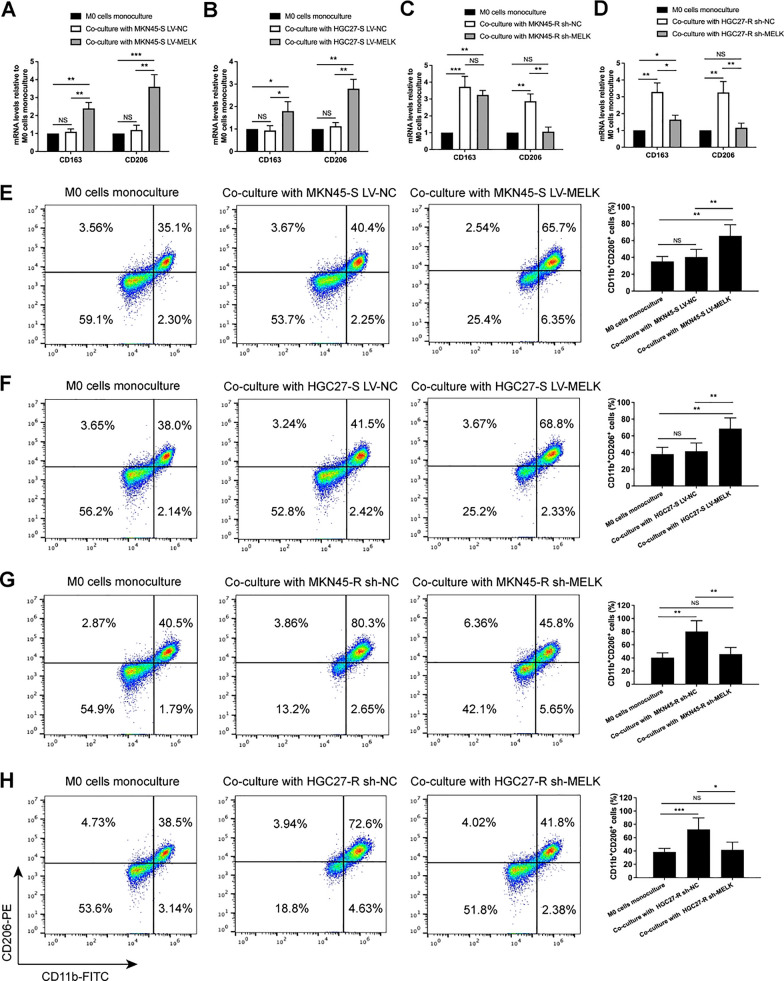
Our previous study demonstrated that chemoresistant GC cells could induce macrophages polarization towards the M2 phenotype, further promoting the chemoresistance of GC cells [21]. The present study indicated that MELK increased the 5-Fu-resistance of GC cells, promoting us to investigate whether MELK could induce M2 polarization of macrophage. TPH-1 cells-derived macrophages were co-cultured with parental GC cells overexpressing MELK, chemoresistant GC cells with MELK knockdown, or their corresponding negative control cells in a non-contacting transwell system for 48 h. The expression of M2 macrophages markers CD163 and CD206 was detected by qRT-PCR. Compared to the negative control cells, co-culturing with parental GC cells overexpressing MELK markedly increased CD163 and CD206 expression in macrophages (Fig. 4A, B). In contrast, co-culturing with chemoresistant GC cells silencing MELK significantly decreased CD163 and CD206 expression in macrophages (Fig. 4C, D). Furthermore, the percentages of CD11b+CD206+ macrophages (M2 macrophages) after co-culturing with GC cells were measured by flow cytometry. Compared to the negative control group, the percentages of CD11b+CD206+ macrophages increased after co-culturing with parental GC cells overexpressing MELK (Fig. 4E, F), whereas the percentages decreased after co-culturing with chemoresistant GC cells that silenced MELK (Fig. 4G, H). These results indicate that MELK induces macrophages polarization towards M2 phenotype.
Fig. 4.
Co-culturing with GC cells overexpressing MELK induced macrophages polarization to M2 phenotype. A–D CD163 and CD206 in macrophages were detected by qRT-PCR in mRNA levels, and the results were normalized by GAPDH mRNA levels. E–H The percentages of CD11b+ CD206+ cells (M2 macrophages) were measured through flow cytometry analysis. Data were presented as mean ± SD of three independent experiments. NS not significantly, *p < 0.05, **p < 0.01, ***p < 0.001
MELK induced macrophages polarization to M2 phenotype via CSF-1/JAK2/STAT3 pathway
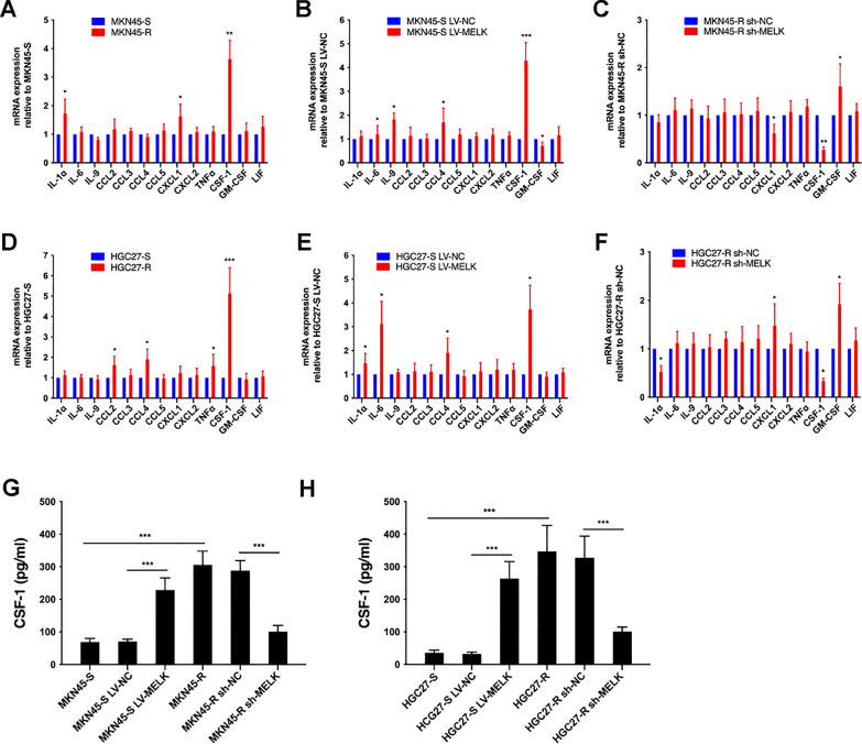
After observing that non-contacting co-culture with GC cells overexpressing or silencing MELK influenced M2 polarization of macrophage, we hypothesized that the distinct abilities of GC cells to induce M2 polarization were attributable to variations in secreted cytokines levels. Following a comprehensive review of relevant literature, thirteen cytokines (IL-1α, IL-6, IL-9, CCL2, CCL3, CCL4, CCL5, CXCL1, CXCL2, TNF-α, CSF-1, GM-CSF and LIF) were selected for analysis via qRT-PCR. As indicated in Fig. 5A–F, among the thirteen cytokines, only the transcriptional level of CSF-1 remained consistent with the trend observed in previous results (Fig. 4). The mRNA levels of CSF-1 elevated in chemoresistant GC cells compared to parental cells (Fig. 5A, D), and they increased in parental GC cells overexpressing MELK compared to negative control cells (Fig. 5B, E). Conversely, they decreased in chemoresistant GC cells with MELK knockdown compared to negative control cells (Fig. 5C, F). CSF-1 levels in the culture supernatants of GC cells were measured using ELISA. Similarly, CSF-1 levels in the culture supernatants aligned with the results of qRT-PCR. Specifically, CSF-1 levels in the culture supernatants of chemoresistant GC cells were significantly higher than those in parental GC cells. Overexpression of MELK augmented CSF-1 protein levels in parental GC cells, whereas silencing of MELK in chemoresistant GC cells diminished CSF-1 protein levels (Fig. 5G, H).
Fig. 5.
CSF-1 deriving from GC cells was associated with the expression of MELK. A–F qRT-PCR detected the mRNA levels of thirteen cytokines in GC cells, the change trend of CSF-1 was consistent with previous results and the difference was statistically significant. G, H ELISA detected CSF-1 in culture supernatants of GC cells. Data were presented as mean ± SD of three independent experiments. *p < 0.05, **p < 0.01, ***p < 0.001
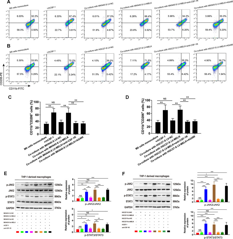
Flow cytometry analysis indicated that incubation in the presence of CSF-1 (100 ng/ml) or co-culture with parental GC cells overexpressing MELK significantly increased the percentages of CD11b+CD206+ cells in macrophages. Additionally, the presence of anti-CSF-1R (0.5 μg/ml) mitigated the effect of MELK overexpression on M2 polarization (Fig. 6A–D). Several studies have verified that the JAK2/STAT3 signaling axis was involved in the process of macrophages M2 polarization [23–26]. Based on these findings, we further investigated whether MELK induced macrophages polarization towards M2 phenotype through the JAK2/STAT3 pathway. As depicted in Fig. 6E and F, the expression of phosphorylated JAK2 and STAT3 significantly increased in macrophages when incubated with CSF-1, co-cultured with chemoresistant GC cells, or in parental GC cells overexpressing MELK. Treatment with anti-CSF-1R markedly weakened the ability of chemoresistant GC cells or parental GC cells overexpressing MELK to activate this signaling pathway. Furthermore, the JAK2/STAT3 signal pathway inhibitor, AG490, significantly attenuated the effect of parental GC cells with MELK overexpression on inducing M2 polarization (Fig. 6A, B).
Fig. 6.
MELK induced macrophages polarization towards M2 phenotype via CSF-1/JAK2/STAT3 pathway. A–D Flow cytometry detected the percentages of CD11b+CD206+ cells in macrophages incubating with CSF-1, co-culturing with parental GC cells with MELK overexpression in the presence of anti-CSF-1R, AG490 or not. E, F Macrophages were incubated with CSF-1, co-cultured with parental GC cells overexpressing MELK and their negative control cells, and co-cultured with chemoresistant GC cells silencing MELK and their negative control cells in the presence of anti-CSF-1R or not, then the JAK2/STAT3 pathway proteins in these macrophages were detected by western blot. Data were presented as mean ± SD of three independent experiments. NS not significantly, *p < 0.05, **p < 0.01, ***p < 0.001
Clinical relevance of the MELK with CSF-1 and CD206
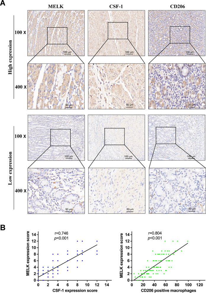
To explore the correlation between MELK and the cytokine CSF-1, as well as stromal macrophages, we conducted staining on tumor tissues using antibodies against CSF-1and the M2 macrophage marker CD206. As depicted in Fig. 7A, there was a correlation observed between high or low expression levels of MELK and corresponding levels of CSF-1 expression, along with varying infiltration degree of CD206 positive macrophages. Spearman rank-order correlation analysis demonstrated a statistically significant relationship (Fig. 7B). These results further supported the notion that MELK promotes M2 polarization of macrophages through the cytokine CSF-1.
Fig. 7.
The correlation of MELK with CSF-1 and CD206. A Representative images of immunohistochemical staining for MELK, CSF-1 and CD206 in human GC tissues. B The correlation of MELK with CSF-1 and CD206 in GC tissue was analyzed through Spearman rank-order correlation
Expression of MELK, CSF-1 and CD206 were correlated with the prognosis of GC patients
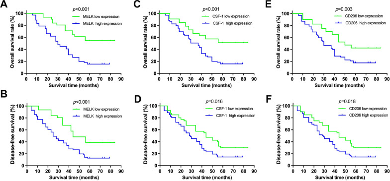
The immunoreactive intensity of MELK, CSF-1 and CD206 was individually categorized as high and low. Patients were followed up until death or more than 5 years, and the average period of follow-up was 36.97 ± 22.37 months (mean ± SD; range, 4 to 84 months). The overall survival (OS) rate and the disease-free survival (DFS) rate were calculated based on the expression of MELK, CSF-1 and CD206. As shown in Fig. 8, patients with high expression of MELK, CSF-1 or CD206 experienced a worse outcome of OS and DFS compared with those with low expression of the three molecules. The data above indicated that MELK, CSF-1 and CD206 expression were correlated with the prognosis of GC patients who underwent 5-Fu based neoadjuvant chemotherapy followed by radical gastrectomy.
Fig. 8.
Correlation of MELK, CSF-1 and CD206 expression with the prognosis of GC patients. A, B Overall survival (OS) and disease-free survival (DFS) analysis based on MELK in our cohort. C, D OS and DFS analysis based on CSF-1. (E and F) OS and DFS analysis based on CD206 (high expression vs. low expression)
Discussion
Over the past decade, upregulation of MELK has been observed in various types of malignant carcinomas and has been correlated with the occurrence and progression through diverse signaling pathways and cellular functions [3, 7–11]. As the roles of MELK in tumorigenesis and tumor progression have been elucidated, it has been increasingly emerged as a potential therapeutic target for tumor therapy. In human GC, elevated MELK expression has been found in cancer tissues and cell lines, with higher expression often associated with poorer clinical outcomes, including advanced stages, distant metastasis, and peritoneal spreading [3, 11, 16, 27]. Consistently, the present study confirms upregulated MELK expression in two GC cell lines and clinical specimens from patients with GC compared to that in normal gastric mucosal epithelial cell line and normal gastric mucosal tissues. Notably, we observed higher MELK expression levels in chemoresistant GC cells and tumor specimens compared to their parental counterparts and non-chemoresistant tumor specimens. Given the unclear function of MELK in the development of chemoresistance in GC, we hypothesized that MELK might contribute to the chemoresistance of GC. Subsequent in vitro and in vivo experiments demonstrated that MELK overexpression significantly increased 5-Fu-resistance in parental GC cells, while MELK knockdown dramatically suppressed the chemoresistance. These findings suggest that MELK can induce 5-Fu-resistance in GC cells.
Patients with GC frequently experience recurrence, mainly due to the development of chemoresistance [2, 28, 29]. Numerous studies have attempted to elucidate the molecular mechanisms of chemoresistance in various cancer cells. These mechanisms include epigenetic alterations, epithelial-mesenchymal transition, drug efflux, enhanced DNA repair and impaired apoptosis [30–32]. In recent years, emerging research has illustrated the interaction between cancer cells and immune cells within the TME, which contributes to tumor progression, metastasis and chemoresistance [33, 34]. Accumulating evidence indicated that tumor associated macrophages (TAMs), especially those exhibiting alternative activation (M2 phenotype), play a significant role in dedicating tumor chemoresistance [20, 30]. An analysis of 7135 patients with breast cancer from multiple independent large cohorts, revealed a positive correlation between high MELK expression and immune cells infiltration, including TAMs [35]. Another study demonstrated that MELK could function as an important component of immune response genes and predict the efficacy of immune checkpoint inhibitors in lung cancer [36]. In addition, high expression of MELK could induce macrophages polarization towards M2 phenotype through the activation of miR-34a/JAK2/STAT3 signaling pathway in the TME of uterine leiomyosarcoma [37]. These findings suggests that MELK might be closely associated with the TME and serves as a critical role in tumor progression. Our previous study demonstrated that chemoresistant GC cells were more effective in inducing macrophages polarization towards M2 phenotype compared to parental GC cells [21]. In the current investigation, we revealed that co-culturing parental GC cells overexpressing MELK with macrophages in a non-contacting transwell system could induce macrophages polarization towards M2 phenotype. In contrast, MELK knockdown attenuated the ability of chemoresistant GC cells to promote M2 polarization. Combined with our previous results, this accumulated data confirms that upregulation of MELK expression could not only promote 5-Fu-resistance in GC cells but was also accompanied by M2 macrophages polarization.
The present study indicated that CSF-1 deriving from GC cells overexpressing or silencing MELK served as a pivotal bridge in the cellular interaction between cancer cells and macrophages. CSF-1, located on human chromosome 1 (1p13.3), encodes a 554 amino acid protein, also known as macrophage colony-stimulating factor (M-CSF). CSF-1 plays a crucial role in regulating macrophage survival, differentiation, and function. The CSF-1 receptor, CSF-1R or CD115, is a tyrosine kinase encoded by the proto-oncogene c-fms and is predominantly expressed on the monocytic lineage [38]. Targeting CSF-1/CSF-1R signal transduction has garnered considerable attention from researchers. Pyonteck et al. reported that CSF-1R blockade could reduce M2 macrophages polarization in treated gliomas and slow the intracranial growth of glioma xenografts [39]. The monoclonal antibody RG7155, an inhibitor of CSF-1R, was found to decrease M2 macrophages in patients with various types of solid malignancies while increasing the CD8 + /CD4 + T cell ratio [40]. Consistently, our findings indicate that the blockade of CSF-1R attenuated the effect of MELK overexpression on M2 polarization.
Supported by the literature, the polarization of M2 macrophages is closely associated with the activation or inhibition of the JAK2/STAT3 signaling pathway. IL-6 could induce macrophages differentiation into the pro-tumor M2 phenotype via activating STAT3 phosphorylation and then promote GC cells proliferation and migration [41]. The inhibitor of JAK2 suppressed the JAK2/STAT3 signaling pathway in macrophages and induced macrophages polarization towards M1 phenotype. Further investigation indicated that the JAK2 inhibitor could reverse the phenotype of macrophages from M2- to M1-type [42]. In breast cancer, the redox-active drug MnTE-2-PyP5+ could decrease phospho-STAT3 levels to inhibit M2 polarization, thus stimulating the immune system [43]. In the present study, the JAK2/STAT3 signal pathway inhibitor AG490 was found to significantly attenuate the effect of parental GC cells with MELK overexpression on inducing M2 polarization. Clinically, MELK expression positively correlated with CSF-1 and the infiltration of CD206 positive macrophages. These results were confirmed by the above data from in the vitro experiments. Furthermore, survival analysis revealed that patients with high expression of MELK, CSF-1, or CD206 had worse OS and DFS than those with low expression of these three molecules.
Conclusion
In summary, this study uncovered that the upregulation of MELK expression could confer chemoresistance of GC. More importantly, MELK overexpression induced macrophage polarization to M2 phenotype via the CSF-1/JAK2/STAT3 signal axis, which in turn further promoted the chemoresistance of GC cells. These findings suggest that targeting MELK and inhibiting the CSF-1/JAK2/STAT3 signal pathway may represent potential therapeutic strategy for patients with GC, especially chemoresistant GC.
Supplementary Information
Supplementary Material 1. Table S1. Baseline characteristics of patients.
Supplementary Material 2. Table S2. Full-length MELK gene sequence.
Supplementary Material 3. Table S3. The sequences of shRNA targeting MELK.
Supplementary Material 4. Table S4. The forward and reverse primer sequences for the targeted genes.
Supplementary Material 5. Text S1. The reason for using 15 μg/ml of 5-FU and the methods for counting cell colonies.
Acknowledgements
Not applicable.
Abbreviations
- GC
Gastric cancer
- MELK
Maternal embryonic leucine zipper kinase
- qRT-PCR
Quantitative real-time polymerase chain reaction
- CSF-1
Colony-stimulating factor 1
- M-CSF
Macrophage colony-stimulating factor
- TME
Tumor microenvironment
- TAMs
Tumor-associated macrophages
- CAP
College of American Pathologists
- CCK-8
Cell counting kit-8
- SD
Standard deviation
- OS
Overall survival
- DFS
Disease-free survival
Author contributions
Conceptualization of the study concept was carried out by PS and JY. Data curation was conducted by PS, TY and YZ. Formal analysis and confirmation of the data were conducted by PS, TY and WK. Funding acquisition was achieved by JY. Research methodology was the responsibility of PS, HH and YL. Project administration was conducted by JY. Supervision was undertaken by WK, YL and JY. Visualization was conducted by PS, CC and MC. Writing of the original draft was carried out by PS and YZ. Writing, review and editing were carried out by PS, YL and JY. All authors read and approved the final version of the manuscript.
Funding
This research was supported by National High Level Hospital Clinical Research Funding (Grant No. 2022-PUMCH-B-005). The funders had no role in study design, data collection and analysis, decision to publish, or preparation of the manuscript.
Availability of data and materials
The datasets used and/or analyzed during the current study are available from the corresponding author on reasonable request.
Declarations
Ethics approval and consent to participate
The present study was approved by the Institutional Review Board of Peking Union Medical College Hospital and the written informed consent was obtained from all patients. The ethical approval code of this study is JS-2587. All animal studies were carried out according to the National Institute of Health Guide for the Care and Use of Laboratory Animals with the approval of Peking Union Medical College Hospital Animal Experiments Committee (ethical approval number: XHDW-2022-114).
Consent for publication
Not applicable.
Competing interests
The authors declare no competing interests.
Footnotes
Publisher's Note
Springer Nature remains neutral with regard to jurisdictional claims in published maps and institutional affiliations.
References
- 1.Sung H, Ferlay J, Siegel RL, et al. Global Cancer Statistics 2020: GLOBOCAN estimates of incidence and mortality worldwide for 36 cancers in 185 countries. CA Cancer J Clin. 2021;71(3):209–49. 10.3322/caac.21660 [DOI] [PubMed] [Google Scholar]
- 2.Biagioni A, Skalamera I, Peri S, et al. Update on gastric cancer treatments and gene therapies. Cancer Metastasis Rev. 2019;38(3):537–48. 10.1007/s10555-019-09803-7 [DOI] [PubMed] [Google Scholar]
- 3.Tang BF, Yan RC, Wang SW, et al. Maternal embryonic leucine zipper kinase in tumor cells and tumor microenvironment: an emerging player and promising therapeutic opportunity. Cancer Lett. 2023;560: 216126. 10.1016/j.canlet.2023.216126 [DOI] [PubMed] [Google Scholar]
- 4.Beullens M, Vancauwenbergh S, Morrice N, et al. Substrate specifificity and activity regulation of protein kinase MELK. J Biol Chem. 2005;280(48):40003–11. 10.1074/jbc.M507274200 [DOI] [PubMed] [Google Scholar]
- 5.Badouel C, Chartrain I, Blot J, et al. Maternal embryonic leucine zipper kinase is stabilized in mitosis by phosphorylation and is partially degraded upon mitotic exit. Exp Cell Res. 2010;316(13):2166–73. 10.1016/j.yexcr.2010.04.019 [DOI] [PubMed] [Google Scholar]
- 6.Davezac N, Baldin V, Blot J, et al. Human pEg3 kinase associates with and phosphorylates CDC25B phosphatase: a potential role for pEg3 in cell cycle regulation. Oncogene. 2002;21(50):7630–41. 10.1038/sj.onc.1205870 [DOI] [PubMed] [Google Scholar]
- 7.Pitner MK, Taliaferro JM, Dalby KN, et al. MELK: a potential novel therapeutic target for TNBC and other aggressive malignancies. Expert Opin Ther Targets. 2017;21(9):849–59. 10.1080/14728222.2017.1363183 [DOI] [PubMed] [Google Scholar]
- 8.Ganguly R, Hong CS, Smith LG, et al. Maternal embryonic leucine zipper kinase: key kinase for stem cell phenotype in glioma and other cancers. Mol Cancer Ther. 2014;13(6):1393–8. 10.1158/1535-7163.MCT-13-0764 [DOI] [PMC free article] [PubMed] [Google Scholar]
- 9.Jurmeister S, Ramos-Montoya A, Sandi C, et al. Identification of potential therapeutic targets in prostate cancer through a cross-species approach. EMBO Mol Med. 2018;10(3): e8274. 10.15252/emmm.201708274 [DOI] [PMC free article] [PubMed] [Google Scholar]
- 10.Ding X, Duan H, Luo H. Identification of core gene expression signature and key pathways in colorectal cancer. Front Genet. 2020;11:45. 10.3389/fgene.2020.00045 [DOI] [PMC free article] [PubMed] [Google Scholar]
- 11.Calcagno DQ, Takeno SS, Gigek CO, et al. Identification of IL11RA and MELK amplification in gastric cancer by comprehensive genomic profiling of gastric cancer cell lines. World J Gastroenterol. 2016;22(43):9506–14. 10.3748/wjg.v22.i43.9506 [DOI] [PMC free article] [PubMed] [Google Scholar]
- 12.Wu S, Chen X, Hu C, et al. Up-regulated maternal embryonic leucine zipper kinase predicts poor prognosis of hepatocellular carcinoma patients in a Chinese Han population. Med Sci Monit. 2017;23:5705–13. 10.12659/MSM.907600 [DOI] [PMC free article] [PubMed] [Google Scholar]
- 13.Joshi K, Banasavadi-Siddegowda Y, Mo XK, et al. MELK-dependent FOXM1 phosphorylation is essential for proliferation of glioma stem cells. Stem Cells. 2013;31(6):1051–63. 10.1002/stem.1358 [DOI] [PMC free article] [PubMed] [Google Scholar]
- 14.Park YY, Jung SY, Jennings NB, et al. FOXM1 mediates Dox resistance in breast cancer by enhancing DNA repair. Carcinogenesis. 2012;33(10):1843–53. 10.1093/carcin/bgs167 [DOI] [PMC free article] [PubMed] [Google Scholar]
- 15.Chunyu G, Banasavadi-Siddegowda YK, Joshi K, Nakamura Y, Kurt H, Gupta S, Nakano I. Tumor-specific activation of the c-JUN/MELK pathway regulates glioma stem cell growth in a p53-dependent manner. Stem Cells. 2013;31(5):870–81. 10.1002/stem.1322. 10.1002/stem.1322 [DOI] [PMC free article] [PubMed] [Google Scholar]
- 16.Du T, Qu Y, Li JF, et al. Maternal embryonic leucine zipper kinase enhances gastric cancer progression via the FAK/Paxillin pathway. Mol Cancer. 2014;13:100. 10.1186/1476-4598-13-100 [DOI] [PMC free article] [PubMed] [Google Scholar]
- 17.Ostuni R, Kratochvill F, Murray PJ, et al. Macrophages and cancer: from mechanisms to therapeutic implications. Trends Immunol. 2015;36(4):229–39. 10.1016/j.it.2015.02.004 [DOI] [PubMed] [Google Scholar]
- 18.Cassetta L, Pollard JW. Targeting macrophages: therapeutic approaches in cancer. Nat Rev Drug Discov. 2018;17(12):887–904. 10.1038/nrd.2018.169 [DOI] [PubMed] [Google Scholar]
- 19.Ginhoux F, Schultze JL, Murray PJ, et al. New insights into the multidimensional concept of macrophage ontogeny, activation and function. Nat Immunol. 2016;17(1):34–40. 10.1038/ni.3324 [DOI] [PubMed] [Google Scholar]
- 20.Boutilier AJ, Elsawa SF. Macrophage polarization states in the tumor microenvironment. Int J Mol Sci. 2021;22(13):6995. 10.3390/ijms22136995 [DOI] [PMC free article] [PubMed] [Google Scholar]
- 21.Su PF, Jiang L, Zhang YJ, et al. Crosstalk between tumor-associated macrophages and tumor cells promotes chemoresistance via CXCL5/PI3K/AKT/mTOR pathway in gastric cancer. Cancer Cell Int. 2022;22(1):290. 10.1186/s12935-022-02717-5 [DOI] [PMC free article] [PubMed] [Google Scholar]
- 22.Westerhoff M, Osecky M, Langer R. Varying practices in tumor regression grading of gastrointestinal carcinomas after neoadjuvant therapy: results of an international survey. Mod Pathol. 2020;33(4):676–89. 10.1038/s41379-019-0393-7 [DOI] [PubMed] [Google Scholar]
- 23.Zhong Y, Gu LJ, Ye YZ, et al. JAK2/STAT3 axis intermediates microglia/macrophage polarization during cerebral ischemia/reperfusion injury. Neuroscience. 2022;496:119–28. 10.1016/j.neuroscience.2022.05.016 [DOI] [PubMed] [Google Scholar]
- 24.Zeng HR, Zhao B, Zhang D, et al. Viola yedoensis Makino formula alleviates DNCB-induced atopic dermatitis by activating JAK2/STAT3 signaling pathway and promoting M2 macrophages polarization. Phytomedicine. 2022;103: 154228. 10.1016/j.phymed.2022.154228 [DOI] [PubMed] [Google Scholar]
- 25.Zhang WT, Yang FH, Zheng ZT, et al. Sulfatase 2 affects polarization of M2 macrophages through the IL-8/JAK2/STAT3 pathway in bladder cancer. Cancers (Basel). 2022;15(1):131. 10.3390/cancers15010131 [DOI] [PMC free article] [PubMed] [Google Scholar]
- 26.Li YM, Shi YK, Zhang XY, et al. FGFR2 upregulates PAI-1 via JAK2/STAT3 signaling to induce M2 polarization of macrophages in colorectal cancer. Biochim Biophys Acta Mol Basis Dis. 2023;1869(4): 166665. 10.1016/j.bbadis.2023.166665 [DOI] [PubMed] [Google Scholar]
- 27.Li S, Li ZY, Guo T, et al. Maternal embryonic leucine zipper kinase serves as a poor prognosis marker and therapeutic target in gastric cancer. Oncotarget. 2016;7(5):6266–80. 10.18632/oncotarget.6673 [DOI] [PMC free article] [PubMed] [Google Scholar]
- 28.Liu Y, Ao X, Wang Y, et al. Long non-coding RNA in gastric cancer: mechanisms and clinical implications for drug resistance. Front Oncol. 2022;12: 841411. 10.3389/fonc.2022.841411 [DOI] [PMC free article] [PubMed] [Google Scholar]
- 29.Wei L, Sun JJ, Zhang NS, et al. Noncoding RNAs in gastric cancer: implications for drug resistance. Mol Cancer. 2020;19(1):62. 10.1186/s12943-020-01185-7 [DOI] [PMC free article] [PubMed] [Google Scholar]
- 30.Chen SH, Chang JY. New insights into mechanisms of cisplatin resistance: from tumor cell to microenvironment. Int J Mol Sci. 2019;20(17):4136. 10.3390/ijms20174136 [DOI] [PMC free article] [PubMed] [Google Scholar]
- 31.De Las RJ, Brozovic A, Izraely S, et al. Cancer drug resistance induced by EMT: novel therapeutic strategies. Arch Toxicol. 2021;95(7):2279–97. 10.1007/s00204-021-03063-7 [DOI] [PMC free article] [PubMed] [Google Scholar]
- 32.Li LY, Guan YD, Chen XS, et al. DNA repair pathways in cancer therapy and resistance. Front Pharmacol. 2021;11: 629266. 10.3389/fphar.2020.629266 [DOI] [PMC free article] [PubMed] [Google Scholar]
- 33.Wang M, Zhao J, Zhang L, et al. Role of tumor microenvironment in tumorigenesis. J Cancer. 2017;8:761–73. 10.7150/jca.17648 [DOI] [PMC free article] [PubMed] [Google Scholar]
- 34.Bhavsar C, Momin M, Khan T, et al. Targeting tumor microenvironment to curb chemoresistance via novel drug delivery strategies. Expert Opin Drug Deliv. 2018;15(7):641–63. 10.1080/17425247.2018.1424825 [DOI] [PubMed] [Google Scholar]
- 35.Oshi M, Gandhi S, Huyser MR, et al. MELK expression in breast cancer is associated with infiltration of immune cell and pathological compete response (pCR) after neoadjuvant chemotherapy. Am J Cancer Res. 2021;11(9):4421–37. [PMC free article] [PubMed] [Google Scholar]
- 36.Pabla S, Conroy JM, Nesline MK, et al. Proliferative potential and resistance to immune checkpoint blockade in lung cancer patients. J Innunother Cancer. 2019;7(1):27. 10.1186/s40425-019-0506-3 [DOI] [PMC free article] [PubMed] [Google Scholar]
- 37.Zhang ZW, Sun CG, Li CC, et al. Upregulated MELK leads to doxorubicin chemoresistance and M2 macrophage polarization via the miR-34a/JAK2/STAT3 pathway in uterine leiomyosarcoma. Front Oncol. 2020;10:453. 10.3389/fonc.2020.00453 [DOI] [PMC free article] [PubMed] [Google Scholar]
- 38.Jeannin P, Paolini L, Adam C, et al. The roles of CSFs on the functional polarization of tumor-associated macrophages. FEBS J. 2018;285(4):680–99. 10.1111/febs.14343 [DOI] [PubMed] [Google Scholar]
- 39.Pyonteck SM, Akkari L, Schuhmacher AL, et al. CSF-1R inhibition alters macrophage polarization and blocks glioma progression. Nat Med. 2013;19(10):1264–72. 10.1038/nm.3337 [DOI] [PMC free article] [PubMed] [Google Scholar]
- 40.Ries CH, Cannarile MA, Hoves S, et al. Targeting tumor-associated macrophages with anti-CSF-1R antibody reveals a strategy for cancer therapy. Cancer Cell. 2014;25(6):846–59. 10.1016/j.ccr.2014.05.016 [DOI] [PubMed] [Google Scholar]
- 41.Fu XL, Duan W, Su CY, et al. Interleukin 6 induces M2 macrophage differentiation by STAT3 activation that correlates with gastric cancer progression. Cancer Immunol Immunother. 2017;66(12):1597–608. 10.1007/s00262-017-2052-5 [DOI] [PMC free article] [PubMed] [Google Scholar]
- 42.He W, Zhu Y, Mu RY, et al. A Jak2-selective inhibitor potently reverses the immune suppression by modulating the tumor microenvironment for cancer immunotherapy. Biochem Pharmacol. 2017;145:132–46. 10.1016/j.bcp.2017.08.019 [DOI] [PubMed] [Google Scholar]
- 43.Griess B, Mir S, Datta K, et al. Scavenging reactive oxygen species selectively inhibits M2 macrophage polarization and their pro-tumorigenic function in part, via STAT3 suppression. Free Radic Biol Med. 2020;147:48–60. 10.1016/j.freeradbiomed.2019.12.018 [DOI] [PMC free article] [PubMed] [Google Scholar]
Associated Data
This section collects any data citations, data availability statements, or supplementary materials included in this article.
Supplementary Materials
Supplementary Material 1. Table S1. Baseline characteristics of patients.
Supplementary Material 2. Table S2. Full-length MELK gene sequence.
Supplementary Material 3. Table S3. The sequences of shRNA targeting MELK.
Supplementary Material 4. Table S4. The forward and reverse primer sequences for the targeted genes.
Supplementary Material 5. Text S1. The reason for using 15 μg/ml of 5-FU and the methods for counting cell colonies.
Data Availability Statement
The datasets used and/or analyzed during the current study are available from the corresponding author on reasonable request.


