



ABSTRACT
Pneumocystis jirovecii pneumonia (PjP) poses a serious risk to individuals with compromised immune systems, such as individuals with HIV/AIDS or undergoing immunosuppressive therapies for cancer or solid organ transplants. Severe PjP triggers excessive lung inflammation, resulting in lung function decline and consequential alveolar damage, potentially culminating in acute respiratory distress syndrome. Non-HIV patients face a 30%–60% mortality rate, emphasizing the need for a deeper understanding of inflammatory responses in PjP. Prior research emphasized macrophages in Pneumocystis infections, neglecting neutrophils’ role in tissue damage. Consequently, the overemphasis on macrophages led to an incomplete understanding of the role of neutrophils and inflammatory responses. In the current investigation, our RNAseq studies on a murine surrogate model of PjP revealed heightened activation of the NLRP3 inflammasome and NETosis cell death pathways in their lungs. Immunofluorescence staining confirmed neutrophil extracellular trap (NET) presence in the lungs of the P. murina-infected mice, validating our findings. Moreover, isolated neutrophils exhibited NETosis when directly stimulated with P. murina. Isolated NETs compromised P. murina viability in vitro, highlighting the potential role of neutrophils in controlling fungal growth and promoting inflammation during P. murina pneumonia through NLRP3 inflammasome assembly and NETosis. These pathways, essential for inflammation and pathogen elimination, bear the risk of uncontrolled activation leading to excessive tissue damage and persistent inflammation. This pioneering study is the first to identify the formation of NETs and inflammasomes during Pneumocystis infection, paving the way for comprehensive investigations into treatments aimed at mitigating lung damage and augmenting survival rates for individuals with PjP.
IMPORTANCE
Pneumocystis jirovecii pneumonia (PjP) affects individuals with weakened immunity, such as HIV/AIDS, cancer, and organ transplant patients. Severe PjP triggers lung inflammation, impairing function and potentially causing acute respiratory distress syndrome. Non-HIV individuals face a 30%–60% mortality rate, underscoring the need for deeper insight into PjP’s inflammatory responses. Past research focused on macrophages in managing Pneumocystis infection and its inflammation, while the role of neutrophils was generally overlooked. In contrast, our findings in P. murina-infected mouse lungs showed neutrophil involvement during inflammation and increased expression of NLRP3 inflammasome and NETosis pathways. Detection of neutrophil extracellular traps further indicated their involvement in the inflammatory process. Although beneficial in combating infection, unregulated neutrophil activation poses a potential threat to lung tissues. Understanding the behavior of neutrophils in Pneumocystis infections is crucial for controlling detrimental reactions and formulating treatments to reduce lung damage, ultimately improving the survival rates of individuals with PjP.
KEYWORDS: Pneumocystis, infectious disease, neutrophils, opportunistic fungi, immunity, Pneumocystis pneumonia, Pneumocystis murina, NETosis, inflammation
INTRODUCTION
Pneumocystis jirovecii pneumonia (PjP) is a leading cause of mortality in hospitalized individuals with HIV/AIDS. However, the incidence of PjP has increased in cancer patients and individuals who have received organ transplants requiring immune-suppressing treatments. Among all hospitalizations for PjP, malignancy stands as the most prevalent predisposing factor, accounting for 46.0%–55.7% of cases, followed by HIV at 17.8% (1–4). Within immunocompetent hosts, signaling cascades initiated by CD4+ T cells and B cells efficiently trigger P. jirovecii clearance with minimal inflammation (5). Conversely, immunosuppressed hosts experience an influx of various immune cells into their lungs, including T lymphocytes, polymorphonuclear neutrophils, and other leukocytes (6–9). This can lead to a profound inflammatory response which leads to considerable morbidity and mortality. Severe PjP is characterized by a neutrophilic inflammatory response presenting with decreased pulmonary function, alveolar damage, and respiratory failure (10, 11). Furthermore, bronchial alveolar lavage (BAL) neutrophilia has been shown to be a predictor of poor prognosis and increased mortality in PjP (12).
Previous research has primarily centered on macrophage polarization and its role in clearance of Pneumocystis from infected hosts. Both classically activated M1 and alternatively activated M2 macrophages are involved in Pneumocystis clearance. Immunocompetent mice show preferential activation of the M2 phenotype during P. murina exposure, while immunosuppressed hosts show enhanced M1 polarization (13). M1-polarized macrophages are effective fungicidal cells and produce a substantial cytokine and chemokine response (14). These secretions not only eliminate infectious organisms but also signal for further recruitment of immune cells.
Pattern recognition receptors, such as C-type lectin receptors dectin-1/2 and toll-like receptors (TLRs), expressed by innate immune cells bind to pathogen-associated molecular patterns and damage-associated molecular patterns (15). Dectin-1/2 and TLR2 have been identified as important receptors in antigen recognition to P. murina in mice, leading to immune responses such as pro-inflammatory cytokine release and fungal clearance (16–18).
Neutrophils play a crucial role in host defenses against pathogenic organisms. During Candida albicans and Aspergillus fumigatus infections in mice, neutrophils aid in controlling fungal growth by phagocytosis and reactive oxygen species (ROS) generation for effective pathogen killing (19, 20). Beyond phagocytosis and ROS generation, neutrophils employ additional antimicrobial activity, including degranulation, cytokine production, and NETosis. NETosis results in the expelling of DNA to form neutrophil extracellular traps (NETs) that ensnare and kill pathogens (21, 22). These NETs are decorated with various proteins, such as histones, neutrophil elastase (NE), calprotectin, myeloperoxidase (MPO), and other antimicrobial proteins (23, 24).
PjP is associated with an accumulation of neutrophils in the lungs (8, 9). Additionally, levels of interleukin-8, a chemotactic and activating agent for neutrophils, within BAL fluid are directly associated with the clinical severity of pneumonia and serve as a prognostic marker for mortality risk and the likelihood of significant respiratory compromise (25). A previous study by Swain et al. found that neutrophils and reactive oxygen species do not contribute to pulmonary tissue damage nor play a major role in clearance of Pneumocystis (26). Consequently, despite the significant influx of neutrophils into the lungs during Pneumocystis infection, their role in inflammation has been both overlooked and understudied, resulting in a significant knowledge gap.
This study revealed an up-regulation in the expression of genes associated with the NLRP3 inflammasome and NETosis in the lungs of mice infected with P. murina. These processes are essential for the elimination of pathogens from the host system. However, overactivation of these pathways leads to heightened inflammation and tissue damage, emphasizing the need for a thorough understanding of their regulation in the context of Pneumocystis infection. This investigation intended to elucidate the roles of neutrophil populations during PjP to establish the groundwork for therapeutic strategies aimed at mitigating excessive inflammatory responses and alveolar damage.
RESULTS
Signaling pathways involved with inflammatory processes increase during P. murina infection
To gain a better understanding of the changes in host immune response during the development of P. murina infection in mice, lung samples were analyzed from both uninfected and P. murina-infected immunosuppressed mice at 5- and 7-week intervals after initial exposure to P. murina. Mice with a 7-week infection exhibited a higher fungal burden than those with 5-week infections (Fig. 1A). Differential gene expression analysis revealed that gene expression patterns across different samples were comparable between biological groups (Fig. 1B). Control samples from both the 5-week (5wC) and 7-week (7wC) groups clustered at the bottom, while infected samples from the 5-week (5wI) group clustered in the middle, and those from the 7-week (7wI) group clustered at the top of the heatmap. A gene set enrichment analysis indicated a significant increase in signaling pathways associated with inflammatory processes as P. murina infection progressed in mice (Fig. 1C; Table 1).
Fig 1.

Signaling pathways associated with inflammatory processes are up-regulated as Pneumocystis murina infection progressed in mice. Immunosuppressed mice were exposed to uninfected or previously P. murina-infected mice. (A) After 5 or 7 weeks of exposure, the fungal burden in the lungs was enumerated. Dashed line, limit of microscopic enumeration. ***, P < 0.001. ****, P < 0.0001. (B) Heatmap of sample-to-sample distances. 5wC and 7wC uninfected samples cluster near the bottom, and 5wI clusters in the middle, while 7wI clusters at the top of the heatmap. (C) Heatmap comparison analysis of gene set enrichment analysis on KEGG pathways relating to signaling. Data are represented as logFC.
TABLE 1.
Functional gene set enrichment analysesb
| Pathway | KEGG ID | NumGenesa | P-value | FDR |
|---|---|---|---|---|
| Antigen processing and presentation | mmu04612 | 77/91 | 1.37E−17 | 3.25E−16 |
| Cytokine-cytokine receptor interaction | mmu04060 | 222/270 | 3.21E−23 | 3.06E−21 |
| Hematopoietic cell lineage | mmu04640 | 83/95 | 2.26E−12 | 2.81E−11 |
| Proteasome | mmu03050 | 45/45 | 9.61E−11 | 9.82E−10 |
| NF-kappa B signaling pathway | mmu04064 | 89/103 | 2.38E−15 | 4.53E−14 |
| Osteoclast differentiation | mmu04380 | 116/130 | 1.45E−10 | 1.38E−09 |
| Chemokine signaling pathway | mmu04062 | 162/192 | 2.30E−08 | 1.68E−07 |
| Cell adhesion molecules | mmu04514 | 140/169 | 4.80E−15 | 8.57E−14 |
| NOD-like receptor signaling pathway | mmu04621 | 152/169 | 1.54E−08 | 1.22E−07 |
NumGenes, number genes mapped to the total number of genes in the set.
Enriched pathways based on differential expression.
NOD-like signaling and NETosis pathways are increased during P. murina infection
Many of the inflammatory signaling pathways during Pneumocystis infections have been extensively studied, such as antigen processing and presentation, cytokine-cytokine receptor interaction, hematopoietic cell lineage, NF-kappa B signaling, chemokine signaling pathway, and cell adhesion molecules (27–31). For that reason, we chose to focus on the understudied NOD-like receptor signaling pathway. In the lungs of mice with both 5- and 7-week infections, there was an observed increase in the expression of key genes associated with the NOD-like receptor signaling pathway that were not present in non-infected mice. These genes included NOD-like protein receptor 3 (Nlrp3), apoptosis-associated speck-like protein containing a CARD (Asc/Pycard), and caspase 1 (Casp1) (Fig. 2). These transcripts encode proteins that form the NLRP3 inflammasome complex, wherein NLRP3 binds to the scaffold protein ASC, which in turn binds to CASP1. The proteolytic enzyme CASP1 component of the NLRP3 inflammasome complex cleaves the pro-inflammatory cytokine interleukin 1 beta (IL-1β) into its mature form. Additionally, CASP1 also cleaves gasdermin D (GSDMD), which inserts into the plasma membrane to facilitate the release of IL-1β to further propagate a proinflammatory response and pyroptotic cell death (32).
Fig 2.
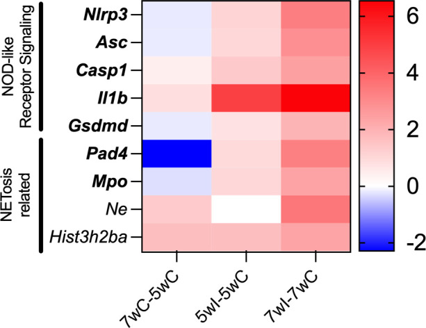
NOD-like receptor signaling and NETosis-related transcripts are up-regulated as P. murina infection progressed in mice. Data are represented as log2 fold change between the displayed groups. Data are represented as logFC. Bold, FDR-adjusted P < 0.05.
The mRNA expression of both Il-1β and Gsdmd increased in the lungs of mice with 5-week and 7-week infections (Fig. 2). While proinflammatory cytokine IL-1 is a crucial mediator for host resistance and immune cell recruitment during P. murina infection (33), inflammasome assembly details have yet been described. Additionally, increased peptidylarginine deiminase 4 (Pad4) expression was detected in P. murina-infected mice. PAD4 plays a role in chromatin decondensation and expulsion of NETs in a regulated cell death pathway called NETosis (34). Furthermore, PAD4 stimulates NLRP3-inflammasome formation, leading to prolonged inflammation through a positive feedback loop by driving the production of pro-inflammatory cytokines and chemokines (35). This is supported by similar findings that bronchial cells exposed to NETs demonstrated increased secretion of pro-inflammatory cytokine IL-1β (36).
NET formation in P. murina-infected lung tissue
Given the increased expression of genes related to NOD-like receptor signaling and the NETosis pathway through differential expression analysis, we sought to confirm the presence of NETs during Pneumocystis murina pneumonia (PmP). Increased expression of NE and MPO, which decorate the extracellular DNA in NETs, is significantly increased in the lungs of P. murina-infected mice (Fig. 3A and B). Immunohistochemistry analysis revealed expression of NE and MPO, suggesting the presence of NETs within P. murina-infected lung tissue (Fig. 3C).
Fig 3.

NETosis-related proteins are present in the lungs of Pneumocystis murina-infected mice. (A) ELISA reveals increased expression of NET components, NE and (B) MPO, in the lung tissue from P. murina-infected (Pm+) mice over uninfected (UI) mice. *t-test, P < 0.05. (C) Immunohistochemistry of the lungs of P. murina-infected mice. The NETosis markers, NE and MPO, are expressed throughout the thickened alveolar spaces of Pm+ mice. Scale, 100 µm.
P. murina stimulates NETosis in neutrophils in vitro
Considering that Pneumocystis infections result in lung neutrophilia and NETs were observed within the lungs of P. murina-infected mice, we investigated whether P. murina could directly induce neutrophils to undergo NETosis and produce NETs. In experiments using bone marrow-derived neutrophils, we found that P. murina stimulated the release of NETs in neutrophils, as evidenced by the extracellular DNA released into the supernatant in a dose-dependent manner (Fig. 4A). Additionally, since NETs carry various proteins, we conducted a sandwich ELISA to detect MPO-DNA complexes within the NETs, which showed that P. murina stimulated the release of DNA-MPO complexes from neutrophils (Fig. 4B).
Fig 4.

Pnuemocystis murina stimulates NET production in bone marrow-derived neutrophils. Bone marrow-derived neutrophils were stimulated with vehicle, phorbol myristate acetate (PMA; 25 nM; positive control), and P. murina at different multiplicities of infection (1, 2, or 5). Controls were established using the same concentrations of P. murina organisms without neutrophils.(A) Culture supernatant was assessed for extracellular DNA released from neutrophils. (B) Culture supernatant assessed for MPO-DNA complexes by ELISA. One-way ANOVA. *, P < 0.05 compared to vehicle.
Immunofluorescence analysis showed the presence of NET structures, as seen by expelled DNA decorated with NE, CitH3, and MPO (Fig. 5). These data indicate that P. murina can directly stimulate the production of NETs in vitro.
Fig 5.

Pneumocystis murina directly stimulates NET production in bone marrow-derived neutrophils. Immunofluorescence of the bone marrow-derived neutrophils stimulated with vehicle, phorbol myristate acetate (PMA; 25 nM), or P. murina. The NET production was seen in PMA- and P. murina-treated neutrophils as shown by neutrophil elastase (green), citrullinated histone H3 (yellow), MPO (magenta) expression. DAPI, blue. Scale, 50 µm.
NETs are detrimental to P. murina viability
NETs are known to play a role in pathogen trapping and killing. Given that P. murina can directly stimulate the production of NETs in neutrophils in vitro, we investigated whether NETs were toxic to P. murina. After treating P. murina with NETs isolated from phorbol myristate acetate (PMA)-treated neutrophils, we observed decreased viability of P. murina (Fig. 6). These data indicate that NETs negatively impacted the viability of P. murina.
Fig 6.
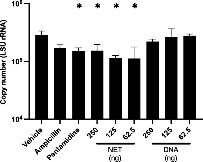
NETs are detrimental to Pneumocystis murina viability. P. murina (n = 3; 5 × 10⁷ nuclei) were inoculated into 96-well plates (Costar 3548, Corning, New York). Isolated NETs from PMA (25 nM)-stimulated neutrophils or salmon sperm DNA samples, used as control for non-complexed DNA, were added to the wells. Plates were incubated at 5% CO2, 37°C. After 24 hours, P. murina was quantified by large subunit (LSU) rRNA copy number by qPCR. One-way ANOVA. *, P < 0.05 compared to vehicle.
DISCUSSION
P. jirovecii pneumonia remains a significant concern, particularly in patients with HIV/AIDS and those undergoing immunosuppressive treatments for conditions such as hematological cancers. Immunosuppressed hosts with PjP experience an influx of various immune cells, including T lymphocytes, polymorphonuclear neutrophils, and other leukocytes, resulting in profound inflammation and considerable morbidity and mortality. Traditionally, the focus of research on host responses to Pneumocystis has been centered around macrophage responses and role in pathogen clearance. However, our findings highlight the overlooked roles of neutrophils and the NLRP3 inflammasome in the host immune response during P. murina infection.
Neutrophils, despite their abundance in infected lungs, have been largely overlooked in the context of immune response to Pneumocystis. These cells participate in the inflammatory response during P. murina infection, as indicated by the increased expression of NLRP3 inflammasome-related genes observed in the lungs (Fig. 2). Moreover, our study identified NE and citrullinated histone H3, NET markers of NETosis, in the lungs of P. murina-infected mice (Fig. 3), indicating that this process was active during PmP. Our in vitro experiments further demonstrated that P. murina can directly stimulate NETosis in isolated neutrophils (Fig. 4 and 5).
Neutrophils are recognized as a critical line of defense against various pathogens. Although previous studies had questioned their significance in Pneumocystis infections and the clearance of Pneumocystis, our results indicate that neutrophils may contribute to the host’s inflammatory response by initiating NLRP3-inflammasome assembly and undergoing NETosis. The increased expression of NLRP3 inflammasome-related genes and the presence of NETs suggest that neutrophils are actively involved in the immune response against P. murina.
Importantly, uncontrolled inflammasome activation and NETs can drive excessive inflammation and tissue damage. Previous research has demonstrated that NETs can exacerbate lung injury and disrupt barrier function in mice, while disruption of NET formation or NET degradation led to decreased tissue damage and increased survival in mice (37–39). Our study highlights the importance of understanding the role of neutrophils and NET formation during Pneumocystis infection, as they may be driving increased inflammation, immune cell recruitment, and tissue damage.
Here, we demonstrated the NETs were detrimental to P. murina viability in vitro (Fig. 6). Similar deleterious impacts of NETs on the fungal viability are shown in Candida albicans (40), Paracoccidioides brasiliensis (41), and Scedosporium apiospermum (42). While NETs exhibit some control over Aspergillus fumigatus infection, they are insufficient for complete eradication of the fungus (43). Moreover, biofilms provide a protective extracellular matrix which can resist the killing effect of NETs (44). C. albicans form protective biofilms that inhibit NET formation and secrete nucleases to degrade NETs (45). Pneumocystis forms biofilms (46), which may confer protection against complete eradication of the fungi by NETs. Pneumocystis biofilms may be utilizing extracellular DNA as a scaffold to form a biofilm, similar to C. albicans and A. fumigatus, where DNA serves as a crucial component in biofilm matrices (47–49). Furthermore, the integrity of these biofilms can be disrupted by DNase, potentially increasing susceptibility to antifungal therapies (49, 50).
In conclusion, our research provides valuable insights into the role of neutrophils, particularly their involvement in NLRP3 inflammasome activation and NETosis, in the host immune response during P. murina pneumonia. By elucidating the participation of neutrophils and their ability to form NETs in response to P. murina, we expand our understanding of the intricate inflammatory dynamics at play during Pneumocystis infection. These findings offer potential avenues for therapeutic interventions aimed at modulating the immune response in individuals at risk of or affected by PjP. Further investigations are warranted to delve into the precise mechanisms underlying neutrophil-mediated inflammation, inflammasome activation, and NETosis in PjP as well as to explore the potential therapeutic strategies that may arise from these discoveries.
MATERIALS AND METHODS
Animals
Male C3H/HeNCrl (5 weeks old; Charles River, Raleigh, NC) mice were housed under barrier conditions with autoclaved food and bedding in sterilized cages equipped with sterile microfilter lids. Mice were immunosuppressed with dexamethasone (4 mg/L) in acidified drinking water, available ad libitum. The mice were infected by co-housing with P. murina-infected mice. Infection was allowed to progress for 5 weeks (to represent a moderate infection; 5wI) and 7 weeks (high infection; 7wI). Time-matched immunosuppressed, uninfected mice were used as controls (moderate and high control; 5wC and 7wC). Mice were euthanized humanely, and their lungs removed for quantification of fungal burdens (n = 3), or the lungs were flash frozen in liquid nitrogen, ground using a mortar and pestle, and then stored at −80°C for RNA sequencing (n = 5). To quantify fungal burden, lungs were homogenized in PBS using gentleMACS (Miltenyi Biotec, Auburn, CA, USA), then stained with a modified Diff-Quik staining (51) to visualize the nuclei for microscopic enumeration. The microscopic counts were log transformed, and values were compared by the one-way analysis of variance.
RNA sequencing
The following steps were performed at UC Genomics, Epigenomics, and Sequencing Core, Department of Environmental Health, University of Cincinnati, Cincinnati, OH. RNA was extracted from lung tissue using mirVana miRNA Isolation Kit (Invitrogen, Carlsbad, CA, USA). The Ribo-Zero Gold (Human/Mouse/Rat) and (Yeast) kit (Illumina, San Diego, CA, USA) were used to deplete rRNA using 300 ng total RNA as input. The isolated RNA was RNase III fragmented and adaptor ligated using PrepX mRNA Library Kit (Takara, Mountain View, CA, USA), then converted into cDNA using Superscript III reverse transcriptase (Lifetech, Grand Island, NY, USA). Resulting cDNA was purification using Agencourt AMPure XP beads (Beckman Coulter, Indianapolis, IN, USA). Barcode index was added using a universal and index-specific primer with PCR to each ligated cDNA sample, and the amplified library was enriched by AMPure XP beads purification.
The quality and yield of the purified library were analyzed by Bioanalyzer (Agilent, Santa Clara, CA, USA) using DNA high-sensitivity chip. Libraries were quantified by qPCR measured by Kapa Library Quantification Kit (Kapa Biosystems, Woburn, MA, USA) using ABI’s 9700HT real-time PCR system (Lifetech, Grand Island, NY, USA). Libraries at the final concentration of 12.0 pM were clustered onto a flow cell using Illumina’s TruSeq SR Cluster Kit v3 and sequenced for 50 cycles using TruSeq SBS Kit on Illumina HiSeq system.
RNAseq analysis
Raw reads were trimmed for quality and barcode removal using fastp (52). Trimmed reads were aligned to the M. musculus genome (accession GCF_000001635.27) and quantified using salmon (53). DEseq2, using an absolute fold change >1.5 and FDR-adjusted P-value of 0.05, was used to identify differentially expressed genes between groups (54). Ensemble of Gene Set Enrichment Analyses was performed to identify pathways with enriched gene expression with an absolute fold change >1.0 and FDR-adjusted P-value of 0.05 (55).
ELISA
Protein was extracted from uninfected and P. murina-infected lung tissue (n = 3) using radio-immunoprecipitation assay buffer. Protein was quantified by BCA Protein Assay Kit (Thermo Scientific, Rockford, IL, USA) and normalized to 2 mg/mL. MPO and NE were quantified from the protein lysate using Mouse MPO ELISA Kit (Abcam ab155458, Cambridge, UK) and Mouse Neutrophil Elastase ELISA Kit (Abcam ab252356, Cambridge, UK) following manufacturer instructions.
Immunostaining
Lung tissue (n = 3) was fixed in neutral buffered formalin for 24 hours at room temperature. The tissue was then dehydrated and stored in 70% ethanol. Fixed lungs were embedded with paraffin, then sliced at 10 um, and mounted on positive-charged slides. Slides were deparaffinized using a xylene series, followed by ethanol exchange, then rehydrated in water. Epitope retrieval was performed using citrate buffer at 91°C for 90 minutes. Anti-NE (1:500; Abcam ab68672, Cambridge, UK) or Anti-MPO (1:100; Abcam ab9535, Cambridge, UK) was incubated with the sections for 1 hour, washed, and then probed with anti-rabbit-HRP (1:1,000; Roche Diagnostics UltraMap 05269717001, Indianapolis, IN). Colorimetric signal was developed using diaminobenzidine and then counterstained with hematoxylin.
Neutrophils (4 × 10⁴; performed in triplicate) were grown on 18-mm glass coverslips (Electron Microscopy Sciences; Hatfield, PA, USA) in a 12-well dish (CytoOne, Ocala, FL, USA). After NETosis experiments, cells were fixed in 3.7% formaldehyde in PBS for 15 minutes and permeabilized with 0.1% Triton-X for 10 minutes.
Neutrophil samples were blocked in 10% goat serum for 1 hour, then incubated with anti-Neutrophil Elastase-AlexaFluor 488 (1:100; Santa Cruz Biotechnology, Dallas, TX), anti-Citrullinated Histone H3 (1:100; Abbomax, San Jose, California), or anti-Myeloperoxidase (1:100; Santa Cruz Biotechnology, Dallas, TX) in blocking buffer for 1 hour. Secondary goat anti-rabbit antibody conjugated with AlexaFluor 594 (1:1,000; Invitrogen, Carlsbad, CA) and goat anti-mouse IgG antibody conjugated with AlexaFluor 647 (1:1,000; Invitrogen, Carlsbad, CA) in blocking buffer were added and incubated for 2 hours in the dark. Cells were washed between incubations with 0.1% Tween 20 in PBS (PBST) three times for 15 minutes. DNA was stained with DAPI (1 ug/mL). Images taken on a Nikon A1 Confocal Microscope at Cincinnati Children’s Hospital Medical Center or on a Leica Stellaris confocal microscope at the University of Cincinnati Live Microscopy Core.
Neutrophil isolation
Bone marrow was obtained from the humerus, femur, and tibia of immunocompetent C3H/HeNCrl male mice (n = 3 groups of pooled bone marrow from two mice). Red blood cells were lysed in 0.08% ammonium chloride for 10 minutes on wet ice and then washed with RPMI 1640 (Gibco, Grand Island, NY, USA) twice. Neutrophils were isolated by positive selection using Anti-Ly-6G MicroBeads UltraPure (Miltenyi Biotec, Auburn, CA, USA) and resuspended in RPMI 1640.
NETosis assay
Neutrophils (1 × 10⁶) were plated onto a 24-well dish (CytoOne, Ocala, FL, USA). Cells were then treated with either RPMI 1640 vehicle, phorbol myristate acetate (25 nM in RPMI 1640; Millipore; Burlington, MA, USA), or P. murina at multiplicities of infection of 1, 2, or 5. Cells were incubated at 37°C 5% CO2 for 4 hours to allow NETosis to occur (56). Neutrophils were then gently washed twice with PBS. Cells were resuspended in 500 µL PBS by using a cell scraper to lift the neutrophils from the plate surface, then centrifuged at 300 × g to separate cell debris from NET material. These experiments were performed in triplicate using isolated neutrophils from three independent groups, as described above. The NET-containing supernatants were collected for downstream analysis.
Quantification of extracellular DNA
Extracellular DNA release was quantified from the supernatant of NETosis-stimulated neutrophils using the QuantiFluor dsDNA system (Promega, Madison, WI) per manufacturer instructions. Briefly, samples and dsDNA standards were stained with QuantiFlour dsDye, and fluorescence (504nmEx/531nmEm) was measured using a BioTek Synergy HTX plate reader.
Detection of MPO-DNA complexes
NET release from the supernatant of NETosis-stimulated neutrophils was determined by detecting complexes of DNA and MPO. High-binding 96-well plates (Corning 2592, Kennebunk, ME, USA) were coated overnight with capture antibody, anti-MPO (1:500; Invitrogen; Carlsbad, CA, USA), in 50 mM carbonate buffer, pH 9.4 at 4°C. After that time, plates were washed three times with PBS with 0.1% Tween 20. Wells were blocked with StartingBlock PBS Blocking Buffer (Thermo Scientific, Rockford, IL, USA) for 2 hours at room temperature, then washed three times with PBST. Cell supernatant from each treatment was diluted 1:10, and 100 µL was added to the wells and incubated at 4°C for 24 hours, then washed three times in PBST. Detection antibody, anti-DNA-HRP (1:100; Zymo Research, Irvine, CA, USA), was added to the plate and incubated for 24 hours. After washing three times in PBST, colorimetric signal was detected using TMB (3,3',5,5' tetramethylbenzidine; Thermo Scientific, Rockford, IL, USA). Absorbance (650 nm) was measured using a BioTek Synergy HTX plate reader.
NET toxicity
Isolated NETs from the supernatant of PMA-stimulated neutrophils were collected and quantified as shown above. Salmon sperm DNA was used as a control for non-complexed DNA. P. murina (5 × 10⁷ nuclei) were inoculated into 96-well plates (Costar 3548; Corning, NY, USA). PBS vehicle, DNA, or NET samples were added to the wells in triplicates. Plates were incubated at 5% CO2, at 37°C. After 24 hours, total RNA was extracted from the samples using TRIZOL reagent (Life Technologies Inc.; Rockville, MD, USA) and Direct-zol-96 MagBead RNA (Zymo Research; Irvine, CA, USA), then reverse transcribed into cDNA using SuperScript IV VILO (Invitrogen, Carlsbad, CA,USA). P. murina organisms were quantified by large subunit (LSU) rRNA copy number, as described previously (57). Briefly, quantitative PCR was performed using TaqMan Fast Advanced Master Mix (Applied Biosystems, Waltham, Massachusetts, USA) following manufacturer instructions. Primers: LSU rRNA (ATGAGGTGAAAAGTCGAAAGGG; TGATTGTCTCAGATGAAAAACCTCTT; 6FAM-AACAGCCCAGAATAATGAATAAAGTTCCTCAATTGTTAC-TAMRA).
ACKNOWLEDGMENTS
This work was supported by HHS NIH R01HL146266 and VA I01 BX004441. M.T.C. is a Senior Research Career Scientist supported by IK6BX005232 Department of Veterans Affairs. We thank Margaret Collins for her assistance with the MPO-DNA ELISA. Confocal microscopy performed at the University of Cincinnati Live Microscopy Core was supported by NIH S10OD030402.
Contributor Information
Steven G. Sayson, Email: Steven.Sayson@uc.edu.
Kirsten Nielsen, University of Minnesota Medical School, Minneapolis, Minnesota, USA.
Jay Kolls, Tulane University School of Medicine, New Orleans, Louisiana, USA.
DATA AVAILABILITY
Fastq files have been deposited to NCBI Sequence Read Archive under BioProject Accession: PRJNA1076530.
ETHICS APPROVAL
These studies were performed in accordance with the Guide for the Care and Use of Laboratory Animals, eighth edition (National Academies Press, Washington, DC, USA), in AAALAC-accredited laboratories under the supervision of veterinarians. In addition, all procedures were conducted in compliance with the Institutional Animal Care and Use Committee at the Veterans Affairs Medical Center, Cincinnati, OH, USA.
REFERENCES
- 1. Kanj A, Samhouri B, Abdallah N, Chehab O, Baqir M. 2021. Host factors and outcomes in hospitalizations for Pneumocystis jirovecii pneumonia in the United States. Mayo Clin Proc 96:400–407. doi: 10.1016/j.mayocp.2020.07.029 [DOI] [PubMed] [Google Scholar]
- 2. Kolbrink B, Scheikholeslami-Sabzewari J, Borzikowsky C, von Samson-Himmelstjerna FA, Ullmann AJ, Kunzendorf U, Schulte K. 2022. Evolving epidemiology of Pneumocystis pneumonia: findings from a longitudinal population-based study and a retrospective multi-center study in Germany. Lancet Reg Health Eur 18:100400. doi: 10.1016/j.lanepe.2022.100400 [DOI] [PMC free article] [PubMed] [Google Scholar]
- 3. Brown GD, Denning DW, Gow NAR, Levitz SM, Netea MG, White TC. 2012. Hidden killers: human fungal infections. Sci Transl Med 4:165rv13. doi: 10.1126/scitranslmed.3004404 [DOI] [PubMed] [Google Scholar]
- 4. Xue T, Kong X, Ma L. 2023. Trends in the epidemiology of Pneumocystis pneumonia in immunocompromised patients without HIV infection. J Fungi (Basel) 9:812. doi: 10.3390/jof9080812 [DOI] [PMC free article] [PubMed] [Google Scholar]
- 5. Lund FE, Schuer K, Hollifield M, Randall TD, Garvy BA. 2003. Clearance of Pneumocystis carinii in mice is dependent on B cells but not on P. carinii-specific antibody. J Immunol 171:1423–1430. doi: 10.4049/jimmunol.171.3.1423 [DOI] [PubMed] [Google Scholar]
- 6. Beck JM, Warnock ML, Curtis JL, Sniezek MJ, Arraj-Peffer SM, Kaltreider HB, Shellito JE. 1991. Inflammatory responses to Pneumocystis carinii in mice selectively depleted of helper T lymphocytes. Am J Respir Cell Mol Biol 5:186–197. doi: 10.1165/ajrcmb/5.2.186 [DOI] [PubMed] [Google Scholar]
- 7. Meissner NN, Lund FE, Han S, Harmsen A. 2005. CD8 T cell-mediated lung damage in response to the extracellular pathogen Pneumocystis is dependent on MHC class I expression by radiation-resistant lung cells. J Immunol 175:8271–8279. doi: 10.4049/jimmunol.175.12.8271 [DOI] [PubMed] [Google Scholar]
- 8. Mason GR, Hashimoto CH, Dickman PS, Foutty LF, Cobb CJ. 1989. Prognostic implications of bronchoalveolar lavage neutrophilia in patients with Pneumocystis carinii pneumonia and AIDS. Am Rev Respir Dis 139:1336–1342. doi: 10.1164/ajrccm/139.6.1336 [DOI] [PubMed] [Google Scholar]
- 9. Lee JY, Park HJ, Kim YK, Yu S, Chong YP, Kim SH, Sung H, Lee SO, Kim MN, Lim CM, Kim YS, Koh Y, Woo JH, Choi SH. 2015. Cellular profiles of bronchoalveolar lavage fluid and their prognostic significance for non-HIV-infected patients with Pneumocystis jirovecii pneumonia. J Clin Microbiol 53:1310–1316. doi: 10.1128/JCM.03494-14 [DOI] [PMC free article] [PubMed] [Google Scholar]
- 10. Sadaghdar H, Huang ZB, Eden E. 1992. Correlation of bronchoalveolar lavage findings to severity of Pneumocystis carinii pneumonia in AIDS. Evidence for the development of high-permeability pulmonary edema. Chest 102:63–69. doi: 10.1378/chest.102.1.63 [DOI] [PubMed] [Google Scholar]
- 11. Hahn PY, Limper AH. 2003. The role of inflammation in respiratory impairment during Pneumocystis carinii pneumonia. Semin Respir Infect 18:40–47. doi: 10.1053/srin.2003.50004 [DOI] [PubMed] [Google Scholar]
- 12. Azoulay E, Parrot A, Flahault A, Cesari D, Lecomte I, Roux P, Saidi F, Fartoukh M, Bernaudin JF, Cadranel J, Mayaud C. 1999. AIDS-related Pneumocystis carinii pneumonia in the era of adjunctive steroids: implication of BAL neutrophilia. Am J Respir Crit Care Med 160:493–499. doi: 10.1164/ajrccm.160.2.9901019 [DOI] [PubMed] [Google Scholar]
- 13. Nandakumar V, Hebrink D, Jenson P, Kottom T, Limper AH. 2017. Differential macrophage polarization from Pneumocystis in immunocompetent and immunosuppressed hosts: potential adjunctive therapy during pneumonia. Infect Immun 85:e00939-16. doi: 10.1128/IAI.00939-16 [DOI] [PMC free article] [PubMed] [Google Scholar]
- 14. Kadomoto S, Izumi K, Mizokami A. 2021. Macrophage polarity and disease control. Int J Mol Sci 23:144. doi: 10.3390/ijms23010144 [DOI] [PMC free article] [PubMed] [Google Scholar]
- 15. Newton K, Dixit VM. 2012. Signaling in innate immunity and inflammation. Cold Spring Harb Perspect Biol 4:a006049. doi: 10.1101/cshperspect.a006049 [DOI] [PMC free article] [PubMed] [Google Scholar]
- 16. Saijo S, Fujikado N, Furuta T, Chung S, Kotaki H, Seki K, Sudo K, Akira S, Adachi Y, Ohno N, Kinjo T, Nakamura K, Kawakami K, Iwakura Y. 2007. Dectin-1 is required for host defense against Pneumocystis carinii but not against Candida albicans. Nat Immunol 8:39–46. doi: 10.1038/ni1425 [DOI] [PubMed] [Google Scholar]
- 17. Kottom TJ, Hebrink DM, Jenson PE, Marsolek PL, Wüthrich M, Wang H, Klein B, Yamasaki S, Limper AH. 2018. Dectin-2 is a C-type lectin receptor that recognizes Pneumocystis and participates in innate immune responses. Am J Respir Cell Mol Biol 58:232–240. doi: 10.1165/rcmb.2016-0335OC [DOI] [PMC free article] [PubMed] [Google Scholar]
- 18. Zhang C, Wang SH, Lasbury ME, Tschang D, Liao CP, Durant PJ, Lee CH. 2006. Toll-like receptor 2 mediates alveolar macrophage response to Pneumocystis murina. Infect Immun 74:1857–1864. doi: 10.1128/IAI.74.3.1857-1864.2006 [DOI] [PMC free article] [PubMed] [Google Scholar]
- 19. Reeves EP, Lu H, Jacobs HL, Messina CGM, Bolsover S, Gabella G, Potma EO, Warley A, Roes J, Segal AW. 2002. Killing activity of neutrophils is mediated through activation of proteases by K+ flux. Nature 416:291–297. doi: 10.1038/416291a [DOI] [PubMed] [Google Scholar]
- 20. Braem SGE, Rooijakkers SHM, van Kessel KPM, de Cock H, Wösten HAB, van Strijp JAG, Haas P-JA. 2015. Effective neutrophil phagocytosis of Aspergillus fumigatus is mediated by classical pathway complement activation. J Innate Immun 7:364–374. doi: 10.1159/000369493 [DOI] [PMC free article] [PubMed] [Google Scholar]
- 21. Brinkmann V, Reichard U, Goosmann C, Fauler B, Uhlemann Y, Weiss DS, Weinrauch Y, Zychlinsky A. 2004. Neutrophil extracellular traps kill bacteria. Science 303:1532–1535. doi: 10.1126/science.1092385 [DOI] [PubMed] [Google Scholar]
- 22. Remijsen Q, Kuijpers TW, Wirawan E, Lippens S, Vandenabeele P, Vanden Berghe T. 2011. Dying for a cause: netosis, mechanisms behind an antimicrobial cell death modality. Cell Death Differ 18:581–588. doi: 10.1038/cdd.2011.1 [DOI] [PMC free article] [PubMed] [Google Scholar]
- 23. Saffarzadeh M, Juenemann C, Queisser MA, Lochnit G, Barreto G, Galuska SP, Lohmeyer J, Preissner KT. 2012. Neutrophil extracellular traps directly induce epithelial and endothelial cell death: a predominant role of histones. PLoS One 7:e32366. doi: 10.1371/journal.pone.0032366 [DOI] [PMC free article] [PubMed] [Google Scholar]
- 24. Urban CF, Ermert D, Schmid M, Abu-Abed U, Goosmann C, Nacken W, Brinkmann V, Jungblut PR, Zychlinsky A. 2009. Neutrophil extracellular traps contain calprotectin, a cytosolic protein complex involved in host defense against Candida albicans. PLoS Pathog 5:e1000639. doi: 10.1371/journal.ppat.1000639 [DOI] [PMC free article] [PubMed] [Google Scholar]
- 25. Benfield TL, Vestbo J, Junge J, Nielsen TL, Jensen AB, Lundgren JD. 1995. Prognostic value of interleukin-8 in AIDS-associated Pneumocystis carinii pneumonia. Am J Respir Crit Care Med 151:1058–1062. doi: 10.1164/ajrccm/151.4.1058 [DOI] [PubMed] [Google Scholar]
- 26. Swain SD, Wright TW, Degel PM, Gigliotti F, Harmsen AG. 2004. Neither neutrophils nor reactive oxygen species contribute to tissue damage during Pneumocystis pneumonia in mice. Infect Immun 72:5722–5732. doi: 10.1128/IAI.72.10.5722-5732.2004 [DOI] [PMC free article] [PubMed] [Google Scholar]
- 27. Hoving JC. 2018. Pneumocystis and interactions with host immune receptors. PLoS Pathog 14:e1006807. doi: 10.1371/journal.ppat.1006807 [DOI] [PMC free article] [PubMed] [Google Scholar]
- 28. Charpentier E, Ménard S, Marques C, Berry A, Iriart X. 2021. Immune response in Pneumocystis infections according to the host immune system status. J Fungi (Basel) 7:625. doi: 10.3390/jof7080625 [DOI] [PMC free article] [PubMed] [Google Scholar]
- 29. Wang J, Gigliotti F, Maggirwar S, Johnston C, Finkelstein JN, Wright TW. 2005. Pneumocystis carinii activates the NF-κB signaling pathway in alveolar epithelial cells. Infect Immun 73:2766–2777. doi: 10.1128/IAI.73.5.2766-2777.2005 [DOI] [PMC free article] [PubMed] [Google Scholar]
- 30. Yu ML, Limper AH. 1997. Pneumocystis carinii induces ICAM-1 expression in lung epithelial cells through a TNF-α-mediated mechanism. Am J Physiol 273:L1103–11. doi: 10.1152/ajplung.1997.273.6.L1103 [DOI] [PubMed] [Google Scholar]
- 31. Kottom TJ, Kennedy CC, Limper AH. 2008. Pneumocystis PCINT1, a molecule with integrin-like features that mediates organism adhesion to fibronectin. Mol Microbiol 67:747–761. doi: 10.1111/j.1365-2958.2007.06093.x [DOI] [PubMed] [Google Scholar]
- 32. Wang Z, Zhang S, Xiao Y, Zhang W, Wu S, Qin T, Yue Y, Qian W, Li L. 2020. NLRP3 inflammasome and inflammatory diseases. Oxid Med Cell Longev 2020:4063562. doi: 10.1155/2020/4063562 [DOI] [PMC free article] [PubMed] [Google Scholar]
- 33. Chen W, Havell EA, Moldawer LL, McIntyre KW, Chizzonite RA, Harmsen AG. 1992. Interleukin 1: an important mediator of host resistance against Pneumocystis carinii . J Exp Med 176:713–718. doi: 10.1084/jem.176.3.713 [DOI] [PMC free article] [PubMed] [Google Scholar]
- 34. Jorch SK, Kubes P. 2017. An emerging role for neutrophil extracellular traps in noninfectious disease. Nat Med 23:279–287. doi: 10.1038/nm.4294 [DOI] [PubMed] [Google Scholar]
- 35. Münzer P, Negro R, Fukui S, di Meglio L, Aymonnier K, Chu L, Cherpokova D, Gutch S, Sorvillo N, Shi L, Magupalli VG, Weber ANR, Scharf RE, Waterman CM, Wu H, Wagner DD. 2021. NLRP3 inflammasome assembly in neutrophils is supported by PAD4 and promotes netosis under sterile conditions. Front Immunol 12:683803. doi: 10.3389/fimmu.2021.683803 [DOI] [PMC free article] [PubMed] [Google Scholar]
- 36. Hudock KM, Collins MS, Imbrogno M, Snowball J, Kramer EL, Brewington JJ, Gollomp K, McCarthy C, Ostmann AJ, Kopras EJ, Davidson CR, Srdiharan A, Arumugam P, Sengupta S, Xu Y, Worthen GS, Trapnell BC, Clancy JP. 2020. Neutrophil extracellular traps activate IL-8 and IL-1 expression in human bronchial epithelia. Am J Physiol Lung Cell Mol Physiol 319:L137–L147. doi: 10.1152/ajplung.00144.2019 [DOI] [PMC free article] [PubMed] [Google Scholar]
- 37. Liu S, Su X, Pan P, Zhang L, Hu Y, Tan H, Wu D, Liu B, Li H, Li H, Li Y, Dai M, Li Y, Hu C, Tsung A. 2016. Neutrophil extracellular traps are indirectly triggered by lipopolysaccharide and contribute to acute lung injury. Sci Rep 6:37252. doi: 10.1038/srep37252 [DOI] [PMC free article] [PubMed] [Google Scholar]
- 38. Lefrançais E, Mallavia B, Zhuo H, Calfee CS, Looney MR. 2018. Maladaptive role of neutrophil extracellular traps in pathogen-induced lung injury. JCI Insight 3:e98178. doi: 10.1172/jci.insight.98178 [DOI] [PMC free article] [PubMed] [Google Scholar]
- 39. Surolia R, Li FJ, Wang Z, Kashyap M, Srivastava RK, Traylor AM, Singh P, Dsouza KG, Kim H, Pittet JF, Zmijewski JW, Agarwal A, Athar M, Ahmad A, Antony VB. 2021. Netosis in the pathogenesis of acute lung injury following cutaneous chemical burns. JCI Insight 6:e147564. doi: 10.1172/jci.insight.147564 [DOI] [PMC free article] [PubMed] [Google Scholar]
- 40. Karkowska-Kuleta J, Smolarz M, Seweryn-Ozog K, Satala D, Zawrotniak M, Wronowska E, Bochenska O, Kozik A, Nobbs AH, Gogol M, Rapala-Kozik M. 2021. Proteinous components of neutrophil extracellular traps are arrested by the cell wall proteins of Candida albicans during fungal infection, and can be used in the host invasion. Cells 10:2736. doi: 10.3390/cells10102736 [DOI] [PMC free article] [PubMed] [Google Scholar]
- 41. Bachiega TF, Dias-Melicio LA, Fernandes RK, de Almeida Balderramas H, Rodrigues DR, Ximenes VF, de Campos Soares ÂMV. 2016. Participation of dectin-1 receptor on nets release against Paracoccidioides brasiliensis: role on extracellular killing. Immunobiology 221:228–235. doi: 10.1016/j.imbio.2015.09.003 [DOI] [PubMed] [Google Scholar]
- 42. Luna-Rodríguez CE, González GM, Montoya AM, Treviño-Rangel R de J, Sánchez-González A. 2020. Production of neutrophil extracellular traps (NETs) in response to Scedosporium apiospermum in a murine model of pulmonary infection. Microb Pathog 149:104349. doi: 10.1016/j.micpath.2020.104349 [DOI] [PubMed] [Google Scholar]
- 43. Liang C, Lian N, Li M. 2022. The emerging role of neutrophil extracellular traps in fungal infection. Front Cell Infect Microbiol 12:900895. doi: 10.3389/fcimb.2022.900895 [DOI] [PMC free article] [PubMed] [Google Scholar]
- 44. Ríos-López AL, González GM, Hernández-Bello R, Sánchez-González A. 2021. Avoiding the trap: mechanisms developed by pathogens to escape neutrophil extracellular traps. Microbiol Res 243:126644. doi: 10.1016/j.micres.2020.126644 [DOI] [PubMed] [Google Scholar]
- 45. He Y, Liu J, Chen Y, Yan L, Wu J. 2022. Neutrophil extracellular traps in Candida albicans infection. Front Immunol 13:913028. doi: 10.3389/fimmu.2022.913028 [DOI] [PMC free article] [PubMed] [Google Scholar]
- 46. Cushion MT, Collins MS, Linke MJ. 2009. Biofilm formation by Pneumocystis spp. Eukaryot Cell 8:197–206. doi: 10.1128/EC.00202-08 [DOI] [PMC free article] [PubMed] [Google Scholar]
- 47. Baillie GS, Douglas LJ. 2000. Matrix polymers of Candida biofilms and their possible role in biofilm resistance to antifungal agents. J Antimicrob Chemother 46:397–403. doi: 10.1093/jac/46.3.397 [DOI] [PubMed] [Google Scholar]
- 48. Zarnowski R, Westler WM, Lacmbouh GA, Marita JM, Bothe JR, Bernhardt J, Lounes-Hadj Sahraoui A, Fontaine J, Sanchez H, Hatfield RD, Ntambi JM, Nett JE, Mitchell AP, Andes DR. 2014. Novel entries in a fungal biofilm matrix encyclopedia. mBio 5:e01333-14. doi: 10.1128/mBio.01333-14 [DOI] [PMC free article] [PubMed] [Google Scholar]
- 49. Rajendran R, Williams C, Lappin DF, Millington O, Martins M, Ramage G. 2013. Extracellular DNA release acts as an antifungal resistance mechanism in mature Aspergillus fumigatus biofilms. Eukaryot Cell 12:420–429. doi: 10.1128/EC.00287-12 [DOI] [PMC free article] [PubMed] [Google Scholar]
- 50. Martins M, Henriques M, Lopez-Ribot JL, Oliveira R. 2012. Addition of DNase improves the in vitro activity of antifungal drugs against Candida albicans biofilms. Mycoses 55:80–85. doi: 10.1111/j.1439-0507.2011.02047.x [DOI] [PMC free article] [PubMed] [Google Scholar]
- 51. Cushion MT, Ashbaugh A, Hendrix K, Linke MJ, Tisdale N, Sayson SG, Porollo A. 2018. Gene expression of Pneumocystis murina after treatment with anidulafungin results in strong signals for sexual reproduction, cell wall integrity, and cell cycle arrest, indicating a requirement for ascus formation for proliferation. Antimicrob Agents Chemother 62:e02513-17. doi: 10.1128/AAC.02513-17 [DOI] [PMC free article] [PubMed] [Google Scholar]
- 52. Chen S, Zhou Y, Chen Y, Gu J. 2018. fastp: an ultra-fast all-in-one FASTQ preprocessor. Bioinformatics 34:i884–i890. doi: 10.1093/bioinformatics/bty560 [DOI] [PMC free article] [PubMed] [Google Scholar]
- 53. Patro R, Duggal G, Love MI, Irizarry RA, Kingsford C. 2017. Salmon provides fast and bias-aware quantification of transcript expression. Nat Methods 14:417–419. doi: 10.1038/nmeth.4197 [DOI] [PMC free article] [PubMed] [Google Scholar]
- 54. Love MI, Huber W, Anders S. 2014. Moderated estimation of fold change and dispersion for RNA-seq data with DESeq2. Genome Biol 15:550. doi: 10.1186/s13059-014-0550-8 [DOI] [PMC free article] [PubMed] [Google Scholar]
- 55. Alhamdoosh M, Law CW, Tian L, Sheridan JM, Ng M, Ritchie ME. 2017. Easy and efficient ensemble gene set testing with EGSEA. F1000Res 6:2010. doi: 10.12688/f1000research.12544.1 [DOI] [PMC free article] [PubMed] [Google Scholar]
- 56. Damascena HL, Silveira WAA, Castro MS, Fontes W. 2022. Neutrophil activated by the famous and potent PMA (phorbol myristate acetate). Cells 11:2889. doi: 10.3390/cells11182889 [DOI] [PMC free article] [PubMed] [Google Scholar]
- 57. Zheng M, Shellito JE, Marrero L, Zhong Q, Julian S, Ye P, Wallace V, Schwarzenberger P, Kolls JK. 2001. CD4+ T cell-independent vaccination against Pneumocystis carinii in mice. J Clin Invest 108:1469–1474. doi: 10.1172/JCI13826 [DOI] [PMC free article] [PubMed] [Google Scholar]
Associated Data
This section collects any data citations, data availability statements, or supplementary materials included in this article.
Data Availability Statement
Fastq files have been deposited to NCBI Sequence Read Archive under BioProject Accession: PRJNA1076530.