Abstract
Portable, low-field magnetic resonance imagers can aid the clinical assessment of stroke. They may also help democratize access to scarce medical imaging resources.
After the U.S. Food and Drug Administration approved magnetic resonance imaging (MRI) scanners in the mid-1980s, the medical imaging community has exhibited an insatiable appetite to improve MRI image quality, primarily by increasing magnetic field strength. Many large university hospitals and research centers now routinely use scanners with magnetic fields as high as 7T, and high-field MRI research conferences include sessions auguring the development of 20-T clinical MRI scanners. From a public health perspective, although 3- and 7-T scanners can produce exquisite brain images, this capability comes at a cost. High-field scanners in radiology departments or outpatient radiology facilities require specialized personnel to operate, use notable electrical power and costly cryogens (used to cool the magnets), and are expensive to purchase (approximately $1 million per tesla) and to maintain.
Recently, low-field MRI scanners have become available that are portable, are cryogen-free, are easy to use, provide rapid patient loading and unloading, have minimal power requirements, and have relatively low purchase prices and maintenance costs. For some indications, including ischemic stroke, these MRI scanners are a welcomed addition to the clinical armamentarium, as they have the potential to improve some aspects of clinical care over the current standard of care. For one, they offer rapid “point-of-care” imaging diagnosis. Owing to their reduced cost and portability, these scanners could be deployed in a myriad of new settings, such as at-large public gatherings (e.g., sporting events or rock concerts), rural health care centers, emergency rooms, and assisted living facilities. Future innovations in motion correction, noise remediation, and image data upload capabilities suggest the eventual use of these scanners in ambulances or even on the battlefield.
MRI scanners for underserved populations
In this issue of Science Advances, Yuen et al. (1) demonstrate the use of a new class of clinical MRI scanners, a low-field (0.064 T) portable MRI, to diagnose and assess patients with ischemic stroke. As a first-line diagnostic tool, this technology shows great promise not only for stroke assessment but also for many other indications, e.g., hydrocephalus and inflammation. The technology could also be used in clinical studies. The portability and ease of use of this innovative class of medical devices could help equalize medical imaging access, making it more widely available in a variety of resource-limited environments.
In many parts of the world, the requisite infrastructure does not exist to support even conventional MRI scanners. The deployment of portable, low-field scanners in lower- and middle-income countries would bring needed imaging capabilities to largely underserved populations. Even in the United States, health care disparities in radiological services abound. For instance, stroke centers are generally concentrated in more densely populated areas. Many people in rural areas do not live within driving distance to a stroke center and may have to be transported by helicopter to them. If urgent care facilities or local hospital emergency rooms offered portable low-field brain MRI as readily as they offer ultrasound imaging, for instance, then it would help bridge this gap in patient access to emergency medical care. Overall, deploying portable, low-cost, and easy-to-use brain imaging systems could democratize the delivery of critical medical imaging services and resources.
Low-field head scanners could also serve a range of patients who could not be scanned with conventional or high-field MRI scanners. Patients on life support, such as those in the ICU, cannot be scanned via conventional MRI because some medical devices used in critical care settings cannot be safely brought into an MRI scanner room. Yuan et al. (1) also showed that the brains of patients with COVID-19 on life support could be scanned with low-field MRI. Another potential application is assessment of hypoxic ischemia of newborns in a hospital setting.
For larger patients who often do not fit comfortably or at all in the MRI scanner bore, a low-field scanner’s open head cradle can make the entry easier, and for those who are claustrophobic, it could lead to less anxiety. The scanner could be wheeled to the bedside, avoiding laying in a long tube in a conventional or high-field scanner residing in a radiology suite. An increased number of available portable, low-field scanners could also provide more timely and comprehensive follow-up assessments.
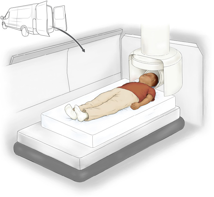
Another promising area for portable low-field MRI is, in research, specifically, in pediatric populations. Children have been chronically underserved in radiological research. This is not due to any malintent. Because children should not be exposed to unnecessary ionizing radiation, many x-ray–based imaging research studies are not viable. Investigational review boards sometimes have not allowed children to be research subjects on some high-field MRI scanners. Portable, low-field scanners, with open designs, would expand pediatric brain imaging research opportunities with an expected higher reward-to-risk ratio than at higher field strengths. Deoni et al. (2) recently demonstrated (see Fig. 1) that a portable low-field MRI system could be used to scan pediatric participants in a converted high-profile delivery van. The possibilities for using these devices for neurodevelopmental studies in low-resource settings abound.
Fig. 1. Scanning a subject in a portable, low-field MRI system installed in a delivery van modified to accommodate the MRI scanner.
These scanners can often be operated using electrical power from a conventional 120-V, 60-Hz power outlet. Credit: Ashley Mastin/Science Advances.
A special aid for stroke assessment
Stroke is also an apt application for low-field MRI scanning because exquisite image resolution and quality, as well as “quantitation,” of brain image data are often not essential for an effective differential diagnosis required in stroke assessment. An attending radiologist or neurologist wants to determine whether the stroke is ischemic (caused by a blocked blood vessel) or hemorrhagic (caused by bleeding) and what part(s) of the brain are affected by the stroke. This is actionable information that leads to recommending a subsequent treatment regime. The sooner these clinical determinations are made, the better the outcome. As the old adage goes in the stroke field, “Time is brain.”
Low-field portable MRI of stroke is not without competition in the low-cost, rapid diagnostic imaging arena. Computerized tomography (CT) perfusion scans can also provide a “go—no go” determination for whether mechanical removal of an embolus should be performed and can even identify the core of lesion and the penumbra or “territory” affected by the stroke. However, CT often requires using contrast agents that makes this process more complex. Still, a head-to-head trial showing the relative effectiveness of portable MRI and CT would be worthwhile.
Improving portable MRI scanners
While all of these promising advances bode well for patient care, the demise of high-field MRI is certainly not imminent. High-field MRI is essential to radiology and will continue to be, as it continues to offer benefits for additional indications. However, portable low-field MRI scanners will likely play an increasing role in the delivery of emergency medical care, as their speed of image acquisition increases and image quality improves. Further decreases in weight and cost, for example using Halbach magnet arrays, are feasible (3).
One way to hasten these needed developments is for portable, low-field MRI manufacturers to provide image acquisition and processing software, hardware specifications, and pulse sequences to the user community, i.e., MRI scientists, inventors, students, radiologists, emergency room physicians, and engineers. These groups could help develop and improve the MRI acquisition and processing pipeline including the experimental design, pulse sequences, radio frequency transmit-and-receive electronics, image data acquisition and reconstruction, and postprocessing (including artifact remediation and image display). Innovations in each area could have a material impact on the technology’s ultimate clinical uses. A groundswell of interest in developing “open source” portable, low-field MRI scanners may put added pressure on manufacturers to improve performance and lower cost.
All in all, portable low-field scanners, while currently unable to produce images comparable to high-field MRI scanners, still offer compelling benefits that will increasingly find an important niche in radiology and radiological research, as well as in other areas of clinical practice. Their success and dissemination in the U.S. market may depend on a number of complex factors, such as issues of reimbursement, but, overall, the future appears bright.
REFERENCES
- 1.Yuen M., Prabhat A. M., Mazurek M. H., Chavva I. R., Crawford A., Cahn B. A., Beekman R., Kim J. A., Gobeske K. T., Petersen N. H., Falcone G. J., Gilmore E. J., Hwang D. Y., Jasne A. S., Amin H., Sharma R., Matouk C., Ward A., Schindler J., Sansing L., de Havenon A., Aydin A., Wira C., Sze G., Rosen M. S., Kimberly W. T., Sheth K. N., Portable, low-field magnetic resonance imaging enables highly accessible and dynamic bedside evaluation of ischemic stroke. Sci. Adv. 8, eabm3952 (2022). [DOI] [PMC free article] [PubMed] [Google Scholar]
- 2.Deoni S. C. L., Medeiros P., Deoni A. T., Burton P., Beauchemin J., D’Sa V., Boskamp E., By S., McNulty C., Mileski W., Welch B. E., Huentelman M., Residential MRI: Development of a mobile anywhere-everywhere MRI lab. Sci. Rep. 1–23 (2022). [DOI] [PMC free article] [PubMed] [Google Scholar]
- 3.O’Reilly T., Teeuwisse W. M., de Gans D., Koolstra K., Webb A. G., In vivo 3D brain and extremity MRI at 50 mT using a permanent magnet Halbach array. Magn. Reson. Med. 85, 495–505 (2020). [DOI] [PMC free article] [PubMed] [Google Scholar]


