Abstract
Background
Modular acetabular components for total hip arthroplasty (THA) provide intraoperative flexibility; however, polyethylene liner dissociation may occur. This study aimed to examine the incidence and causes of liner dissociation associated with a specific acetabular component design at a single centre.
Materials and methods
A retrospective analysis of 7027 patients who underwent primary THA was performed to identify isolated liner dislocations. Patient demographics, clinical presentations, surgical and implant details, and both radiographic and computed tomography (CT) findings were analysed. Patients with liner dislocation were matched to a control group via 2:1 propensity score matching, and a logistic regression analysis was employed to identify associated risk factors.
Results
A total of 32 patients (0.45%) experienced liner dislocation at a mean 71.47 ± 60.10 months post surgery. Significant factors contributing to dislocations included the use of a conventional compared with a highly crosslinked polyethylene component (p = 0.049) and screw fixation (p = 0.028). Radiographic and CT analysis highlighted the importance of proper component orientation, revealing that patients experiencing dislocations demonstrated significantly lower acetabular cup anteversion angles (p = 0.001) compared with the control group. Impingement and malposition, identified in 41% and 47% of the cases, respectively, further underscored the multifactorial nature of dislocation risks.
Conclusions
While the overall rate of polyethylene liner dislocation was low, the findings of this study highlight the importance of appropriate cup placement to decrease the risk of dissociation. It further substantiates the influence of impingement and malposition in liner displacement, with increased mechanical stress exerted on the locking mechanism under adverse conditions and the potential risk increase due to screw placement.
Keywords: Total hip arthroplasty, Liner dislocation, Polyethylene, Modularity, Component positioning, Case–control study
Introduction
Total hip arthroplasty (THA) is one of the most common surgical interventions worldwide [1]. As the global population ages, projections suggest that, by 2050, an even larger segment will require this surgical intervention, underscoring its essential role in preserving mobility and enhancing quality of life [2]. Modularity in THA offers significant intraoperative flexibility, allowing surgeons to adjust the length, offset, and anteversion of the components. Yet, while this modularity enhances versatility, it can also introduce potential failure modes. Specifically, wear on the backside and disassociation of the polyethylene liner from the acetabular shell have been observed [3]. Several manufacturers with modular designs, including Pinnacle® (DePuy, Johnson & Johnson), Harris-Galante® (Zimmer), Trident® (Stryker), and G7® cup (Zimmer-Biomet), have encountered issues related to liner dissociation [3, 4]. Despite the 10-year survival rate of up to 99.2% using the Pinnacle acetabular system [5–7], there are several reported instances of cup/liner dissociation [8–14].
The incidence of cup/liner dissociation in this system is reported between 0.17% and 2.40%, requiring revision surgery associated with increased resource utilization and morbidity to the patient that make this entity a matter of pressing concern [8, 9]. Different liner options, such as neutral, lipped, lateralized, and the 10° face changing variants, have shown differential rates of dissociation. Notably, neutral and face-changing liners appear to have a propensity for higher dissociation rates compared with their lipped counterparts [15]. However, comprehensive studies focussing on the underlying reasons for the dissociation of the liner remain scant [8]. Given the potential morbidity associated with liner dissociation, which necessitates surgical revision, a more in-depth exploration is necessary.
This retrospective observational study aims to further explore this concern by investigating the frequency and underlying causes of dissociation of the liner in a cohort of patients who have undergone a THA with this acetabular system at a single institution since 2010. The study hypothesized that acetabular component position influences the risk of polyethylene liner dissociation.
Materials and methods
Study design and setting
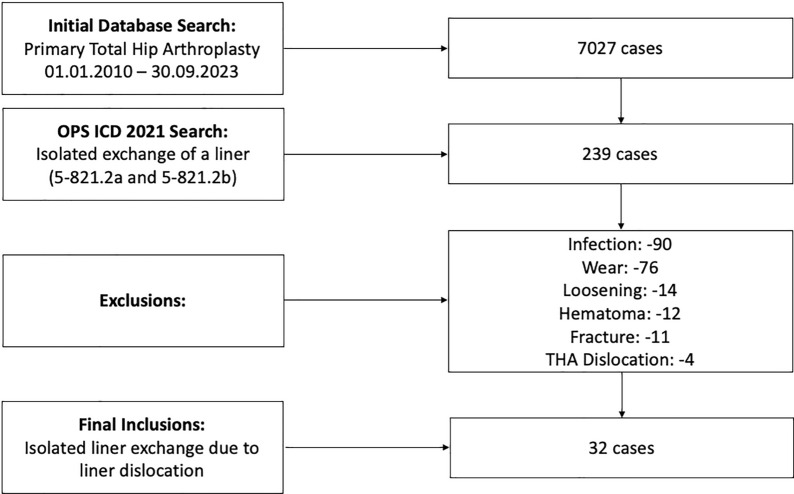
A retrospective observational study was conducted at a single institution to assess the incidence and underlying causes of Pinnacle (DePuy, Johnson & Johnson) cup/liner dissociation. Between 2010 and 2023, the period covered by our electronic records, a total of 7027 primary THA procedures were identified in which this acetabular system was implanted. Using the OPS ICD 2021 codes 5–821.2a and 5–821.2b, a total of 239 THA liner exchanges were identified and further analysed. From this group, 207 cases were excluded after reviewing the ICD-10-GM codes that documented the reasons for liner exchange. The excluded cases involved liner exchange due to infections, component loosening, hematomas, hip dislocations without evidence of liner dissociation, and liner wear. Only patients who had undergone liner exchange specifically due to dissociation of the polyethylene liner, confirmed by image analysis and operative note review, were included in the final analysis. After this selection process, the final cohort consisted of 32 patients suitable for analysis and a comprehensive set of data was collected from the internal clinical database (ORBIS, Agfa healthcare) including patient demographics, surgical variables, and clinical findings (Fig. 1).
Fig. 1.
Flowchart illustrating the database search process divided into two distinct categories: primary total hip arthroplasty (THA) and THA revision refined using specific OPS codes for isolated exchange of the liner
The suspected cause of the dislocation, and the period between symptom onset and the surgical intervention were documented. The type of primary implantation, the surgical approach employed, head and neck lengths, specifics about the dislocated liner, the age of the implant at the time of liner exchange, intraoperative findings, and other crucial details about the implant components were also assessed.
Measurements
Anteroposterior pelvic radiographs were retrieved from the last postoperative follow-up examination before the dislocation of the liner and, when available, from extremity rotational CT scans, taken at the time of dislocation. X-ray evaluations were not conducted on the radiographs obtained at the time of dislocation, as the displaced head would have influenced the leg length discrepancy and obscured the acetabular cup rim (Fig. 2). The measurements were obtained using radiographs with a graduated sphere of 25 mm and processed via Medicad software (Hemtec, Landshut, Germany).
Fig. 2.
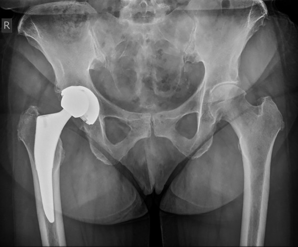
Anteroposterior X-ray of the pelvis showing a dislocation of the femoral head caused by liner dissociation from the acetabular cup. The image clearly depicts the contact of the dislocated femoral head with the acetabular cup
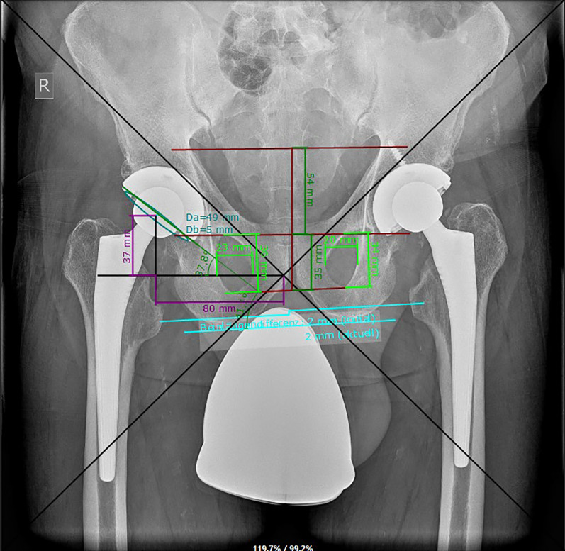
To evaluate the acetabular anteversion and inclination on both the anterior pelvis plane (APP) and the vertical functional coronary plane (FCP) (Fig. 3), we utilized an algorithmic model developed by Schwarz et al. [16, 17]. This model helps accurately assess the orientation of the acetabular cup on pelvic radiographs by correcting for potential errors caused by the positioning of the X-ray beam. Specifically, it adjusts for deviations in the central beam that can lead to inaccurate measurements of cup anteversion and inclination. The algorithm takes into account the effects of pelvic tilt and rotation, which can distort the appearance of the cup on radiographs. By applying mathematical corrections, it compensates for these distortions, allowing for a more accurate assessment of the cup’s orientation [16, 17]. Experimental results using a dummy pelvis demonstrated that the corrected measurements had an average absolute difference of only 0.4° for anteversion and 0.3° for inclination, compared with other methods that showed much larger errors [16]. Additionally, the algorithm was tested with various pelvic positions, confirming its robustness across different scenarios [17].
Fig. 3.
The acquired values were input into an algorithm that calculated the acetabular cup’s anteversion and inclination on the anterior pelvis plane (APP) and the vertical functional coronary plane (FCP)
Furthermore, an algorithm by Kanazawa et al. was employed to determine the rotation of the pelvis [18]. This algorithm quantifies three-dimensional (3-D) pelvic rotation using the width and height ratio of the obturator foramina under various pelvic tilts, thus enabling an estimation of pelvic rotation on plain anteroposterior radiographs. The algorithm’s validity was confirmed by comparing the calculated rotations with known values on reconstructed pelvises from CT scans. The authors demonstrated a strong linear correlation between the width-to-height ratio (WR) of the obturator foramina and axial pelvic rotation, with correlation coefficients ranging from 0.94 to 0.98, regardless of sagittal pelvic tilt up to 10° anteriorly [18].
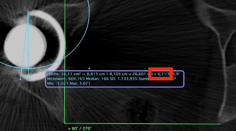
For the rotational CT scans of the lower extremities, we utilized OsirixLite software (Pixmeo, Bernex, Switzerland) to assess the images. In the CT scans, we determined the acetabular cup anteversion on the APP, the stem anteversion, and the combined anteversion. To measure the CT scans, we adopted the prevalent method in the current literature [19, 20]. The cup’s anteversion was determined using the angle between a line that connects the cup’s lateral anterior and posterior edges and a perpendicular line linking two symmetrical points on each side of the pelvis. The stem’s anteversion, which reflects the relationship between the condylar axis and the stem neck, was measured using a line drawn between the posterior portions of the medial and lateral femoral condyles and another line that joins the centre of the femoral head to the femoral component’s neck centre. The combined anteversion was obtained by summing the angle of the cup and the femoral component’s anteversion (Fig. 4). All measurements were conducted by a single senior surgical resident (S.P.) and subsequently assessed, verified, and confirmed by a senior surgeon (G.M.).
Fig. 4.
Rotational CT scans of the lower extremities were assessed using the software OsirixLite (Pixmeo, Bernex, Switzerland). In the provided example, the cup’s anteversion is measured
Statistical analysis
Continuous variables are described using mean values accompanied by their standard deviation (SD). Categorical data are presented as absolute counts (n) and associated percentages (%).
A matched control group was established via a 2:1 propensity score matching method, implemented using XLSTAT (Lumivero, Denver, USA). The 32 patients with cup/liner dissociation were aligned with 64 corresponding controls from the original pool of 7027 hip arthroplasty cases. The matching was based on age and body mass index (BMI). We employed the Mahalanobis distance within a calliper of 0.2 standard deviations of the propensity score’s logit. This statistical method was used to control for confounding variables, ensuring that the matched groups were comparable and reducing the risk of confounding bias.
Continuous variables were analysed using independent t-tests, while categorical variables were examined using chi-square tests. Logistic regression was performed to identify independent risk factors significantly associated with liner dislocation. All statistical tests were two-sided, with a significance level set at p < 0.05.
IBM SPSS Statistics (IBM Corp, New York, USA) version 29 was employed for the statistical analyses. The study adhered to institutional ethical standards and the Declaration of Helsinki (protocol no. 23-3384-104, 19 May 2023).
Results
Demographics and clinical presentation
Of the 7027 patients who underwent primary THA during the study period, 32 experienced dislocation of the liner, leading to a 0.45% complication rate. The average age at the time of revision surgery for polyethylene liner exchange was approximately 72 years (SD 9.95 years). All patients with a dislocated liner reported a squeaking noise from their hip, and in half (50.0%), no discernible cause for the dislocation was identified. Detailed demographics, clinical presentations, and causes can be found in Table 1.
Table 1.
Summary of demographics, symptoms, and causes for Pinnacle liner dissociation in the patient court
| Demographics | Dislocated liners (n = 32) |
|---|---|
| Age (y) | |
| Range | 53–88 |
| Mean (± SD) | 71.66 (± 9.95) |
| BMI (kg/m2) | |
| Range | 20.20–44.01 |
| Mean (± SD) | 29.5 (± 4.96) |
| Sex | |
| Women | 13 (40.6%) |
| Men | 19 (59.4%) |
| Pain | |
| Yes | 19 (59.4%) |
| No | 13 (40.6%) |
| Squeaking noises | |
| Yes | 32 (100%) |
| No | 0 (0%) |
| Duration of symptoms (days) | |
| Range | 1–84 |
| Mean (± SD) | 18.72 (± 25.77) |
| Cause of dislocation | |
| Squatting | 4 (12.5%) |
| Bucking | 5 (15.6%) |
| Lifting | 2 (6.3%) |
| Stumbling | 4 (12.5%) |
| Leg external rotation | 1 (3.1%) |
| None apparent cause | 16 (50.0%) |
Surgical and implant data
A significant association between the type of liner used and the risk of dislocation was found. Patients with conventional polyethylene (Marathon) were approximately 2.8 times more likely to experience dislocation compared with those with highly crosslinked polyethylene (Altrx) (odds ratio [OR] = 2.797, 95% confidence interval [CI] = 1.002 to 7.807, p = 0.049). The employment of fixation screws was also found to be associated with an increased risk of liner dislocations (OR = 11.667, 95% CI = 1.301 to 104.647, p = 0.028). Surgical details and component specifics are outlined in Table 2.
Table 2.
Surgical details (a) and implant specifics (b) for patients with Pinnacle liner dissociation compared with the control group
| Dislocated liners (n = 32) | Control group (n = 64) | p-Value | |
|---|---|---|---|
| (a) Surgical details | |||
| Side | |||
| Right | 20 (62.5%) | 33 (51.6%) | 0.639 |
| Left | 12 (37.5%) | 31 (48.4%) | |
| ASA | |||
| 1 | 3 (9.4%) | 9 (14.1%) | 0.348 |
| 2 | 22 (68.8%) | 34 (53.1%) | |
| 3 | 7 (21.9%) | 21 (32.8%) | |
| Time from THA (m) | |||
| Range | 0–203 | – | – |
| Mean (± SD) | 68.41 (± 60.13) | – | |
| Surgical approach | |||
| MI anterolateral (lateral decubitus) | 29 (90.6%) | 56 (87.5%) | 0.652 |
| Lateral (supine position) | 3 (9.4%) | 8 (12.5%) | |
| (b) Implant specifics | |||
| Cup diameter (mm) | |||
| 48 | 3 (9.4%) | 2 (3.1%) | 0.386 |
| 50 | 2 (6.3%) | 13 (20.3%) | |
| 52 | 10 (31.3%) | 17 (26.6%) | |
| 54 | 5 (15.6%) | 13 (20.3%) | |
| 56 | 5 (15.63%) | 10 (15.6%) | |
| 58 | 4 (12.50%) | 3 (4.7%) | |
| 60 | 3 (9.4%) | 5 (7.8%) | |
| 62 | 0 (0.0%) | 1 (1.6%) | |
| Screw fixation | |||
| Yes | 5 (15.7%) | 1 (1.6%) | 0.028 |
| No | 27 (84.4%) | 63 (98.4%) | |
| Liner model | |||
| Marathon | 23 (71.9%) | 37 (57.8%) | 0.049 |
| Altrx | 6 (18.8%) | 27 (42.2%) | |
| N/A | 3 (9.4%) | – | |
| Liner diameter (mm) | |||
| 28 | 2 (6.3%) | 1 (1.6%) | 0.699 |
| 32 | 30 (93.8%) | 63 (98.4%) | |
| 4-mm offset liner | |||
| Yes | 2 (6.3%) | 6 (9.4%) | 0.644 |
| No | 30 (93.8%) | 58 (90.6%) | |
| 10° face changing liner | |||
| Yes | 2 (6.3%) | 0 (0.0%) | 0.999 |
| No | 30 (93.8%) | 64 (100.0%) | |
| Head length (mm) | |||
| 1 | 12 (37.5%) | 35 (54.7%) | 0.054 |
| 5 | 12 (37.5%) | 19 (29.7%) | |
| 9 | 6 (18.8%) | 10 (15.6%) | |
| 13 | 1 (3.1%) | 0 (0.0%) | |
| 17 | 1 (3.1%) | 0 (0.0%) | |
| Head material | |||
| Metal | 31 (96.9%) | 56 (97.5%) | 0.170 |
| Ceramic | 1 (3.1%) | 8 (12.5%) | |
| Stem Model | |||
| Corail | 19 (59.4%) | 42 (65.6%) | 0.835 |
| Tri-Lock | 9 (28.1%) | 22 (34.4%) | |
| Link | 1 (3.1%) | 0 (0.0%) | |
| Proxima | 2 (6.3%) | 0 (0.0%) | |
| Fjord | 1 (3.1%) | 0 (0.0%) | |
| High-offset | |||
| Yes | 15 (46.9%) | 32 (50.0%) | 0.773 |
| No | 17 (53.1%) | 32 (50.0%) | |
Statistically significant values (p < 0.05) are highlighted in bold
m months, SD standard deviation, MI minimal invasive
N/A: data not available
Radiographic and CT measurements
Comparison of acetabular cup inclination angles revealed minimal disparity averaging 45.39° ± 6.24° in the dislocated liner cohort versus 44.33° ± 5.85° in the control group, a difference not reaching statistical significance (p = 0.418). However, 14 out of 30 acetabular cups (47%) in patients with liner dissociation were positioned outside the Lewinnek safe zones, which are defined as an inclination of 40 ± 10° and an anteversion of 15 ± 10° [21]. In comparison, 25 out of 64 acetabular cups (39%) in the control group were outside these safe zones. This difference was not statistically significant (p = 0.486).
Notably, a significant discrepancy was observed in the acetabular cup anteversion angles, as determined using the Lewinnek measurement method. The group with dislocated liners exhibited a mean anteversion angle of 10.38° (± 7.75), markedly lower than the 16.65° (± 8.15) observed in the control group (OR = 0.903, 95% CI = 0.848 to 0.962, p = 0.001).
CT measurements, available only in the case group, showed an average cup anteversion of 15.31 ± 14.57° and a combined anteversion of 34.00 ± 16.34°. Two patients’ X-rays and CT rotational analysis for 13 patients with dislocated liner were not available (Table 3).
Table 3.
X-ray (a) and CT (b) radiographic measurements: ranges, averages, and standard deviations
| (a) X-ray measurements | Dislocated liners (n = 30)1 | Control group (n = 64) | p-Value |
|---|---|---|---|
| Leg length difference (mm) | |||
| Range | −16 to 21 | −11 to 18 | 0.894 |
| Mean (± SD) | 1.07 (± 7.52) | 0.86 (± 6.91) | |
| Cup inclination (°) | |||
| Range | 31.40 to 60.60 | 29.40 to 57.90 | 0.418 |
| Mean (± SD) | 45.39 (± 6.24) | 44.33 (± 5.85) | |
| Cup anteversion (°) | |||
| Range | 2.29 to 30.95 | 0.00 to 35.90 | 0.001 |
| Mean (± SD) | 10.38 (± 7.75) | 16.65 (± 8.15) | |
| APP cup anteversion (°) | |||
| Range | −12.70 to 50.98 | −5.40 to 32.00 | 0.274 |
| Mean (± SD) | 10.69 (± 11.91) | 13.26 (± 9.90) | |
| APP cup inclination (°) | |||
| Range | 32.46 to 58.69 | 27.70 to 59.80 | 0.308 |
| Mean (± SD) | 43.70 (± 6.46) | 45.15 (± 6.40) | |
| FCP cup anteversion (°) | |||
| Range | −3.53 to 48.52 | −3.20 to 37.50 | 0.174 |
| Mean (± SD) | 12.68 (± 11.24) | 15.66 (± 9.08) | |
| FCP cup inclination (°) | |||
| Range | 33.18 to 58.69 | 28.50 to 59.20 | 0.313 |
| Mean (± SD) | 44.21 (± 6.36) | 45.62 (± 6.26) | |
| (b) CT measurements | Dislocated liners (n = 18)2 |
|---|---|
| Cup anteversion (°) | |
| Range | −13.70 to 34.10 |
| Mean (± SD) | 15.31 (± 14.57) |
| Stem anteversion (°) | |
| Range | −23.20 to 43.20 |
| Mean (± SD) | 17.48 (± 18.22) |
| Combined anteversion (°) | |
| Range | −8.50 to 60.20 |
| Mean (± SD) | 34.00 (± 16.34) |
No CTs were available in the control group
1X-ray data were not available for two patients
2Lower extremity rotational CT was available in 17 cases. For one patient, only a CT scan of the pelvis was available
SD standard deviation, APP anterior pelvis plane, FCP vertical functional coronary plane
Intraoperative findings
During surgery, the most frequent dislocation direction of the liner was caudal, occurring in eight cases (25.0%). Deformations and broken liners were identified in eight (25.0%) and six (18.8%) patients, respectively (Fig. 5). Metallosis was observed in 17 cases (53.1%), and impingement was evident in 13 cases (40.6%) (Table 4).
Fig. 5.
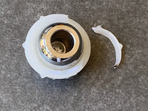
In this particular case the replaced liner shows damage on one edge, with no signs of wear or ovalization. The image has been edited to obscure the serial numbers
Table 4.
Distribution and frequencies of dislocation direction, liner alterations, metallosis, and presence of impingement
| Intraoperative findings | Dislocated Liners (n = 32) |
|---|---|
| Dislocation direction | |
| Ventral | 5 (15.6%) |
| Dorsal | 5 (15.6%) |
| Caudal | 8 (25.0%) |
| N/D | 14 (43.8%) |
| Liner deformation | |
| Yes | 8 (25.00%) |
| No | 2 (6.3%) |
| N/D | 22 (68.8%) |
| Broken liner | |
| Yes | 6 (18.8%) |
| No | 9 (28.1%) |
| N/D | 17 (53.1%) |
| Metallosis | |
| Yes | 17 (53.1%) |
| No | 12 (37.5%) |
| N/D | 17 (53.1%) |
| Impingement | |
| Yes | 13 (40.6%) |
| No | 19 (59.4%) |
N/D not described
Revision surgery outcomes
Three patients experienced re-dislocation after their first revision surgery, during which an identical liner was substituted to address the initial dislocation. These patients required a second revision surgery to manage the subsequent dislocation.
Among the patient cohort, four cases during the first revision surgery necessitated not only the replacement of the liner but also the reimplantation of the metal cup due to significant damage. For all other revision surgeries, the procedures generally involved only exchanges of the liner and head components.
Discussion
The current study underscores that, while the incidence of liner dissociation is low (0.45%), it remains a serious complication in THA patients requiring revision surgery, supporting earlier findings [8–15]. Perkins et al.’s review of literature from 2009 to 2020 found 53 cases of liner dislocations across different studies, with an incidence rate ranging from 0.17% to 2.40%. They also reported 26 instances of dissociation within their own group of 212 patients; all identified cases necessitated surgical revision owing to dissociation [8]. Their analysis suggests that implant malposition, impingement during movement, and potential issues with the locking mechanism of cups are significant factors contributing to dislocation occurrences [8]. Ensuring proper implant positioning and considering potential issues with the locking mechanism during the initial surgery could reduce the incidence of these complications.
However, while malposition alone may not directly lead to liner dislocation, the findings of the current study indicate that acetabular component anteversion may affect dislocation risk, since a higher degree of Lewinnek anteversion was associated with a reduced risk of liner dislocation. This protective mechanism is likely owing to the reduction in impingement that occurs during gait and activities requiring maximal leg flexion, since increased anteversion of the cup has been associated with enhanced range of motion and a reduced risk of impingement, as established by prior studies [22–24]. A lower anteversion angle of the acetabular cup could increase mechanical stress at the extreme range of motion, putting disproportionate stress on the liner against the head and elevating the likelihood of dislocation due to extreme movements acting as leverage. Surgeons should strive for optimal acetabular component positioning, particularly ensuring appropriate anteversion angles to minimize impingement and mechanical stress on the liner.
Beckmann et al. noted that factors such as impingement, malposition (especially during early dissociation), age-related deterioration of the liner, and increased ranges of motion in later stages play vital roles in liner dissociation [25]. These factors often lead to heightened stress and torsion on the locking mechanism, resulting in liner movement and potential dissociation, particularly if coincident damage occurs at the impingement site. However, excessively high anteversion angles can conversely increase wear and deterioration of the polyethylene liner if exceeding 25° [26]. Although recent literature challenges the established “safe zones” proposed by Lewinnek as definitive benchmarks for acetabular implant positioning, they continue to serve as clinical references due to their historical importance and the lack of universally accepted alternatives [26–29].
Perkins et al. [8] noted in their systematic review that 46% out of 53 dissociated liners were lipped, suggesting an increased risk of impingement during normal walking cycles. However, the current study observed a lower frequency of specific liner types, including 4-mm offset and 10° face changing liners, within the current cohort, without a significant correlation to disassociation events. The choice of liner type should consider the potential risk of impingement and dissociation, even though no significant correlation was found in this study, highlighting the complexity of implant selection.
While malposition is often cited as the primary cause of liner dislocation [8], various hypotheses exist concerning the reasons behind the dislocation of modular polyethylene liners [13, 14]. The study by Beckmann et al. emphasized the essential role of anti-rotation tabs (ARTs) in the liner locking mechanism [25]. Their mechanical tests revealed that the absence of ARTs in the Marathon liner resulted in a lower lever-out force required for separation of the cup and liner in this system compared with other tested constructs. This finding aligns with observations in this study, which indicated an increased risk of dislocation associated with the Marathon compared with Altrx liner. Notably, during revision surgeries for disassociations, it was often discovered that the Marathon liners lacked at least three ARTs, a situation that contributes to liner rotation and displacement [11, 12, 14, 15, 25, 30, 31]. However, the lack of detailed records on the integrity of the liner’s ARTs in the current study’s documentation limits further analysis.
The potential influence of implant design features, such as the prominence of screw heads, on liner dissociation has been also previously noted [9]. Although this risk is inherent in modular designs, this acetabular cup appears to exhibit a disproportionately higher rate of this complication.
The findings of this study confirm that screw fixation represents a risk factor for liner disassociation, and avoiding it where possible could help reduce the incidence of this complication.
The current study represents the largest single-centre cohort of liner dissociation compared with the existing literature [3, 8, 13], however; it is not without limitations. The retrospective design of this study introduces inherent biases and limits our ability to fully control for all confounding factors. Therefore, it is challenging to draw definitive causal relationships between the observed factors and liner dissociation. Potential underestimation of this complication owing to loss to follow-up or database inaccuracies, especially with varying ICD coding, remains also a concern. Given our reliance on data from a single institution, it is possible that additional patients experienced liner dislocations but sought care at other facilities, leading to an underreporting of the true incidence. Furthermore, despite this single-centre study with standardized THA implantation procedures, individual surgeon variation may have been present. Limited access to detailed surgical reports and rotational CT analysis for all cases further constrains the study. To mitigate this, an algorithm designed to enhance the accuracy of anteversion and inclination calculations using pelvic plain AP radiographs was used, albeit recognizing the limitations in direct comparison with CT measurements due to methodological differences. Lastly, the exclusive use of 28 mm and 32 mm heads, as opposed to the 36 mm heads utilized in other centres for cups 52 mm and larger, may affect the generalizability of our findings and should be considered in the interpretation of the results.
Conclusions
While liner dissociation is infrequent, it requires surgical revision in all affected cases.
Diagnosis is typically straightforward, as evidenced by the lateralization of the femoral head on X-rays and audible hip squeaking. Early diagnosis is fundamental to minimize potential damage to the acetabular cup, subsequently reducing the complexity of revision surgeries.
Key factors that contribute to the higher risk of liner dissociation include the use of conventional in comparison with highly crosslinked polyethylene. Appropriate acetabular cup anteversion may decrease the risk of liner disassociation, while using screws may increase the risk. It is important to interpret these findings with caution owing to the exclusive use of smaller femoral heads (28 and 32 mm) and a single implant design, which may limit their broader applicability. Moreover, the retrospective nature of this study introduces inherent biases, which must be considered when interpreting the results.
Acknowledgements
Not applicable.
Author contributions
S.P. and G.M. were responsible for the conception and design of the study. S.P., T.K., J.R., and M.S. were responsible for data acquisition. S.P., T.K., J.R., and M.S. were responsible for data analysis. S.P., J.P., and G.M. were responsible for drafting the manuscript. G.M. and J.P. were responsible for supervision and manuscript review. All authors read and approved the final manuscript.
Funding
Open Access funding enabled and organized by Projekt DEAL.
Availability of data and materials
All data generated or analysed during this study are included in this published article.
Declarations
Ethics approval and consent to participate
The study was conducted upon approval from the Ethics Committee of the University of Regensburg (protocol no. 23-3384-104).
Consent for publication
Not applicable.
Competing interests
The authors declare the following financial interests/personal relationships which may be considered as potential competing interests: J.P. is a paid consultant for Smith & Nephew; has stock or stock options in Eventum Orthopedics; received research support from Osteal Therapeutics; received royalties, financial or material support from VisualDX; is in the editorial/governing board of Journal of Arthroplasty; and serves on the AAHKS research committee. The remaining authors have nothing to disclose.
Footnotes
Publisher’s Note
Springer Nature remains neutral with regard to jurisdictional claims in published maps and institutional affiliations.
References
- 1.Markatos K, Savvidou OD, Foteinou A, Kosmadaki S, Trikoupis I, Goumenos SD, Papagelopoulos PJ (2020) Hallmarks in the history and development of total hip arthroplasty. Surgical innovation 27(6):691–694. 10.1177/1553350620947209 10.1177/1553350620947209 [DOI] [PubMed] [Google Scholar]
- 2.Pabinger C, Lothaller H, Portner N, Geissler A (2018) Projections of hip arthroplasty in OECD countries up to 2050. Hip Int 28(5):498–506. 10.1177/1120700018757940 10.1177/1120700018757940 [DOI] [PubMed] [Google Scholar]
- 3.Cheppalli N, Goel A, Wassef A, Bhandarkar AW, Kakish S (2022) Polyethylene liner dissociation in the early post-operative period following total hip arthroplasty: a case report and literature review. Cureus 14(5):e25119. 10.7759/cureus.25119 10.7759/cureus.25119 [DOI] [PMC free article] [PubMed] [Google Scholar]
- 4.González della Valle A, Ruzo PS, Li S, Pellicci P, Sculco TP, Salvati EA (2001) Dislodgment of polyethylene liners in first and second-generation Harris-Galante acetabular components. A report of eighteen cases. J Bone Joint Surg Am 83(4):553–559. 10.2106/00004623-200104000-00010 10.2106/00004623-200104000-00010 [DOI] [PubMed] [Google Scholar]
- 5.Bedard NA, Callaghan JJ, Stefl MD, Willman TJ, Liu SS, Goetz DD (2014) Fixation and wear with a contemporary acetabular component and cross-linked polyethylene at minimum 10-year follow-up. J Arthroplasty 29(10):1961–1969. 10.1016/j.arth.2014.05.008 10.1016/j.arth.2014.05.008 [DOI] [PubMed] [Google Scholar]
- 6.Johnson & Johnson/DePuy. (2000-2012). Multi-center, prospective, clinical evaluation of pinnacle acetabular implants in total hip arthroplasty. Retrieved from https://clinicaltrials.gov/show/NCT00306930
- 7.Kindsfater K, Lesko J (2017) Survivorship of a modular acetabular cup system: medium- to long-term follow-up. Arthroplast Today 4(3):376–382. 10.1016/j.artd.2017.07.001 10.1016/j.artd.2017.07.001 [DOI] [PMC free article] [PubMed] [Google Scholar]
- 8.Perkins TJ, Kop AM, Whitewood C, Pabbruwe MB (2023) Dissociation of polyethylene liners with the Depuy Pinnacle cup: a report of 26 cases. Hip Int 33(1):28–33. 10.1177/11207000211008459 10.1177/11207000211008459 [DOI] [PubMed] [Google Scholar]
- 9.Yun A, Koli EN, Moreland J, Iorio R, Tilzey JF, Mesko JW, Lee GC, Froimson M (2016) Polyethylene liner dissociation is a complication of the DePuy Pinnacle Cup: a report of 23 cases. Clin Orthop Relat Res 474(2):441–446. 10.1007/s11999-015-4396-5 10.1007/s11999-015-4396-5 [DOI] [PMC free article] [PubMed] [Google Scholar]
- 10.Hill DS, Patel K, Herlekar D (2016) Early catastrophic failure of a PINNACLE MARATHON® polyethylene acetabular liner: a report of a malpositioned PINNACLE DUOFIX HA® acetabular shell resulting in point loading and accelerated wear. Hip Int 26(6):e52–e55. 10.5301/hipint.5000447 10.5301/hipint.5000447 [DOI] [PubMed] [Google Scholar]
- 11.Kagan R, Anderson MB, Peters C, Pelt C, Gililland J (2018) Pinnacle polyethylene liner dissociation: a report of 3 cases. Arthroplasty Today 4(4):441–446. 10.1016/j.artd.2018.08.001 10.1016/j.artd.2018.08.001 [DOI] [PMC free article] [PubMed] [Google Scholar]
- 12.Memon AR, Gwynne-Jones D (2020) Polyethylene liner dissociation with the Pinnacle acetabular component: should we be concerned? Arthroplasty Today 6(1):5–8. 10.1016/j.artd.2019.12.001 10.1016/j.artd.2019.12.001 [DOI] [PMC free article] [PubMed] [Google Scholar]
- 13.Keohane D, Sheridan GA, Harty J, O’Loughlin P (2021) Polyethylene liner dissociation in a DePuy pinnacle cup with a manufacturer analysis of the failed component. BMJ Case Reports 14(2):e238333. 10.1136/bcr-2020-238333 10.1136/bcr-2020-238333 [DOI] [PMC free article] [PubMed] [Google Scholar]
- 14.Napier RJ, Diamond O, O’Neill CKJ, O’Brien S, Beverland DE (2017) The incidence of dissociated liners in 4,751 consecutive total hip arthroplasties using Pinnacle polyethylene acetabular liners. Hip Int 27(6):537–545. 10.5301/hipint.5000512 10.5301/hipint.5000512 [DOI] [PubMed] [Google Scholar]
- 15.Singleton N (2018) Polyethylene liner dissociation with the Depuy Pinnacle cup: a report of 6 cases. OPROJ. 10.31031/OPROJ.2018.03.000573 10.31031/OPROJ.2018.03.000573 [DOI] [Google Scholar]
- 16.Schwarz T, Weber M, Wörner M, Renkawitz T, Grifka J, Craiovan B (2017) Central X-ray beam correction of radiographic acetabular cup measurement after THA: an experimental study. Int J Comput Assist Radiol Surg 12(5):829–837. 10.1007/s11548-016-1489-x 10.1007/s11548-016-1489-x [DOI] [PubMed] [Google Scholar]
- 17.Schwarz TJ, Weber M, Dornia C, Worlicek M, Renkawitz T, Grifka J, Craiovan B (2017) Correction of Pelvic Tilt and Pelvic Rotation in Cup Measurement after THA—an Experimental Study. Korrektur der Beckenverkippung und der Beckenverdrehung bei der Pfannenmessung nach Hüft-TEP-Versorgungen—Eine experimentelle Studie. RoFo Fortschritte auf dem Gebiete der Rontgenstrahlen und der Nuklearmedizin 189(9):864–873. 10.1055/s-0043-110012 10.1055/s-0043-110012 [DOI] [PubMed] [Google Scholar]
- 18.Kanazawa M, Nakashima Y, Arai T, Ushijima T, Hirata M, Hara D, Iwamoto Y (2016) Quantification of pelvic tilt and rotation by width/height ratio of obturator foramina on anteroposterior radiographs. Hip Int 26(5):462–467. 10.5301/hipint.5000374 10.5301/hipint.5000374 [DOI] [PubMed] [Google Scholar]
- 19.Li L, Zhang Y, Lin YY, Li ZX, Chen L, Chen DS, Fan P (2020) A specific anteversion of cup and combined anteversion for total hip arthroplasty using lateral approach. Orthop Surg 12(6):1663–1673. 10.1111/os.12790 10.1111/os.12790 [DOI] [PMC free article] [PubMed] [Google Scholar]
- 20.Fujishiro T, Hayashi S, Kanzaki N, Hashimoto S, Kurosaka M, Kanno T, Masuda T (2014) Computed tomographic measurement of acetabular and femoral component version in total hip arthroplasty. Int Orthop 38(5):941–946. 10.1007/s00264-013-2264-z 10.1007/s00264-013-2264-z [DOI] [PMC free article] [PubMed] [Google Scholar]
- 21.Lewinnek GE, Lewis JL, Tarr R, Compere CL, Zimmerman JR (1978) Dislocations after total hip-replacement arthroplasties. J Bone Joint Surg Am 60(2):217–220 10.2106/00004623-197860020-00014 [DOI] [PubMed] [Google Scholar]
- 22.Habe Y, Hamada H, Uemura K, Takashima K, Ando W, Sugano N (2023) Cup safe zone and optimal stem anteversion in total hip arthroplasty for patients with highly required range of motion. J Orthopaedic Res. 10.1002/jor.25769 10.1002/jor.25769 [DOI] [PubMed] [Google Scholar]
- 23.Widmer KH (2020) The impingement-free, prosthesis-specific, and anatomy-adjusted combined target zone for component positioning in THA depends on design and implantation parameters of both components. Clin Orthop Relat Res 478(8):1904–1918. 10.1097/CORR.0000000000001233 10.1097/CORR.0000000000001233 [DOI] [PMC free article] [PubMed] [Google Scholar]
- 24.Harada S, Hamai S, Motomura G, Ikemura S, Fujii M, Kawahara S, Sato T, Hara D, Nakashima Y (2022) Evaluation of optimal implant alignment in total hip arthroplasty based on postoperative range of motion simulation. Clin Biomech (Bristol, Avon) 92:105555. 10.1016/j.clinbiomech.2021.105555 10.1016/j.clinbiomech.2021.105555 [DOI] [PubMed] [Google Scholar]
- 25.Beckmann NA, Schonhoff M, Bastian JD, Renkawitz T, Jaeger S (2023) Dissociation of liner from cup in THA: does liner damage affect the risk of dissociation? Arch Orthop Trauma Surg 143(5):2747–2754. 10.1007/s00402-022-04529-8 10.1007/s00402-022-04529-8 [DOI] [PMC free article] [PubMed] [Google Scholar]
- 26.Baghdadi J, Alkhateeb S, Roth A, VITAS-Group, Jäger M (2023) Cup positioning and its effect on polyethylene wear of vitamin E- and non-vitamin E-supplemented liners in total hip arthroplasty: radiographic outcome at 5-year follow-up. Arch Orthop Trauma Surg 143(3):1679–1688. 10.1007/s00402-022-04424-2 10.1007/s00402-022-04424-2 [DOI] [PMC free article] [PubMed] [Google Scholar]
- 27.Burapachaisri A, Elbuluk A, Abotsi E, Pierrepont J, Jerabek SA, Buckland AJ, Vigdorchik JM (2020) Lewinnek safe zone references are frequently misquoted. Arthroplasty today 6(4):945–953. 10.1016/j.artd.2020.09.011 10.1016/j.artd.2020.09.011 [DOI] [PMC free article] [PubMed] [Google Scholar]
- 28.Esposito CI, Gladnick BP, Lee YY, Lyman S, Wright TM, Mayman DJ, Padgett DE (2015) Cup position alone does not predict risk of dislocation after hip arthroplasty. J Arthroplasty 30(1):109–113. 10.1016/j.arth.2014.07.009 10.1016/j.arth.2014.07.009 [DOI] [PMC free article] [PubMed] [Google Scholar]
- 29.Abdel MP, von Roth P, Jennings MT, Hanssen AD, Pagnano MW (2016) What safe zone? The vast majority of dislocated THAs are within the Lewinnek safe zone for acetabular component position. Clin Orthop Relat Res 474(2):386–391. 10.1007/s11999-015-4432-5 10.1007/s11999-015-4432-5 [DOI] [PMC free article] [PubMed] [Google Scholar]
- 30.Mesko JW (2009) Acute liner disassociation of a Pinnacle acetabular component. J Arthroplasty 24(5):815–818. 10.1016/j.arth.2008.03.010 10.1016/j.arth.2008.03.010 [DOI] [PubMed] [Google Scholar]
- 31.O’Neill CK, Napier RJ, Diamond OJ, O’Brien S, Beverland DE (2015) Acetabular liner dissociation following total hip arthroplasty: a rare but serious complication that may be easily misinterpreted in the emergency department. Case Rep Emerg Med 2015:802753. 10.1155/2015/802753 10.1155/2015/802753 [DOI] [PMC free article] [PubMed] [Google Scholar]
Associated Data
This section collects any data citations, data availability statements, or supplementary materials included in this article.
Data Availability Statement
All data generated or analysed during this study are included in this published article.


