Abstract
Background
Distal tibial fractures represent common lower limb injuries, frequently accompanied by significant soft tissue damage. The optimal surgical approach for managing these fractures remains a topic of considerable debate. The aim of this study was to perform a comparative analysis of the outcomes associated with retrograde intramedullary tibial nails (RTN) and minimally invasive plate osteosynthesis (MIPO) in the context of treating extra-articular distal tibial fractures.
Methods
A retrospective review was conducted on a cohort of 48 patients who sustained extra-articular distal tibial fractures between December 2019 and December 2021. Patients underwent either RTN or MIPO procedures. Various parameters, including operative duration, intraoperative fluoroscopy exposure, time to union, duration until full weight-bearing, American Orthopedic Foot and Ankle Society (AOFAS) scores, and complications, were recorded and compared between the two treatment groups.
Results
No statistically significant differences were observed in operative duration, time to union, angulation of the distal tibial coronal plane, or AOFAS scores between the RTN and MIPO groups. However, the RTN group had a higher average number of intraoperative fluoroscopy images (8.2 ± 2.3) compared to the MIPO group (4.1 ± 2.0). The RTN group demonstrated shorter average hospital stays (7.1 ± 1.4 days) and a quicker return to full weight-bearing (9.9 ± 1.3 weeks), which were significantly superior to the MIPO group (9.0 ± 2.0 days and 11.5 ± 1.5 weeks, respectively). In terms of complications, the RTN group had one case of superficial infection, whereas the MIPO group exhibited two cases of delayed union and nonunion, two occurrences of deep infection, and an additional three cases of superficial infection.
Conclusions
Both RTN and MIPO are effective treatment options for extra-articular distal tibial fractures. However, RTN may offer superior outcomes in terms of decreased inpatient needs, faster return to full weight-bearing capacity, and a lower rate of complications.
Keywords: Extra-articular distal tibial fractures, Retrograde tibial intramedullary nails, Locking plate, Hospital stay, Full weight-bearing, Complications
Introduction
Distal tibial fractures represent a notable occurrence in clinical practice, constituting roughly 10% of all tibial fractures [1] and 1.5% of all adult fractures [2]. In recent years, there has been an increase in the incidence of distal tibial fractures, primarily attributed to the rising prevalence of high-energy injuries stemming from factors such as traffic accidents and falls from significant heights. The management of distal tibial fractures has posed a challenge due to several complicating factors, including limited local soft tissue coverage, heightened skin tension, anatomical ischemia at the fracture site, and the adjacency of the distal tibia to the ankle joint [3]. Currently, two common techniques for addressing extra-articular distal tibial fractures are antegrade tibial intramedullary nails and locking plates. Nevertheless, both of these modalities exhibit notable limitations. Plate fixation, while widely employed due to its capacity to achieve a proper degree of reduction and offer rigid fixation, proves unsuitable for patients presenting serious injuries of soft tissue or open fractures. Even with the adoption of the minimally invasive plate osteosynthesis (MIPO), there remains a necessity for relatively extensive wound exposure and soft tissue dissection to facilitate fracture reduction. This heightened exposure elevates the complication risk like infection, tissue necrosis, delayed union, and nonunion [4]. While antegrade tibial intramedullary nails offer theoretical advantages like load-sharing characteristics, minimally invasive application, and reduced soft tissue irritation, their utility is hampered by complications including instability, malalignment of fractures, and postoperative anterior knee pain [5].
The RTN emerges as a novel minimally invasive internal fixation device, conceived by Kuhn et al., specifically tailored for addressing distal tibial fractures [6]. Biomechanical assessments have demonstrated that the RTN exhibits biomechanical advantages over the conventional medial distal tibial plate and expertly applied antegrade tibial intramedullary nails concerning fracture fretting and internal fixation stress [7]. Moreover, its clinical application has shown favorable outcomes [8]. Nevertheless, it is noteworthy that the existing body of literature primarily encompasses biomechanical studies, with clinical investigations comparing RTN to other internal fixation methods remaining relatively limited. In the present study, we aimed to provide a comprehensive comparative analysis of two surgical approaches: RTN and MIPO, in the management of extra-articular distal tibial fractures.
Patients and methods
Clinical data
In this retrospective study, we conducted an analysis of consecutive clinical cases, ensuring that we obtained written informed consent from all participants prior to their procedures. All techniques employed during this investigation adhered to the ethical guidelines outlined in the 2013 revision of the Helsinki Declaration and the institutional research committee’s protocols. Our study focused on patients diagnosed with extra-articular distal tibia fractures at our level 1 trauma center during the period from December 2019 to December 2021.
To be included in the study, patients needed to meet specific criteria, which were as follows: (1) Inclusion of AO/OTA 43 A closed fractures or Gustilo I, II, or IIIA open fractures. (2) Location of the tibia fracture line between 4 and 11 cm proximal to the plafond and these fractures accompanied with nondisplaced posterior malleolar fractures. (3) Patients were required to be skeletally mature adults with a minimum 12-month follow-up duration. Patients who fell into the categories of Gustilo IIIB, IIIC open fractures, those with pathological fractures of the tibia, those with distal intra-articular or proximal intra-articular fractures of the tibia, or those with a follow-up period of less than 12 months were excluded from the study.
Pre-operative preparation
Patients presenting with open fractures received immediate debridement, suturing, and either calcaneal traction (for Gustilo I fractures) or temporary external fixation (for Gustilo II or IIIA fractures) on the day of admission. In cases of closed fractures, calcaneal traction with additional immobilization or compression dressing was initiated following admission. In all patients, the affected leg was elevated, and a thorough examination of the affected deep vein was conducted via ultrasound to exclude the presence of thrombosis. Patients were encouraged to engage in ankle pump exercises to mitigate swelling in the affected limb.
Surgical procedure
In cases involving fibula fractures that compromise ankle stability, specifically those occurring within 8 cm above the malleolar fossa, the initial intervention entails open reduction and plate fixation of the fibula through a lateral approach.
RTN Group: Patients underwent either general or epidural anesthesia while lying supine on a fluoroscopic surgical bed. A lower limb traction device was employed to facilitate closed reduction, guided by C-arm fluoroscopy. In cases where closed reduction proved unsuccessful, a small incision was made at the fracture site to assist in the reduction process. A longitudinal incision, measuring 2 ~ 3 cm, was performed at the medial malleolus using sharp dissection and separation techniques. The insertion point of the guide wire was verified through C-arm fluoroscopy to ensure its proper alignment at the midpoint of the medial malleolus in both lateral and anteroposterior radiographs (Fig. 1A) according to previous report [9]. Subsequently, the guide wire was directed parallel to the medial cortex in the anteroposterior radiograph, aligning with the anatomical axis of the distal tibia in the lateral radiograph (Fi. 1A). Following this, a hole reamer (Fig. 1B) and a cannulated awl (Fig. 1C) were employed along the guide wire to create a pathway leading to the medullary canal. The guide wire and awl were then adjusted to introduce prosthesis trials into the medullary canal, facilitating the selection of the appropriate length and diameter of the RTN (Fig. 1D). Once the RTN (Double Medical Technology Inc., Xiamen, China) was assembled on the aiming device, it was inserted into the medullary canal with gentle force and small twisting movements until it was flush with the cortex of the medial malleolus (Fig. 1E). C-arm fluoroscopy was utilized to confirm satisfactory reduction and the correct positioning of the nail. Subsequently, distal and proximal locking screws (Fig. 1F, G) were introduced through the trocar combination, and an end cap was placed at the nail’s extremity (Fig. 1H).
Fig. 1.
Steps in RTN operations. (A) The insertion point of was situated at the midpoint of the medial malleolus and the guide wire was directed parallel to the medial cortex in the anteroposterior radiograph, aligning with the anatomical axis of the distal tibia in the lateral radiograph. (B, C) A hole reamer and a cannulated awl were employed along the guide wire to create a pathway leading to the medullary canal. (D) A prosthesis trial was introduced into the medullary canal. (E) the RTN was assembled on the aiming device and inserted into the medullary canal until it was flush with the cortex of the medial malleolus. (F, G) Distal and proximal locking screws were introduced through the trocar combination. (H) An end cap was placed at the nail’s extremity
MIPO Group: Patients were positioned in a supine manner and underwent closed reduction using a lower limb traction device. If closed reduction failed, a small incision was made at the fracture site to assist in the reduction process. Once the reduction was deemed satisfactory, temporary fixation was achieved using Kirschner wires or reduction forceps. An anatomically contoured locking compression plate (anterior lateral plates or medial plates) of suitable length was selected and inserted through a subcutaneous tunnel between the deep fascia and periosteum of the tibia. Finally, both proximal and distal locking screws were introduced under the guidance of C-arm fluoroscopy.
All surgeries were performed by two separate groups of surgeons who possess experience in these techniques. It should be noted that both of the two implants are not for reduction, but for fixation. The quality of reduction depends on the surgeon before inserting the implants.
Postoperative management
All patients received a 24-hour course of intravenous antibiotics postoperatively, and active and passive functional exercises were initiated in the postoperative period.
Outcome assessment
The following parameters were recorded: operative duration, the count of intraoperative fluoroscopy exposures, and the length of the hospital stay. To monitor the progress of fracture healing, regular follow-up appointments were scheduled, and monthly postoperative X-ray examinations were performed. A period of partial weight-bearing, limited to sole contact or up to 15 kg of pressure [10], was allowed after a 4 ~ 6 week period. The increment in load-bearing was determined in accordance with various factors, including the fracture pattern and location, soft tissue condition, bone quality, and the presence or absence of pain induced by weight-bearing. During the last follow-up, any instance where the distal tibial coronal plane angle exceeded 5° was indicative of ankle inversion or valgus deformity. Functional outcomes were evaluated using the AOFAS score.
Statistical analysis
Statistical analysis was carried out utilizing SPSS version 26.0. Quantitative data are expressed as the mean ± standard error of the mean (SEM), and between-group comparisons were conducted using a two-sample t-test. Qualitative data are represented as the number of cases and corresponding percentages (%), and their analysis was conducted using either the chi-square test or the two-sided Fisher exact test, depending on appropriateness. P < 0.05 denotes statistically significant differences.
Results
The preoperative characteristics of both study groups are summarized in Table 1. The RTN group consisted of 21 patients, including 9 females and 12 males, with an age range of 19 ~ 85 years (mean age: 44.4 ± 17.9 years). The etiology of injuries included traffic accidents (7 patients), falls from heights (5 patients), rotational injury (5 patients), and crush injuries (4 patients). Utilizing the AO/OTA system, the fractures were classified, with 7 cases classified as type 43A1, 6 as type 43A2, and 8 as type 43A3 distal tibial fractures. Among these patients, 15 had closed fractures, and 6 had open fractures (comprising 1 Gustilo III A, I, 2 Gustilo II, and 3 Gustilo I). Additionally, 4 patients presented with associated injuries, including traumatic brain injury (1), hemopneumothorax (1), abdominal injury (1), and other injuries (1). Six patients had comorbidities, including diabetes (2), hypertension (3), and chronic obstructive pulmonary disease (1). Thirteen patients had associated fibular fractures, and 9 patients underwent fixation due to involvement of the ankle joint. The tibial stem fracture line length from the articular surface ranged from 4.5 to 11.0 cm, with an average of 8.8 ± 1.3 cm.
Table 1.
Baseline characteristics of both groups
| Characteristics | RTN group | MIPO group | Total | P Value |
|---|---|---|---|---|
|
Gender Male (%) Female (%) Age (Years) Causes Of Injury Traffic accident (%) Fall from height (%) Sprain (%) Crushes (%) AO/OAT classification 43A1 (%) 43A2 (%) 43A3 (%) Type of fractures Closed fractures (%) Open fractures (%) Gustilo I Gustilo II Gustilo IIIA Fibular fractures (%) Operation (%) No operation (%) Multiple injuries (%) Traumatic brain injury Chest injury Abdominal injury Other injuries Comorbidities (%) Diabetes Hypertension Chronic obstructive pulmonary disease Length of fracture line from joint surface (cm) |
21 12 (57.1%) 9 (42.9%) 44.4 ± 17.9 (19 ~ 85) 7 (33.3%) 5 (23.8%) 5 (23.8%) 4 (19.1%) 7 (33.3%) 6 (28.6%) 8 (38.1%) 15 (71.4%) 6 (28.6%) 3 2 1 13 (62.0%) 9 (69.2%) 4 (30.8%) 4 (19.0%) 1 1 1 1 6 (28.8%) 2 3 1 8.8 ± 1.3 (4.5 ~ 11.0) |
27 15 (55.5%) 12 (44.5%) 43.8 ± 16.5 (19 ~ 76) 10 (37.0%) 7 (26.0%) 5 (18.5%) 5 (18.5%) 9 (33.3%) 8 (29.6%) 10 (37.1%) 19 (70.3%) 8 (29.7%) 4 3 1 18 (66.6%) 12 (66.7%) 6 (33.3%) 5 (18.5%) 1 1 1 2 8 (29.6%) 4 3 1 8.3 ± 1.7 (5.8 ~ 11.0) |
48 27 (56.2%) 21 (43.8%) 44.1 ± 16.9 (19 ~ 85) 17 (35.4%) 12 (25.0%) 10 (20.8%) 9 (18.8%) 16 (33.3%) 14 (29.2%) 18 (37.5%) 34 (70.8%) 14 (29.1%) 7 5 2 31(64.6%) 21 (67.7%) 10 (32.3%) 9 (18.8%) 2 2 2 3 14 (29.2%) 6 6 2 8.5 ± 1.9 (5.8 ~ 11.0) |
0.91 0.90 0.97 0.99 0.96 1.00 0.97 0.82 0.28 |
In the MIPO group, there were 15 male and 12 female patients, aged between 19 and 76 years (mean age: 43.8 ± 16.5 years). The injury causes included 10 cases resulting from traffic accidents, 7 cases attributed to high-impact falls, 5 cases caused by rotational injury, and 5 cases resulting from crush injuries. Based on the AO/OTA classification, there were 9 patients with 43A1 fractures, 8 with 43A2 fractures, and 10 with 43A3 fractures. Eight patients had open fractures, comprising 4 Gustilo I, 3 Gustilo II, and 1 Gustilo III A), and 19 patients had closed fractures. In addition, 18 patients had associated fibular fractures, and 12 patients underwent fixation. Five patients had concurrent injuries, including one case with traumatic brain injury, one case with multiple rib fractures, one case with abdominal injury, and two cases with other injuries. Eight of the patients had comorbidities, including four with diabetes, three with hypertension, and one with chronic obstructive pulmonary disease. The tibial stem fracture line length from the articular surface ranged from 5.8 to 11.0 cm, with an average of 8.3 ± 1.7 cm. Baseline characteristics were compared between the two groups, and no significant differences were observed (Table 1).
Table 2 summarizes the key clinical and follow-up data for both groups. No statistically significant differences were found in operation duration, time to union, the distal tibial coronal plane angulation, and AOFAS score at the final follow-up between the two groups. The average number of intraoperative fluoroscopy exposures in the RTN group was 8.2 ± 2.3, which was higher than that in the MIPO group (4.1 ± 2.0). The average hospital stay duration and time to full weight-bearing in the RTN group were 7.1 ± 1.4 days and 9.9 ± 1.3 weeks, respectively, which were significantly superior to those in the MIPO group (9.0 ± 2.0 days and 11.5 ± 1.5 weeks). In the RTN group, there was 1 case of superficial infection, whereas the MIPO group exhibited 2 cases of nonunion and delayed union, 2 cases of deep infection, and 3 cases of superficial infection. The incidence of complications was significantly higher in the MIPO group.
Table 2.
Comparison of the main clinical data for both groups
| Clinical data | RTN group (n = 21) | MIPO group (n = 27) | P Value |
|---|---|---|---|
|
Operation time (minutes) Number of intraoperative fluoroscopy (times) Hospital stay (days) Average follow up period (months) Weeks to union (weeks) Weeks to full weight bear (weeks) The angulation of the distal tibial coronal plane (°) AOFAS score at final follow up Excellent and Good Fair and Poor Complications Nonunion or delayed union Superficial infection Deep infection Implant failure |
58.6 ± 9.9 8.2 ± 2.3 7.1 ± 1.4 15.7 ± 2.4 14.9 ± 2.3 9.9 ± 1.3 2.6 ± 1.6 84.5 ± 4.7 17 (81.0%) 4 (19.0%) 1 (4.8%) 0 1 0 0 |
60.2 ± 9.9 4.1 ± 2.0 9.0 ± 2.0 15.6 ± 2.8 15.4 ± 2.3# 11.5 ± 1.5 2.0 ± 1.4 82.6 ± 5.8 22 (81.5%) 5 (18.5%) 8 (29.6%) 2 3 2 1 |
0.58 0.000* 0.000* 0.89 0.46 0.001* 0.19 0.24 0.96 0.031* |
* P < 0.05
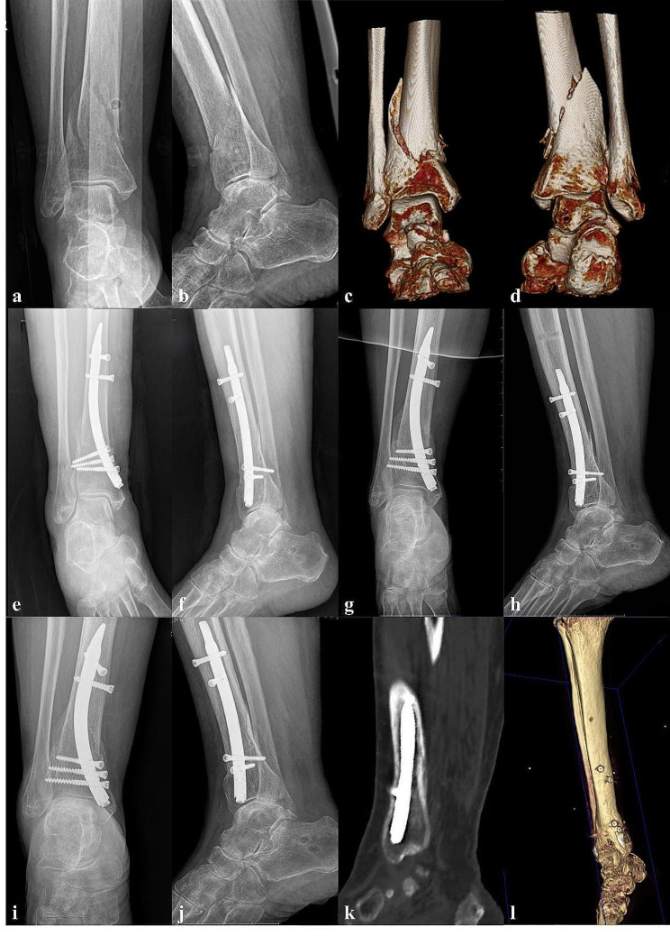
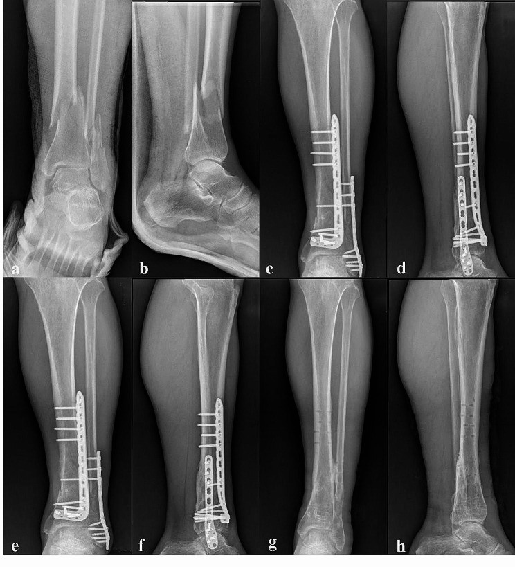
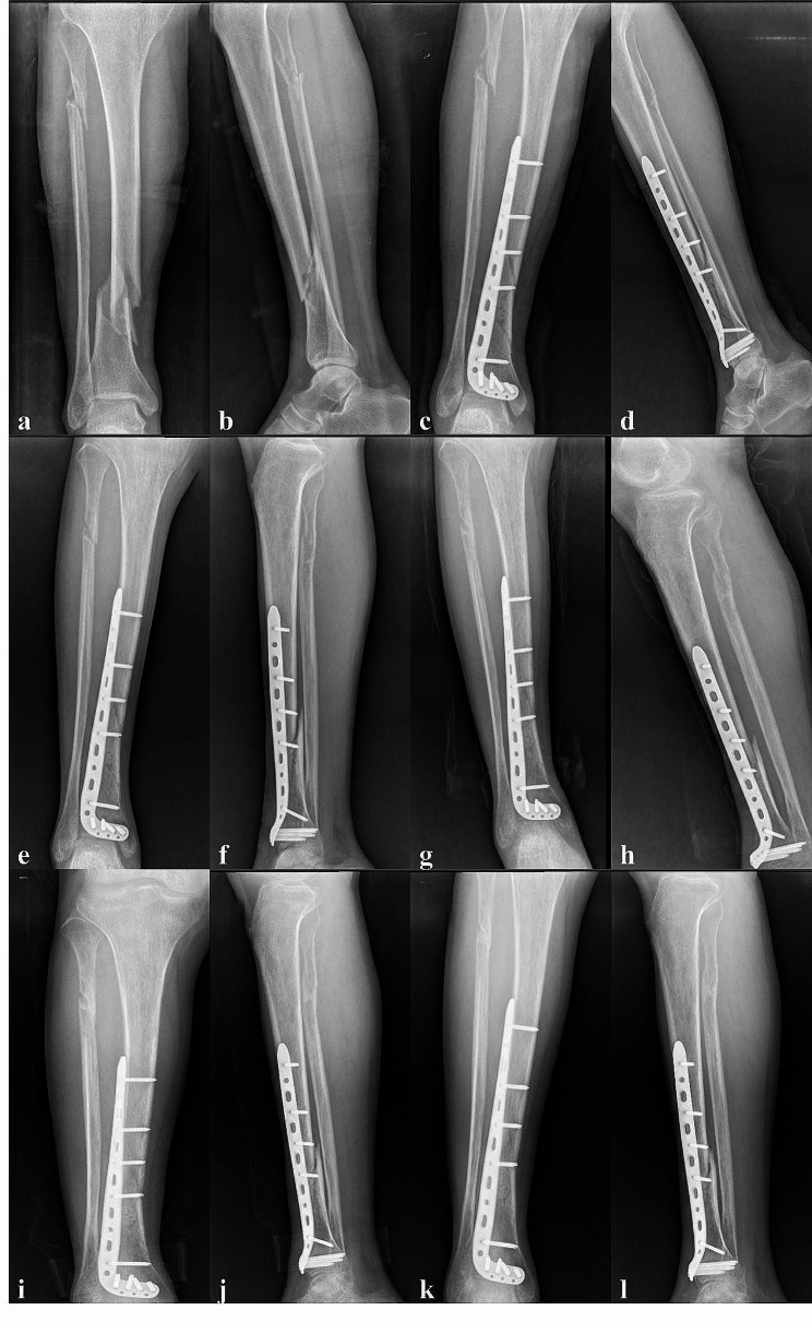
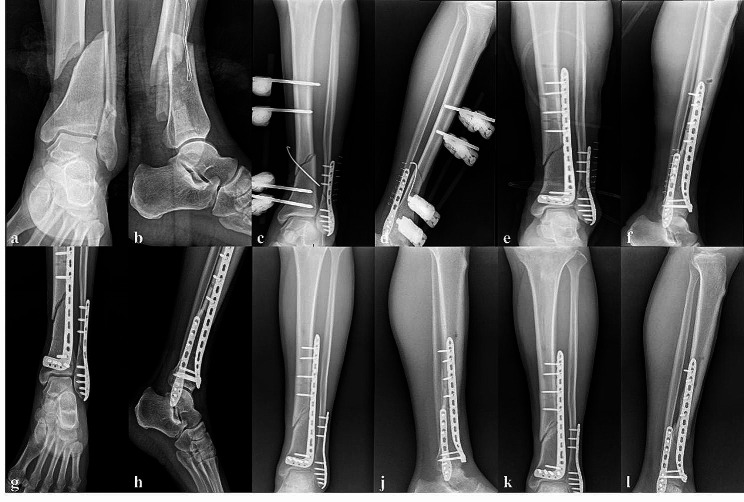
Exemplar cases have been visually illustrated in Figs. 2, 3, 4 and 5 for illustrative purposes.
Fig. 2.
A 85-year-old female with a extra-articular distal tibial fracture received an operation with RNT fixation. (A-D) Preoperative radiography. (E, F) X-ray at 5 days after the operation. (G, H) X-ray at 8 weeks after the operation. (I-L) radiography at 14 weeks after the operation. The fracture was union
Fig. 3.
A 63-year-old male with a distal tibial and fibular fracture received an operation with plates fixation. (A, B) X-ray at admission. (C, D) X-ray at one week after the operation. (E, F) X-ray at 10 week after the operation. (G, H) X-ray after the remove of the internal fixation
Fig. 4.
A 51-year-old male with a extra-articular distal tibial fracture received an operation with distal tibial plate fixation. (A, B) X-ray at admission. (C, D) X-ray at 4 days after the operation. (E, F) X-ray at 6 week after the operation. (G, H) X-ray at 15 week after the operation. (I, J) X-ray at 21 week after the operation. (K, L) X-ray at 37 week after the operation. Diagnosis was made as delayed union of the distal tibial fracture
Fig. 5.
A 54-year-old male with a Gustilo IIIA open fracture received an operation with distal tibial and fibular plate fixation. (A, B) X-ray at admission. (C, D) Immediate debridement, suturing, temporary external fixation and fibular plate fixation was used on the day of admission. (E, F) 7 days after the first operation, tibial plate was used as a substitute for external fixation. (G, H) X-ray at 4 weeks after the operation. (I, J) X-ray at 24 week after the operation. (K, L) X-ray at 48 week after the operation. Diagnosis was made as nonunion of the distal tibial fracture
Discussion
Extra-articular distal tibial fractures, encountered frequently in clinical practice, pertain to fractures occurring within a specific range, situated 4 to 11 cm proximal to the tibial plafond [11]. The distal tibia exhibits unique anatomical characteristics, transitioning from a triangular cross-sectional morphology in the tibial shaft to a circular or quadrangular shape, with a gradual widening of the medullary cavity. Notably, the anteromedial aspect of the distal tibia lacks muscle attachments and suffers from inadequate soft tissue coverage. Additionally, its blood supply relies solely on the trophoblastic vessels in the medullary cavity, elevating the susceptibility to infection, delayed union, and non-union. The surge in high-energy injuries attributed to advancements in industry and transportation has contributed to an elevated incidence of serious soft tissue injuries and open distal tibial fractures, further complicating the treatment of such fractures. Consequently, the distal tibial fractures treatment continues to pose significant challenges and remains a subject of debate. One of the key points is careful soft tissue handling as relevant as the treatment of the fracture itself. It is crucial to spare the extraosseous vessels during surgery as best as possible. Preexisting injury- and surgery-related factors all contribute to disturbed healing and increase the risk of cortical necrosis, delayed or non-union, or infection.
Presently, surgical intervention is the primary approach for managing displaced or unstable distal tibial fractures, with plate fixation and antegrade tibial intramedullary nails being the most common methods. While there is no substantial discrepancy in overall effectiveness between these two modalities [12], plate fixation imposes greater demands on soft tissue conditions and is unsuitable for patients with severe soft tissue injuries, peripheral vascular disease, or uncontrolled diabetes affecting the surgical site. Antegrade intramedullary nailing not only boasts favorable mechanical properties but also offers biological advantages by preserving vascularity at the fracture site and the integrity of surrounding soft tissues, thus earning it the status of the gold standard for treating tibial shaft fractures. When applied to distal tibial fractures, antegrade intramedullary nailing can yield improved function, reduced infection risk, and an acceptable range of motion [13]. Nevertheless, owing to the gradual expansion of the distal tibial medullary cavity and its distinctive anatomical structure, intramedullary nails may not snugly fit in the medullary canal, predisposing the fracture to poor reduction and instability, which can lead to malalignment [14], malunion, and delayed union [15]. Therefore, the application of antegrade intramedullary locking nails for distal tibial fractures often necessitates the use of auxiliary blocking screw techniques, inevitably increasing procedural complexity, operative duration, and the number of intraoperative fluoroscope exposures. Additionally, postoperative anterior knee pain represents the most commonly encountered complication of antegrade intramedullary nailing, with reported incidences as high as 47.4% [16].
In the late 1990s, Hofmann et al. [17] introduced the concept of RTN, which was subsequently designed and implemented in clinical practice by Kuhn et al. [6]. Recent biomechanical studies have highlighted RTN as an innovative surgical alternative for managing distal tibial fractures, demonstrating its potential superiority over plating techniques [18]. Moreover, clinical applications have yielded promising results. In a study involving 9 patients with distal tibial fractures, the application of RTN achieved an impressive 100% excellent-to-good rate in AOFAS scores [8]. At present, most reports comparing RTN with other internal fixation methods primarily focus on biomechanical aspects, with relatively few clinical comparative studies conducted. In this study, we performed a comparison of two surgical procedures—RTN and MIPO—in the extra-articular distal tibial fracture treatment. RTN demonstrated several advantages, including shorter hospital stays, early attainment of full weight-bearing, and a diminished complication incidence. All steps of the RTN procedure need to be confirmed through intraoperative fluoroscopy to ensure accuracy and minimally invasive of the surgery, resulting in higher intraoperative fluoroscopy exposure reported in the RTN group. Building upon the findings of previous research and our own investigations, we assert that RTN presents unique advantages. Firstly, the distal curved configuration of RTN aligns more harmoniously with the distal tibia morphology. Biomechanically, RTN exhibits superior resistance to rotation and axial stability when compared to antegrade tibial intramedullary nails and plates [6, 19]. The successful achievement of fracture healing without distal tibial valgus deformity in all RTN patients underscores the commendable stability provided by this technique. Secondly, RTN simplifies fracture reduction and fixation, offering greater convenience with a high accuracy in its locking nail aiming device, thereby reducing operation time and radiation exposure. Thirdly, the limited length of RTN ensures that the nail does not traverse the tibial shaft’s isthmus, resulting in reduced influence on the bone marrow cavity. This feature can reduce the fat embolism risk, particularly in patients concurrently dealing with lung injuries or multiple traumas [8]. Finally, RTN employs medial ankle as entry point, effectively averting anterior knee pain. Postoperative AOFAS results confirm its minimal impact on the ankle joint. However, RTN may have a risk of distant ankle pain, which needs to be verified with a larger sample size and long-term follow-up.
As a new technology, several considerations merit attention when using RTN for distal tibial fractures: (1) RTN’s limited length renders it unsuitable for patients with middle and lower tibial fractures. (2) The medial malleolus serves as the insertion point for RTN, making it unsuitable for children, adolescents with epiphyses, or individuals with skin infections or ulcerations at the medial malleolus. (3) Particular care needs to be exercised to protect the triangular ligament and medial malleolus during RTN insertion to prevent ligament tearing or medial malleolus fracture. The specific steps should be referred to the RTN surgical procedures including the correct entry point, the direction of the guide wire, how to create a pathway leading to the medullary canal and insert the RTN. According to the steps above, no case of medial malleolar fracture was found in this study fortunately. (4) Due to the wide medullary cavity of the distal tibia, there exists a heightened risk of fracture displacement during RTN insertion. Therefore, we recommend the application of a lower limb traction device throughout the surgery to maintain fracture stability, with adjunctive blocking screw techniques employed when deemed necessary.
Conclusion
Both RTN and MIPO prove to be effective approaches for treating extra-articular distal tibial fractures, yielding favorable clinical outcomes. RTN, in particular, offers some benefits, including minimal invasiveness, a low incidence of complications, and rapid restoration of function, warranting further exploration in clinical practice. Nonetheless, this study exhibits certain limitations: (1) Retrospective Study Design: This study adopts a retrospective design, potentially introducing selection bias into the research methodology. (2) Limited Sample Size and Short Follow-Up Duration: The study’s sample size is relatively small, and the follow-up period is of short duration. Future investigations plan to address these limitations by conducting large-scale prospective comparative studies (RTN, antegrade tibial intramedullary nailing and MIPO plates) to reaffirm the aforementioned findings.
Acknowledgements
We thank all participants in this study for their enthusiasm, tireless work, and sustained support.
Author contributions
JWu and HL wrote the manuscript, analyzed and interpreted the patient data; WX and JZ were responsible for acquisition of data, and analyzed and interpreted the patient data; YX and ZX were responsible for designing the study, and the analysis and interpretation of the data.
Data availability
No datasets were generated or analysed during the current study.
Declarations
Ethics approval and consent to participate
All procedures performed in studies involving human participants were in accordance with the ethical standards of the institutional and/or national research committee and with the Helsinki Declaration and its later amendments or comparable ethical standards. Informed consent was obtained from all individual participants or their legal guardians included in the study.
Consent for publication
All authors have read and approved the final manuscript.
Competing interests
The authors declare no competing interests.
Footnotes
Publisher’s Note
Springer Nature remains neutral with regard to jurisdictional claims in published maps and institutional affiliations.
Hui Liu and Weizhen Xu contributed equally to this paper.
References
- 1.Wang Z, Cheng Y, Xin D, et al. Expert Tibial nails for treating distal tibial fractures with soft tissue damage: a patient series. J Foot Ankle Surg. 2017;56(6):1232–5. 10.1053/j.jfas.2017.07.011 [DOI] [PubMed] [Google Scholar]
- 2.Prasad P, Nemade A, Anjum R, Joshi N. Extra-articular distal tibial fractures, is interlocking nailing an option? A prospective study of 147 cases. Chin J Traumatol. 2019;22(2):103–7. 10.1016/j.cjtee.2018.12.005 [DOI] [PMC free article] [PubMed] [Google Scholar]
- 3.Sitnik A, Beletsky A, Schelkun S. Intra-articular fractures of the distal tibia: current concepts of management. EFORT Open Rev. 2017;2(8):352–61. 10.1302/2058-5241.2.150047 [DOI] [PMC free article] [PubMed] [Google Scholar]
- 4.Joveniaux P, Ohl X, Harisboure A, et al. Distal tibia fractures: management and complications of 101 cases. Int Orthop. 2010;34(4):583–8. 10.1007/s00264-009-0832-z [DOI] [PMC free article] [PubMed] [Google Scholar]
- 5.Liu XK, Xu WN, Xue QY. Intramedullary Nailing Versus minimally invasive plate osteosynthesis for distal tibial fractures: a systematic review and Meta-analysis. Orthop Surg. 2019;11(6):954–65. 10.1111/os.12575 [DOI] [PMC free article] [PubMed] [Google Scholar]
- 6.Kuhn S, Appelmann P, Pairon P, Mehler D, Rommens PM. The Retrograde tibial nail: presentation and biomechanical evaluation of a new concept in the treatment of distal tibia fractures. Injury. 2014;45(Suppl 1):S81–86. 10.1016/j.injury.2013.10.025 [DOI] [PubMed] [Google Scholar]
- 7.Wang YC, Lin WJ, Lin T, et al. Finite element analysis of three internal fixation methods for distal tibial fractures. Zhongguo Zuzhi Gongcheng Yanjiu. 2023;27(36):5760–5. [Google Scholar]
- 8.Peng B, Wan T, Tan W, Guo W, He M. Novel Retrograde Tibial Intramedullary nailing for distal tibial fractures. Front Surg. 2022;9:899483. 10.3389/fsurg.2022.899483 [DOI] [PMC free article] [PubMed] [Google Scholar]
- 9.He M, Jiang Z, Tan W, Li Z, Peng B. Ideal entry point and direction of retrograde intramedullary nailing of the tibia. J Orthop Surg Res. 2023;18(1):472. 10.1186/s13018-023-03921-3 [DOI] [PMC free article] [PubMed] [Google Scholar]
- 10.Li Y, Liu L, Tang X, et al. Comparison of low, multidirectional locked nailing and plating in the treatment of distal tibial metadiaphyseal fractures. Int Orthop. 2012;36(7):1457–62. 10.1007/s00264-012-1494-9 [DOI] [PMC free article] [PubMed] [Google Scholar]
- 11.Vallier HA, Le TT, Bedi A. Radiographic and clinical comparisons of distal tibia shaft fractures (4 to 11 cm proximal to the plafond): plating versus intramedullary nailing. J Orthop Trauma. 2008;22(5):307–11. 10.1097/BOT.0b013e31816ed974 [DOI] [PubMed] [Google Scholar]
- 12.Kariya A, Jain P, Patond K, Mundra A. Outcome and complications of distal tibia fractures treated with intramedullary nails versus minimally invasive plate osteosynthesis and the role of fibula fixation. Eur J Orthop Surg Traumatol. 2020;30(8):1487–98. 10.1007/s00590-020-02726-y [DOI] [PubMed] [Google Scholar]
- 13.Xue XH, Yan SG, Cai XZ, Shi MM, Lin T. Intramedullary nailing versus plating for extra-articular distal tibial metaphyseal fracture: a systematic review and meta-analysis. Injury. 2014;45(4):667–76. 10.1016/j.injury.2013.10.024 [DOI] [PubMed] [Google Scholar]
- 14.Daas S, Jlidi M, Baghdadi N, Bouaicha W, Mallek K, Lamouchi M, Khorbi A. Risk factors for malunion of distal tibia fractures treated by intramedullary nailing. J Orthop Surg Res. 2024;19(1):5. 10.1186/s13018-023-04472-3 [DOI] [PMC free article] [PubMed] [Google Scholar]
- 15.Kc KM, Pangeni BR, Marahatta SB, Sigdel A, Kc A. Comparative study between intramedullary interlocking nailing and minimally invasive percutaneous plate osteosynthesis for distal tibia extra-articular fractures. Chin J Traumatol. 2022;25(2):90–4. 10.1016/j.cjtee.2021.08.004 [DOI] [PMC free article] [PubMed] [Google Scholar]
- 16.Katsoulis E, Court-Brown C, Giannoudis PV. Incidence and aetiology of anterior knee pain after intramedullary nailing of the femur and tibia. J Bone Joint Surg Br. 2006;88(5):576–80. 10.1302/0301-620X.88B5.16875 [DOI] [PubMed] [Google Scholar]
- 17.Hofmann GO, Gonschorek O, BÜhler M, Buehren V. Retrograde Nagelung Der Tibia. Trauma Und Berufskrankheit. 2001;3(2 Supplement):S303–8. 10.1007/PL00014733 [DOI] [Google Scholar]
- 18.Greenfield J, Appelmann P, Wunderlich F, Mehler D, Rommens PM, Kuhn S. Retrograde tibial nailing of far distal tibia fractures: a biomechanical evaluation of double-versus triple-distal interlocking. Eur J Trauma Emerg Surg. 2022;48(5):3693–700. 10.1007/s00068-021-01843-5 [DOI] [PMC free article] [PubMed] [Google Scholar]
- 19.Kuhn S, Appelmann P, Mehler D, Pairon P, Rommens PM. Retrograde tibial nailing: a minimally invasive and biomechanically superior alternative to angle-stable plate osteosynthesis in distal tibia fractures. J Orthop Surg Res. 2014;9:35. 10.1186/1749-799X-9-35 [DOI] [PMC free article] [PubMed] [Google Scholar]
Associated Data
This section collects any data citations, data availability statements, or supplementary materials included in this article.
Data Availability Statement
No datasets were generated or analysed during the current study.


