Abstract
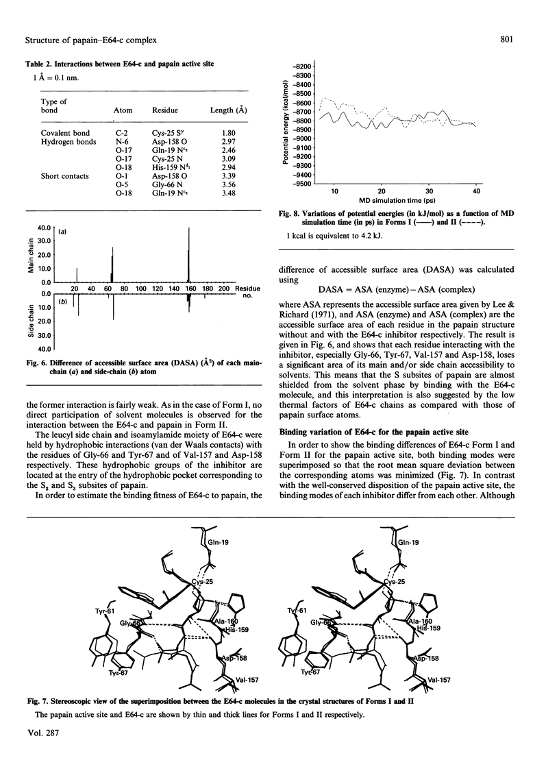
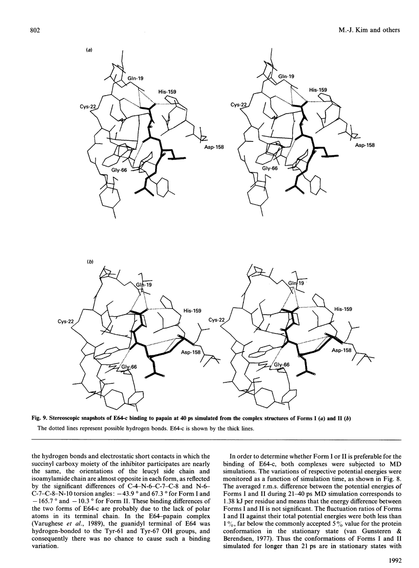
In order to investigate the binding mode of E64-c (a synthetic cysteine proteinase inhibitor) to papain at the atomic level, the crystal structure of the complex was analysed by X-ray diffraction at 1.9 A (1 A is expressed in SI units as 0.1 nm) resolution. The crystal has a space group P2(1)2(1)2(1) with a = 43.37, b = 102.34 and c = 49.95 A. A total of 21,135 observed reflections were collected from the same crystal, and 14811 unique reflections of up to 1.9 A resolution [Fo > 3 sigma(Fo)] were used for the structure solution and refinement. The papain structure was determined by means of the molecular replacement method, and then the inhibitor was observed on a (2 magnitude of Fo-magnitude of Fc) difference Fourier map. The complex structure was finally refined to R = 19.4% including 207 solvent molecules. Although this complex crystal (Form II) was polymorphous as compared with the previously analysed one (Form I), the binding modes of leucine and isoamylamide moieties of E64-c were significantly different from each other. By the calculation of accessible surface area for each complex atom, these two different binding modes were both shown to be tight enough to prevent the access of solvent molecules to the papain active site. With respect to the E64-c-papain binding mode, molecular-dynamics simulations proposed two kinds of stationary states which were derived from the crystal structures of Forms I and II. One of these, which corresponds to the binding mode simulated from Form I, was essentially the same as that observed in the crystal structure, and the other was somewhat different from the crystal structure of Form II, especially with respect to the binding of the isoamylamide moiety with the papain S subsites. The substrate specificity for the papain active site is discussed on the basis of the present results.
Full text
PDF
Selected References
These references are in PubMed. This may not be the complete list of references from this article.
- Barrett A. J., Kembhavi A. A., Brown M. A., Kirschke H., Knight C. G., Tamai M., Hanada K. L-trans-Epoxysuccinyl-leucylamido(4-guanidino)butane (E-64) and its analogues as inhibitors of cysteine proteinases including cathepsins B, H and L. Biochem J. 1982 Jan 1;201(1):189–198. doi: 10.1042/bj2010189. [DOI] [PMC free article] [PubMed] [Google Scholar]
- Berger A., Schechter I. Mapping the active site of papain with the aid of peptide substrates and inhibitors. Philos Trans R Soc Lond B Biol Sci. 1970 Feb 12;257(813):249–264. doi: 10.1098/rstb.1970.0024. [DOI] [PubMed] [Google Scholar]
- Delaissé J. M., Eeckhout Y., Vaes G. In vivo and in vitro evidence for the involvement of cysteine proteinases in bone resorption. Biochem Biophys Res Commun. 1984 Dec 14;125(2):441–447. doi: 10.1016/0006-291x(84)90560-6. [DOI] [PubMed] [Google Scholar]
- Denhardt D. T., Greenberg A. H., Egan S. E., Hamilton R. T., Wright J. A. Cysteine proteinase cathepsin L expression correlates closely with the metastatic potential of H-ras-transformed murine fibroblasts. Oncogene. 1987;2(1):55–59. [PubMed] [Google Scholar]
- Drenth J., Jansonius J. N., Koekoek R., Wolthers B. G. The structure of papain. Adv Protein Chem. 1971;25:79–115. doi: 10.1016/s0065-3233(08)60279-x. [DOI] [PubMed] [Google Scholar]
- Drenth J., Kalk K. H., Swen H. M. Binding of chloromethyl ketone substrate analogues to crystalline papain. Biochemistry. 1976 Aug 24;15(17):3731–3738. doi: 10.1021/bi00662a014. [DOI] [PubMed] [Google Scholar]
- Harris J. O., Olsen G. N., Castle J. R., Maloney A. S. Comparison of proteolytic enzyme activity in pulmonary alveolar macrophages and blood leukocytes in smokers and nonsmokers. Am Rev Respir Dis. 1975 May;111(5):579–586. doi: 10.1164/arrd.1975.111.5.579. [DOI] [PubMed] [Google Scholar]
- Kamphuis I. G., Kalk K. H., Swarte M. B., Drenth J. Structure of papain refined at 1.65 A resolution. J Mol Biol. 1984 Oct 25;179(2):233–256. doi: 10.1016/0022-2836(84)90467-4. [DOI] [PubMed] [Google Scholar]
- Katunuma N., Kominami E. Abnormal expression of lysosomal cysteine proteinases in muscle wasting diseases. Rev Physiol Biochem Pharmacol. 1987;108:1–20. doi: 10.1007/BFb0034070. [DOI] [PubMed] [Google Scholar]
- Katunuma N. Mechanisms and regulation of lysosomal proteolysis. Revis Biol Celular. 1989;20:35–61. [PubMed] [Google Scholar]
- Lee B., Richards F. M. The interpretation of protein structures: estimation of static accessibility. J Mol Biol. 1971 Feb 14;55(3):379–400. doi: 10.1016/0022-2836(71)90324-x. [DOI] [PubMed] [Google Scholar]
- Matsumoto K., Yamamoto D., Ohishi H., Tomoo K., Ishida T., Inoue M., Sadatome T., Kitamura K., Mizuno H. Mode of binding of E-64-c, a potent thiol protease inhibitor, to papain as determined by X-ray crystal analysis of the complex. FEBS Lett. 1989 Mar 13;245(1-2):177–180. doi: 10.1016/0014-5793(89)80216-9. [DOI] [PubMed] [Google Scholar]
- Ramakrishnan C., Ramachandran G. N. Stereochemical criteria for polypeptide and protein chain conformations. II. Allowed conformations for a pair of peptide units. Biophys J. 1965 Nov;5(6):909–933. doi: 10.1016/S0006-3495(65)86759-5. [DOI] [PMC free article] [PubMed] [Google Scholar]
- Schechter I., Berger A. On the size of the active site in proteases. I. Papain. Biochem Biophys Res Commun. 1967 Apr 20;27(2):157–162. doi: 10.1016/s0006-291x(67)80055-x. [DOI] [PubMed] [Google Scholar]
- Varughese K. I., Ahmed F. R., Carey P. R., Hasnain S., Huber C. P., Storer A. C. Crystal structure of a papain-E-64 complex. Biochemistry. 1989 Feb 7;28(3):1330–1332. doi: 10.1021/bi00429a058. [DOI] [PubMed] [Google Scholar]
- Yamamoto D., Matsumoto K., Ohishi H., Ishida T., Inoue M., Kitamura K., Mizuno H. Refined x-ray structure of papain.E-64-c complex at 2.1-A resolution. J Biol Chem. 1991 Aug 5;266(22):14771–14777. doi: 10.2210/pdb1pe6/pdb. [DOI] [PubMed] [Google Scholar]


