Abstract
The mechanistic target of rapamycin complex 1 controls cellular anabolism in response to growth factor signaling and to nutrient sufficiency signaled through the Rag GTPases. Inhibition of mTOR reproducibly extends longevity across eukaryotes. Here we report that mice that endogenously express active mutant variants of RagC exhibit multiple features of parenchymal damage that include senescence, expression of inflammatory molecules, increased myeloid inflammation with extensive features of inflammaging and a ~30% reduction in lifespan. Through bone marrow transplantation experiments, we show that myeloid cells are abnormally activated by signals emanating from dysfunctional RagC-mutant parenchyma, causing neutrophil extravasation that inflicts additional inflammatory damage. Therapeutic suppression of myeloid inflammation in aged RagC-mutant mice attenuates parenchymal damage and extends survival. Together, our findings link mildly increased nutrient signaling to limited lifespan in mammals, and support a two-component process of parenchymal damage and myeloid inflammation that together precipitate a time-dependent organ deterioration that limits longevity.
Subject terms: Nutrient signalling, Experimental models of disease, Inflammation
Ortega-Molina et al. demonstrate that mouse models with mild, genetic overactivation of mTOR signaling develop chronic myeloid inflammation, causing reduced healthspan and lifespan, without an increase in tumor incidence.
Main
The relentless increase in the aged population worldwide imposes medical, socioeconomical and philosophical challenges to mankind. By 2050, the global population aged 65 years or older would have tripled in a century, from 5% to 17% (around 2 billion people)1. While the prevention and management of aging-related disorders will improve, the overall comorbidities that occur with the multi-organ physiological decline of older adults impose a challenge unlikely to be controlled by the treatment of single symptoms and disorders. Instead, to intervene in the systemic frailty and health decline associated with aging, we need to understand the processes that precipitate cellular and organ malfunctioning with age. Genetic and pharmacological approaches in model organisms have taught us that the process of aging can be modulated and longevity can be shortened or extended by actioning on the functions of a discrete number of proteins2,3. Among these is inhibition of the mechanistic target of rapamycin (mTOR) complex 1 (mTORC1), a master regulator of cellular anabolism in response to cellular nutrients and growth factors4,5. Intracellular levels of amino acids, intermediate metabolites of glycolysis, certain lipids and other molecules have dedicated sensors that signal to the Rag GTPases upstream of mTORC1 (refs. 4,6). Under nutrient sufficiency, RagC loads GDP, whereas its obligate heterodimeric partner RagA is GTP-loaded. This nucleotide configuration allows the recruitment of mTORC1 to the outer lysosomal surface, where mTORC1 binds the Ras homolog enriched in brain (Rheb) that leads to kinase activation of mTORC1 in a growth-factor-dependent manner7,8.
Pharmacological and genetic inhibition of TOR extends longevity in yeast9, worms10,11 and flies12,13. In mice, rapamycin14,15 and hypomorphic mTORC1 signaling achieved by genetic means also extends lifespan16,17. Conversely, genetic activation of mTORC1 signaling has been linked to shortened longevity in lower organisms, including yeast and worms, but mouse models of increased mTORC1 activity, such as heterozygous or homozygous deletion of Tsc1 or Tsc2 (refs. 18–20), Pten21–23 or genetic activation of positive regulators of mTORC1, such as Akt, Pi3k, Rheb or Rraga24–28, have been invariably incompatible with aging studies due to the early development of specific deleterious pathologies that precluded the analyses of mechanisms and processes underlying organismal aging in mammals.
We have previously engineered two knock-in mice expressing active, GDP-like mutant variants of RagC (also known as Rragc)29, originally identified in human B cell lymphomas30–32. Heterozygous RragcS74C or RragcT89N mice show accelerated development of lymphomas when bred to a lymphoma-prone strain, and a moderate increase in nutrient signaling to mTORC1. Together with a newly generated knock-in strain of mice, expressing a canonical GDP-like mutation (S74N), we now show that in the absence of cooperating oncogenes, a mild increase in nutrient signaling to mTORC1 does not lead to spontaneous tumorigenesis, and instead, these mice show a shortened lifespan. This genetic system allows for the interrogation of the mechanisms underscoring the connections between the nutrient–mTORC1 axis and longevity. Increased nutrient signaling to mTORC1 leads to autonomous organ damage and prominent myeloid inflammation in response to evoking signals from damaged organs. Late-life blockade of myeloid cell infiltration mitigates several of these phenotypes and extends lifespan.
Results
Rragc-mutant mice have shortened lifespan
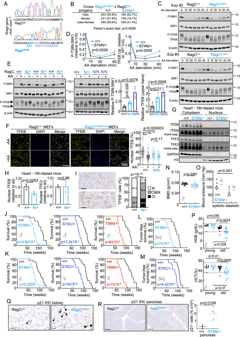
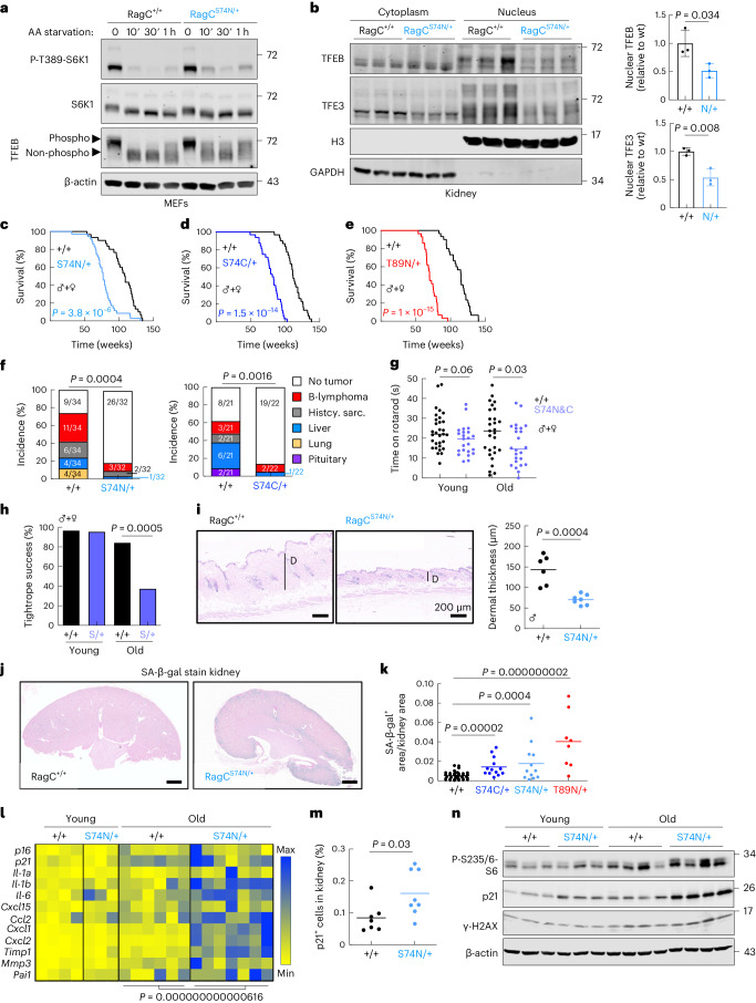
We have previously reported knock-in mice expressing activating mutant variants of Rragc (RragcS74C and RragcT89N)29. These mutations encode amino acid substitutions translating from single-nucleotide changes recurrently found in human B cell lymphomas at the equivalent positions S75 and T90 in human RRAGC30. We have now knocked in a ‘canonical’ GDP-bound, activating RagC mutation, translating into RragcS74N (refs. 33,34) and not found in human lymphomas as two contiguous genetic mutations must occur. We engineered a two-nucleotide substitution in Rragc exon 1, which translates into the serine-to-asparagine change in aminoacidic position 74, together with additional silent mutations (Extended Data Fig. 1a). Heterozygous RragcS74N/+ mice, as seen in RragcS74C/+ and RragcT89N/+ mice were found at sub-Mendelian ratios (Extended Data Fig. 1b). As previously observed in RragcS74C/+ and RragcT89N/+ cells29, primary mouse embryonic fibroblasts (MEFs) from RragcS74N/+ mice show a modest increase in mTORC1 activity, as seen by the phosphorylation status of the mTORC1 targets S6k1 and Tfeb (Fig. 1a and Extended Data Fig. 1c–e), indicating that the effects of these three activating mutations in RagC on mTORC1 signaling are very similar. RragcS74N/S74N mice are not viable28,29, but RragcS74N/S74N MEFs show an increase in phospho-S6K1, plus a remarkable upshift of Tfeb, indicative of a large increase in its phosphorylation (Extended Data Fig. 1e). Immunofluorescence-based staining of Tfeb showed the expected nucleo-cytoplasmic shuttle of Tfeb upon amino acid-starvation/replenishment of Rragc+/+ MEFs, but cytoplasmic retention of Tfeb in RragcS74N/S74N MEFs even under amino acid starvation (Extended Data Fig. 1f). Tissues from heterozygous RragcS74N/+ fasted mice present decreased Tfeb and Tfe3 in the nucleus (Fig. 1b and Extended Data Fig. 1g,h). Increased cytoplasmic retention of Tfeb in RragcS74N/+ mice was validated by quantification of immunohistochemical stain of Tfeb in kidneys (Extended Data Fig. 1i).
Extended Data Fig. 1. Rragc-mutant mice have shortened lifespan.
a. Chromatogram of the Rragcwt and RragcS74N allele. RragcS74N allele encodes two amino acid substitutions (blue bold), plus silent diagnostic mutations (purple). b. Progenies of RragcS74N breeding schemes. c. MEFs from Rragc+/+ and RragcS74N/+ mice were deprived of all amino acids in RPMI medium with dialyzed serum for the indicated time-points. d. Quantification of P-T389-S6K1 and TFEB upper band (phosphorylated) from triplicates from Fig. 1a and Figure S1c (Statistical significance was calculated by two-tailed Student’s t-test for AUC). Data are presented as mean values ± SD. e. (Left) MEFs from Rragc+/+ (n = 4 biologically independent cells), RragcS74N/+ (n = 4 biologically independent cells), and RragcS74N/S74N (n = 2 biologically independent cells), mice were deprived from and re-stimulated with amino acids for 10 min. (Right) Quantification of P-T389-S6K1 and TFEB upper band in -AA samples. Data are presented as mean values ± SD f. (Left) Immunofluorescence of TFEB in Rragc+/+ and RragcS74N/S74N MEFs in RPMI with and without amino acids; (Right) with the percentage of nuclear TFEB per cell (Rragc+/+ + AA n = 112 –AA n = 117; RragcS74N/S74N + AA n = 72 –AA n = 91). Bars represent 100μm g. TFE3 and TFEB levels fractions of hearts from Rragc+/+ (n = 3) and RragcS74N/+ (n = 3) mice after fasting. h. Quantification of TFE3 and TFEB levels in the nucleus. Data are presented as mean values ± SD. i. Representative anti-TFEB IHC (left) and quantification (right) of TFEB-positive cells in the cytoplasm (Ct), nucleus (N) and cytoplasm&nucleus (Ct&N) in renal tubules from young Rragc+/+ (n = 4) and RragcS74N/+ (n = 4) male mice. Scale bar 100 µm. j. Kaplan–Meier survival curves of Rragc+/+ (n = 18) and RragcS74N/+ (n = 20) male mice (left), Rragc+/+ (n = 19) and RragcS74C/+ (n = 13) male mice (center) and Rragc+/+ (n = 18) and RragcT89N/+ (n = 19) male mice (right). k. Kaplan–Meier survival curves of Rragc+/+ (n = 21) and RragcS74N/+ (n = 24) female mice (left), Rragc+/+ (n = 19) and RragcS74C/+ (n = 23) female mice (center) and Rragc+/+ (n = 17) and RragcT89N/+ (n = 22) female mice (right). l. Kaplan–Meier survival curves of Rragc+/+ (n = 9) and RragcS74N/+ (n = 24) mice that were free of detectable malignant tumors at the time of death. m. Kaplan–Meier survival curves of Rragc+/+ (n = 10) and Rragc S74C/+ (n = 17) mice without malignant tumors. In j-m, statistical significance was calculated with the log-rank test. n. Bone mineral density (BMD) measured in 13.5-21.5-month-old Rragc+/+ (n = 17) and RragcS74N/+ (n = 21) male mice. o. Blood pressure in 16-19-month-old Rragc+/+ (n = 6) and RragcS74N/+ (n = 7) male mice. p. Hematological parameters in 3–5- and 18-month-old Rragc+/+ (young, n = 8; old, n = 16) and RragcS74N/+ (young, n = 9; old, n = 14) male mice. q. Representative anti-p21 IHC in kidneys from 18-month-old Rragc+/+ and RragcS74N/+ male mice. Bars represents 20 μm. Arrows indicate p21-positive cells. r. Representative anti-p21 IHC (left) and quantification (right) from pancreas harvested from 18-month-old Rragc+/+ (n = 3) and RragcS74N/+ (n = 5) male mice. Bars represent 200 μm. Statistical significance in e, h, n, p and r was calculated by two-tailed Student’s t-test. Statistical significance in f was calculated by one-way ANOVA and Tukey’s multiple comparisons test. Statistical significance in a and i was calculated by by two-sided Fisher’s exact test. Statistical significance in o was calculated by Two-way Anova.
Fig. 1. Rragc-mutant mice have a shortened lifespan.
a, Rragc+/+ and RragcS74N/+ MEFs were deprived of all amino acids in RPMI with dialyzed serum for 10 min, 30 min and 1 h. Whole-cell protein lysates were immunoblotted for the indicated proteins. b, Tfe3 and Tfeb protein levels in subcellular fractions from kidneys from young Rragc+/+ (n = 3) and RragcS74N/+ (n = 3) mice fasted 16 h. Quantification of Tfe3 and Tfeb levels in the nucleus is relative to histone 3 levels. Data are presented as mean ± s.d. c–e, Kaplan–Meier survival curves of Rragc+/+ (n = 30) and RragcS74N/+ (n = 35) (c); Rragc+/+ (n = 31) and RragcS74C/+ (n = 32) (d); Rragc+/+ (n = 30) and RragcT89N/+ (n = 29) (e) mice. f, Tumor incidence in cohorts from Rragc+/+ (n = 34) and RragcS74N/+ (n = 32) (left) and from Rragc+/+ (n = 21) and RragcS74C/+ (n = 22) (right). Percentage of mice with no tumors, B cell lymphomas, histiocytic sarcomas, hepatocellular carcinomas, lung carcinomas and pituitary tumors. g, Time on rotarod measured in young (4–7.5-mo-old) and old (14.5–20-mo-old) Rragc+/+ (young, n = 31; old, n = 32) and RragcS74N/+ and RragcS74C/+ (young, n = 23; old, n = 24) mice. h, Tightrope assay performed in the same groups of mice as in g. Scale bars represent the percentage of mice that passed the assay. i, Dermal thickness measured in back skin of 18-mo-old Rragc+/+ (n = 6) and RragcS74N/+ (n = 7) males. Scale bars, 200 μm. j, Representative pictures of SA-β-gal+ in kidney. Scale bars, 1,000 μm. k, Quantification of SA-β-gal+ within the kidney of 18-mo-old Rragc+/+ (n = 38), RragcS74C/+ (n = 13), RragcS74N/+ (n = 12) and RragcT89N/+ (n = 8) mice. l, qRT–PCR analysis of genes of the SASP in kidneys of young (4-mo-old) and old (18-mo-old) Rragc+/+ (young, n = 4; old, n = 6) and RragcS74N/+ (young, n = 3; old, n = 7) male mice. m, Quantification of IHC staining of anti-p21 in kidneys collected from 18-mo-old Rragc+/+ (n = 7) and RragcS74N/+ (n = 8) male mice. n, Immunoblot for the indicated proteins from 4-mo-old and 18-mo-old Rragc+/+ and RragcS74N/+ male mice. Statistical significance was assessed by log-rank test (c–e); two-sided chi-squared test (f); two-sided Fisher’s exact test (h); two-tailed Student’s t-test (b,g,i,k,m); and two-way analysis of variance (ANOVA) (l). mo, month.
When bred to the follicular lymphoma-prone strain VavP-Bcl2Tg35, Rragcmut/+ mice rapidly develop lymphomas29, but in the absence of additional genetic modifications, the expression of Rragcmut in heterozygosity lead to a strikingly similar shortened lifespan in all three Rragcmut/+ male and female cohorts (Fig. 1c–e and Extended Data Fig. 1j,k).
We first reasoned that the premature death of Rragcmut/+ mice was a consequence of spontaneous tumor development; however, and surprisingly for full-body expression of bona fide oncogenic mutations, upon postmortem histopathological examination, RragcS74N/+ and RragcS74C/+mice showed reduced, rather than increased, spontaneous tumor incidence (Fig. 1f). When all sacrificed mice with tumors identified post hoc were removed from the Kaplan–Meier survival curves, tumor-free RragcS74N/+ and RragcS74C/+ mice still showed a shortened lifespan compared to tumor-free Rragc+/+ mice (Extended Data Fig. 1l, m), indicating that the premature death of Rragcmut/+ mice is not related to differences in spontaneous tumorigenesis. Instead, we found multiple canonical markers of aging36 in ~16-month-old Rragcmut/+ mice: loss of neuromuscular coordination in rotarod and tightrope tests (Fig. 1g,h), thinning of the dermal layer of the skin (Fig. 1i), loss of bone mineral density (Extended Data Fig. 1n), increased systolic and diastolic pressure (Extended Data Fig. 1o) and decreased hemoglobin and hematocrit (Extended Data Fig. 1p). We also quantified senescence-associated β-galactosidase (SA-β-gal) activity, a readout of cellular senescence (a process strongly associated with aging37), in organs from ~16-month-old wild-type (wt) and Rragcmut/+ mice (Fig. 1j). Automatic quantification of SA-β-gal revealed a significant increase in the kidneys from the three Rragcmut/+ strains (Fig. 1k). Consistently, the expression of markers of senescence and a senescence-associated secretory phenotype (SASP) was also increased (Fig. 1l). In addition, immunohistochemistry (IHC) staining for Cdkn1a (p21) revealed significantly increased abundance of p21-positive cells in the kidney and pancreas from RragcS74N/+ mice (Fig. 1m and Extended Data Fig. 1q,r), a difference confirmed by immunoblot (Fig. 1n). Additional markers of damage associated with aging, such as phosphorylation of H2ax (γ-H2ax; Fig. 1n), were also augmented in RragcS74N/+ samples compared to Rragc+/+ samples of an identical chronological age.
Old Rragc-mutant mice show an inflammaging phenotype
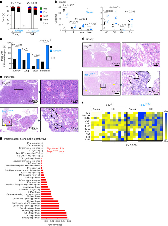
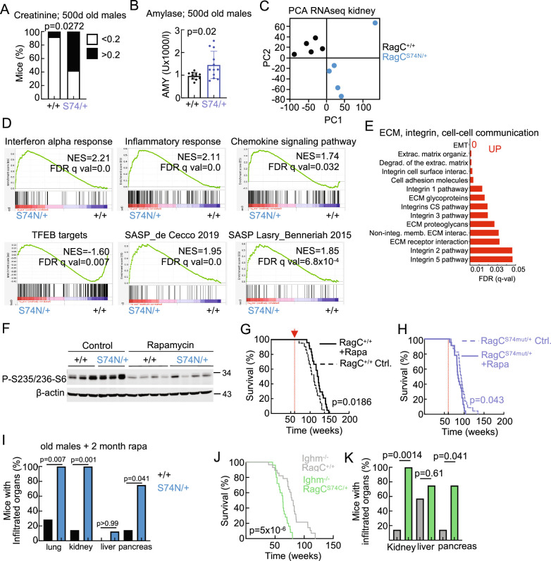
Quantification of peripheral blood mononuclear cells (PBMCs) from Rragc+/+ and Rragcmut/+ mice revealed another typical feature of old individuals, increased mature myeloid cells and decreased mature lymphoid populations38,39 (Fig. 2a,b). Histological examination of tissues from RragcS74N/+ mice showed scattered but widespread foci of inflammation in kidney, pancreas, liver and lung (Fig. 2c–e). Moreover, inflammatory cytokines were significantly elevated in the peripheral blood from old RragcS74N/+ mice (Fig. 2f). RragcS74N/+ mice also presented elevated levels of creatinine and amylase in blood, markers of kidney and pancreas damage, respectively (Extended Data Fig. 2a,b). To identify transcriptomic changes reflecting underlying differences between old Rragc+/+ and old RragcS74N/+ mice, we conducted bulk RNA sequencing (RNA-seq) from kidneys from ~16-month-old Rragc+/+ and Rragc74N/+ mice. Principal-component analysis (PCA) clustered samples by genotype (Extended Data Fig. 2c). Moreover, 1,368 genes were differentially expressed in old RragcS74N/+ versus Rragc+/+ kidneys (792 upregulated and 576 downregulated in RragcS74N/+; Supplementary Table 1). Among the most upregulated signatures in RragcS74N/+ samples were inflammatory signatures such as interferon (IFN)γ and IFNα response, tumor necrosis factor (TNF) signaling and interleukin (IL)-6 signaling (Fig. 2g and Extended Data Fig. 2d). We also found chemokine-related signaling pathways (Fig. 2g and Extended Data Fig. 2d), integrins and extracellular matrix (ECM) remodeling signatures upregulated (Extended Data Fig. 2e). Moreover, SASP signatures40,41 were also enriched in RragcS74N/+ samples (Extended Data Fig. 2d), in accordance with the increased SA-β-gal staining observed in the kidney (Fig. 1j,k). These results strongly suggest the existence of ‘inflammaging’, a basal and systemic myeloid inflammation associated with aging, and collectively support the occurrence of a phenotype resemblant of early onset of aging in mice with a mild increase in nutrient signaling.
Fig. 2. Old Rragc-mutant mice show an inflammaging phenotype.
a, White blood cell (WBC) count performed in young (3–5-mo-old) and old (18-mo-old) Rragc+/+ (young, n = 8; old, n = 9) and RragcS74N/+ (young, n = 12; old, n = 12) males. Each colored stack represents cell-type percentage (Lym, lymphocyte; Neu, neutrophil; Mon, monocyte; Eos, eosinophil; Bas, basophil). Data are presented as mean ± s.d. b, Percentage of the indicated cell populations in the blood of 18-mo-old Rragc+/+ (n = 9) and RragcS74N/+ (n = 9) male mice. B, B cell; T, T cell; NK, natural killer; DC, dendritic cell; Mac, macrophage. c, Incidence of infiltrated inflammatory cells in the indicated tissues from 18-mo-old Rragc+/+ (n = 18) and RragcS74N/+ (n = 21) mice. d, Representative H&E pictures in the same mice as in c, showing inflammatory foci in kidney (dashed lines). e, Representative H&E pictures in the same mice as in c, showing inflammatory foci in pancreas (dashed lines). f, Quantification of inflammatory cytokines in sera from young (4-mo-old) and old (18-mo-old) Rragc+/+ (young, n = 4; old, n = 9) and RragcS74N/+ (young, n = 4; old, n = 9) male mice measured by Legendplex assay using flow cytometry. g, Graphical representation of the false discovery rates (FDRs) from the indicated KEGG, Hallmark, REACTOME and WikiPathways gene sets enriched in kidneys from 18-mo-old RragcS74N/+ (n = 5) versus Rragc+/+ (n = 5) mice. Statistical significance was calculated by two-way ANOVA (a,f); two-tailed Student’s t-test (b); and two-sided Fisher’s exact test (c).
Extended Data Fig. 2. Old Rragc-mutant mice show an inflammaging phenotype.
a. Percentage of mice with increased creatinine levels (>0.2 mg/dL) measured in blood from 18-month-old Rragc+/+ (n = 12) and RragcS74N + S74C/+ (n = 12) male mice. Statistical significance was calculated by two-sided Fisher’s exact test. b. Amylase levels measured in blood from 18-month-old Rragc+/+ (n = 11) and RragcS74N + S74C/+ (n = 12) male mice. Statistical significance was calculated by two-tailed Student’s t-test. Data are presented as mean values ± SD. c. Principal component analysis (PCA) from the RNA-seq performed from kidney samples from 18-month-old Rragc+/+ (n = 5) and RragcS74N/+ (n = 5) mice. d. Gene set enrichment analysis (GSEA) related to inflammatory signatures, TFEB target genes, and genes involved in senescence-associated secretory phenotype in kidneys from 18-month-old RragcS74N/+ (n = 5) versus Rragc+/+ (n = 5) mice. e. Graphical representation of the false discover rates (FDRs) from the indicated KEGG, Hallmark, REACTOME and Wikipathways gene sets enriched in kidneys from RragcS74N/+ (n = 5) versus Rragc+/+ (n = 5) 18-month-old mice. f. Western blot of harvested kidneys from 16-month-old Rragc+/+ and RragcS74N/+ male mice fed with control chow or with rapamycin-supplemented chow for 2 months. g. Kaplan–Meier survival curves of control (n = 21) and rapamycin-treated Rragc+/+ (n = 24) mice. Statistical significance was calculated with the log-rank test. h. Kaplan–Meier survival curves of control (n = 23) and rapamycin-treated RragcS74mut/+ (n = 23) mice. Statistical significance was calculated with the log-rank test. i. Incidence of infiltrated inflammatory cells in the indicated tissues from 16-month-old Rragc+/+ (n = 7) and RragcS74N/+(n = 8) mice treated with rapamycin for 2 months. Statistical significance was calculated by two-sided Fisher’s exact test. j. Kaplan–Meier survival curves of Ighm-/-; Rragc+/+ (n = 28) and Ighm-/-; RragcS74C/+ (n = 25) mice. Statistical significance was calculated with the log-rank test. k. Incidence of infiltrated inflammatory cells in the indicated tissues from Ighm-/-; Rragc+/+ (n = 7) and Ighm-/-; RragcS74C/+ (n = 8) mice analyzed at humane end point. Statistical significance was calculated by two-sided Fisher’s exact test.
RagC participates in a heterodimeric complex with RagA that binds mTORC1 in response to nutrients42,43, so we reasoned that pharmacological inhibition of mTORC1 with rapamycin could correct the phenotypic defects and the shortened longevity of Rragcmut/+ mice. In addition, rapamycin administration to ~1.5-year-old C57BL/6 mice significantly extends longevity14. Thus, we administered encapsulated rapamycin in food to 14-month-old mice, and at the same formulation as used elsewhere14. As expected, rapamycin-fed mice showed decreased phosphorylation of S6 in serines 235 and 236, and a canonical readout of suppressed mTORC1 activity in several organs, regardless of Rragc genotype (Extended Data Fig. 2f). Moreover, rapamycin administration led to a significant extension in longevity in Rragc+/+ mice (Extended Data Fig. 2g), but failed to extend survival in Rragcmut/+ (Extended Data Fig. 2h) mice and to decrease inflammation in organs from Rragcmut/+ (Extended Data Fig. 2i) mice. Collectively, these data suggest that chronic damage occurring before pharmacological inhibition of mTORC1 impaired a life-extending effect of rapamycin in Rragcmut/+ mice. In addition, the selective lack of effect of rapamycin in Rragcmut/+ mice points to rapamycin-resistant functions of an active RagC–mTORC1 axis, such as inhibition of TFEB family members44,45, playing a role in the shortened longevity of Rragcmut/+ mice.
Despite the lack of spontaneous lymphomas in Rragcmut/+ mice (Fig. 1f) and the lymphoid-to-myeloid inflammatory switch (Fig. 2a,b), we conceived that aberrant functions of B cells, or other immune cells, could be driving systemic health decline and death. To test whether aberrant B cell functions were an early driver of the inflammation and early death of Rragcmut/+ mice, we interbred Rragcmut/+ with IghmμMT/μMT mice, genetically deficient for mature B cells46, to generate Rragcmut/+ mice devoid of mature B cells. Overall survival of IghmμMT/μMT mice, regardless of the Rragc genotype, was shorter (Extended Data Fig. 2j) most likely due to their partial immunosuppression. Nevertheless, increased inflammation (Extended Data Fig. 2k) and premature death (Extended Data Fig. 2j) were still present in Rragcmut/+ mice in a B cell-deficient context. Together with the decreased B cell abundance in circulating blood from Rragcmut/+ compared to Rragc+/+ mice (Fig. 2b), these results show that increased inflammation and premature death are not a consequence of detrimental effects of aberrant B cell functions in Rragcmut/+ mice.
Similarities between changes in Rragcmut/+ mice and physiological aging in mice and humans
The relevance of the Rag GTPase–mTORC1 pathway on aging is supported by genetic and pharmacological studies and is considered an important regulator of longevity across eukaryotes2,3,16,17,47,48. Nevertheless, because of the nature of the RagC mutations used herein, identified in patients with cancer and not in progeria patients, we sought to interrogate the relevance of our observations in physiological mouse and human aging. We analyzed the transcriptome of organs from young and old C57BL/6 mice from two independent cohorts: a cohort housed at the Spanish National Cancer Research Centre (CNIO) and an independent cohort house at the National Institute on Aging (NIA). To analyze these two independent datasets, we compiled a tailored list of gene signatures (Supplementary Table 2), including selected canonical gene sets from the Hallmark and KEGG databases, gene lists curated from previous literature on aging, senescence and inflammation40,41 and signatures of TFEB targets49,50. In addition, to specifically interrogate whether cellular processes downstream of activation of RagC are affected in normal aging, we curated lists of differentially expressed genes (DEGs) obtained from transcriptomic analyses (Extended Data Fig. 2c and Supplementary Table 1) of old and young organs from RragcS74N/+ and wt (Fig. 3a) mice.
Fig. 3. Similarities between changes in Rragcmut/+ mice and physiological aging in mice and humans.
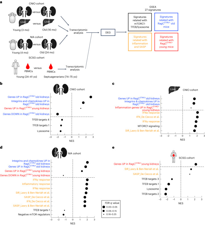
a, Experimental setup for the preranked GSEA performed in old and young wt mice from CNIO (kidney and liver) and NIA (kidney) cohorts, and the SCSG cohort (PBMCs from septuagenarians and young individuals) using 27 curated signatures related to mTORC1–TFEB and lysosomes (in black), signatures related to RragcS74N/+ old mice (blue), signatures related to inflammation and SASP (orange) and signatures related to RragcS74N/+ young mice (red). See Supplementary Table 2 for a detailed description of the signatures. b, Normalized enrichment score (NES) of indicated gene sets when comparing gene expression in old wt versus young wt kidneys from CNIO cohort. c, NES of indicated signatures when comparing gene expression in old wt versus young wt livers from CNIO cohort. d, NES of indicated signatures when comparing gene expression in old wt versus young wt kidney from NIA cohort. e, NES of indicated signatures when comparing gene expression in septuagenarians versus young PBMCs from the SCSG cohort. For b–e, a positive NES reflects enrichment of the gene set at the top of the ranked list and gene sets with a negative NES are overrepresented at the bottom of the gene list. The size of the bubble represents the FDR q value. Pictures of the liver and kidney were created with BioRender.
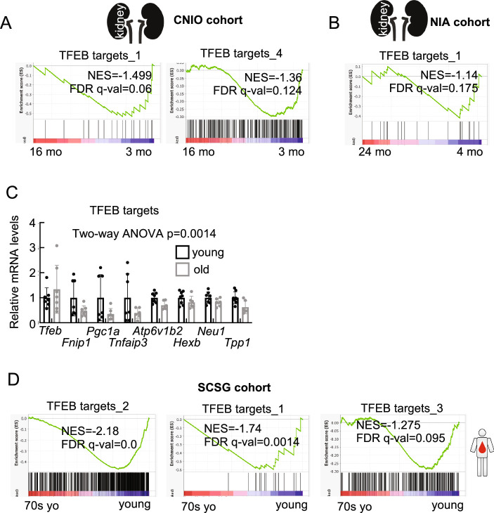
In datasets from both CNIO and NIA, old wt mice show a striking enrichment of upregulated DEGs from RragcS74N/+ young and old mice, whereas genes repressed in old and young RragcS74N/+ mice are downregulated in old wt mice from CNIO and NIA cohorts. (Fig. 3b–d). These associations strongly support that both the early and late changes caused by activation of Rag GTPase signaling in mouse tissues strikingly overlap with the transcriptional changes that occur over time in naturally aged mice. Samples from naturally aged mice also exhibit upregulation of canonical signatures and previously published genes related to senescence, its secretory phenotype, inflammation, interferon (Fig. 3b–d), consistently with similar changes observed in Rragcmut/+ mice (Figs. 1l and 2g and Extended Data Fig. 2d,e) and indicating that processes occurring during physiological aging are exacerbated in, and not exclusive of, RagC-mutant mice. This similarity also applies if the comparison is conducted with genes related to inflammation and to integrins, senescence and chemokines. The direct consequence of activation of Rag GTPase signaling, namely activation of mTORC1 and suppression of TFEB activity and lysosomal biogenesis, which were seen in the transcriptomic analysis of RragcS74N/+ mice (Extended Data Fig. 2d), are observed in samples from naturally aged CNIO and NIA cohorts (Fig. 3b–d and Extended Data Fig. 3a,b). Furthermore, we validated the expression by quantitative PCR with reverse transcription (qRT–PCR) of several TFEB targets and found a significant reduction in the transcript abundance of several TFEB targets in old wt kidneys compared to young wt kidneys (Extended Data Fig. 3c). Collectively, these analyses support that the Rag GTPase–mTORC1–TFEB–lysosome axis is deregulated during aging and suggest that this axis is of relevance for mammalian aging.
Extended Data Fig. 3. Similarities between changes in Rragc-mutant mice and physiological aging in mice and humans.
a. Gene set enrichment analysis (GSEA) related to TFEB target genes in kidneys for CNIO cohort from 16-month-old wt (n = 5) versus 3-month-old (n = 4) mice. b. Gene set enrichment analysis (GSEA) related to TFEB target genes in kidneys for NIA cohort from 24-month-old wt (n = 5) versus 2-month-old (n = 5) mice. c. qRT–PCR analysis of TFEB target in kidney from 3-month-old wt (n = 8) and 16-month-old wt (n = 7) male mice. Statistical significance was calculated by two-way ANOVA. Data are presented as mean values ± SD. d. Gene set enrichment analysis (GSEA) related to TFEB target genes in PBMCs for the Spanish Centenarian Study Group (SCSG) cohort (Borras et al. 2016) from Septuagenarians (74-75 yr old) (n = 10) versus Young (24-41 yr old) (n = 10) individuals.
In addition to the analysis of mouse cohorts, we obtained the transcriptomic datasets from PBMCs from young adult and septuagenarian healthy individuals from the Spanish Centenarian Study Group (SCSG) cohort51. We applied the same curated signature analysis used above in search for overlaps between DEGs from RagC-mutant samples, canonical markers of aging and specific functions downstream of the Rag GTPases (TFEB and lysosome) in the septuagenarian cohort of this human study. The septuagenarian group exhibits a positive enrichment in the inflammatory signatures obtained from previous literature40,41 (Fig. 3e and Extended Data Fig. 3d) and notably, the upregulated genes from RragcS74N/+ kidneys from young mice were enriched in septuagenarians compared to younger individuals. Moreover, samples from septuagenarians exhibit a significant depletion of the lysosome signature from KEGG and a significant depletion of signatures of TFEB targets. These positive and negative associations strongly support that changes in the Rag GTPase–mTORC1–lysosome occur in normal aging of both mice and humans.
Rragc-mutant ‘reverse’ BM chimeras exhibit shortened longevity
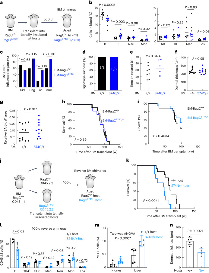
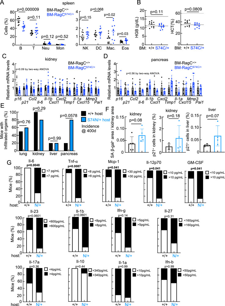
B cells have a negligible effect in the premature death of Rragcmut mice (Extended Data Fig. 2j). Nonetheless, the increased inflammation observed in old RragcS74N/+ mice pointed to a bone marrow (BM)-derived population triggering the accelerated health decline in Rragcmut/+, so we sublethally irradiated wt C57BL/6 hosts and reconstituted their BM with cells obtained from BM of either Rragc+/+ or Rragcmut/+ mice (Fig. 4a), and monitored inflammation, markers of aging and overall survival. At 500 days, Rragcmut/+ BM chimeras had the same decrease in B cell lineage and partial increase in myeloid populations (Fig. 4b and Extended Data Fig. 4a) observed in full-body Rragcmut/+ mice (Fig. 2b), indicating that the lymphoid-to-myeloid switch observed in full-body Rragcmut/+ is BM-cell intrinsic. In contrast, BM chimeras failed to phenocopy the increased parenchymal inflammation (Fig. 4c), the decrease in hemoglobin and hematocrit (Extended Data Fig. 4b), the loss of neuromuscular coordination (Fig. 4d,e) and the dermal thinning (Fig. 4f) observed in full-body Rragcmut/+ mice. In addition, Rragcmut/+ BM chimeras did not show evidence of increased senescence (Fig. 4g and Extended Data Fig. 4c,d). More notably, and in contrast to the shortened lifespan of full-body Rragcmut/+ mice, the median and maximal lifespans of BM-Rragc+/+, BM-RragcS74C/+ and BM-RragcS74N/+ chimeras were identical (Fig. 4h,i). These transplantation experiments demonstrate that BM cells with increased nutrient signaling do not suffice to recapitulate the aging-like phenotype of mice with increased nutrient signaling.
Fig. 4. Rragc-mutant ‘reverse’ BM chimeras exhibit shortened longevity.
a, Experimental setup for the generation of Rragc+/+ and RragcS74C/+ BM chimeras. Aging features and inflammation were measured 18 months after transplantation. b, Percentage of cell populations in the blood of 18-mo-old BM-Rragc+/+ (n = 9) and BM-RragcS74C/+ (n = 10) male mice. B, B cell; T, T cell; Mon, monocyte; NK, natural killer; Mac, macrophage; Eos, eosinophil. c, Incidence of inflammatory cells in tissues from 18-mo-old BM-Rragc+/+ (n = 10) and BM-RragcS74C/+ (n = 11) mice. d, Tightrope assay in 18-mo-old BM-Rragc+/+ (n = 8) and BM-RragcS74C/+ (n = 6) male mice. Bars represent the percentage of mice that passed the test. e, Time on rotarod test measured in the same mice as in c. f, Dermal thickness of back skin of 18-mo-old BM-Rragc+/+ (n = 11) and BM-RragcS74C/+ (n = 11) male mice. g, Quantification of SA-β-gal+ area within the kidney area of 18-mo-old BM-Rragc+/+ (n = 12) and BM-RragcS74C/+ (n = 11) mice. h, Kaplan–Meier survival curves of BM-Rragc+/+ (n = 13) and BM-RragcS74C/+ (n = 12) mice. i, Kaplan–Meier survival curves of BM-Rragc+/+ (n = 9) and BM-RragcS74N/+ (n = 20) mice. j, Experimental setup for the generation of reverse chimeric Rragc+/+ and RragcS74C/+ mice. Readout 14 months after transplant of wt BM into Rragc+/+ and RragcS74N/+ hosts. k, Kaplan–Meier survival curves of reverse BM-Rragc+/+ (n = 14) and BM-RragcS74N/+ (n = 10) mice. l, Percentage of cell populations in the blood of 14-mo-old reverse BM-Rragc+/+ (n = 5) and BM-RragcS74N/+ (n = 5) male mice. m, Quantification of IHC for myeloperoxidase in kidneys (left) and livers (right) collected from 14-mo-old reverse BM-Rragc+/+ (n = 5) and reverse BM-RragcS74N/+ (n = 5) male mice. n, Dermal thickness measured in back skin of 14-mo-old reverse BM-Rragc+/+ (n = 5) and RragcS74N/+ (n = 4) males. Statistical significance was assessed by two-tailed Student’s t-test (b,e–g,l,n); two-sided Fisher’s exact test (c,d); log-rank test (h–k); and two-way ANOVA (m). Data are presented as mean ± s.d. (l–n).
Extended Data Fig. 4. Rragc-mutant ‘reverse’ BM chimeras exhibit shortened longevity.
a. Percentage of the indicated cell populations in the spleen of 18-month-old BM-Rragc+/+ (n = 9) and BM-RragcS74C/+ (n = 10) male mice. (B: B cells, T: T cells, Neu: neutrophils, Mon: monocytes, NK: natural killers, DC: dendritic cells, Mac: macrophages and Eos: eosinophils. b. Hematological parameters (hemoglobin and hematocrit) measured in 18-month-old BM-Rragc+/+ (n = 10) and BM-RragcS74C/+ (n = 11) male mice. c. qRT–PCR analysis of genes of the senescence-associated secretory phenotype (SASP) genes in kidneys from 18-month-old BM-Rragc+/+ (n = 12) and BM-RragcS74C/+ (n = 12) male mice. Data are presented as mean values ± SD. d. qRT–PCR analysis of genes of the senescence-associated secretory phenotype (SASP) in pancreas from 18-month-old BM-Rragc+/+ (n = 12) and BM-RragcS74C/+ (n = 12) male mice. Data are presented as mean values ± SD. e. Incidence of infiltrated inflammatory cells in the indicated tissues from 400 days-old Rragc+/+ (n = 5) and RragcS74N/+(n = 5) male hosts. f. Quantification of SA-β-gal+ area within the kidney area (left), p21 in kidney (center) and liver (right) from 400 days-old Rragc+/+ (n = 5) and RragcS74N/+(n = 5) male hosts. Data are presented as mean values ± SD. g. Quantification of inflammatory cytokines in sera from 390 days-old Rragc+/+ (n = 9) and RragcS74N/+(n = 7) male hosts measured by Legendplex assay using flow cytometry. Statistical significance in a, b and f was calculated by two-tailed Student’s t-test. Statistical significance in c and d was calculated by two-way ANOVA. Statistical significance in e and g was calculated by two-sided Fisher exact test.
We next decided to test whether the shortened lifespan of Rragcmut/+ mice was instead driven by parenchymal damage and so we conducted ‘reverse’ BM chimera reconstitution experiments, in which wt BM cells were used to reconstitute the hematopoietic system of young Rragc+/+ and RragcS74N/+ sublethally irradiated hosts (Fig. 4j). Notably, RragcS74N/+ hosts with wt BM had shortened longevity (Fig. 4k), lymphoid-to-myeloid switch (Fig. 4l), increased myeloid inflammation (MPO+ cells, Fig. 4m and Extended Data Fig. 4e), increased SA-β-gal with increase in systemic inflammatory cytokines (Extended Data Fig. 4f,g) and dermal thinning (Fig. 4n). Collectively, the BM transplantation experiments demonstrate that increased nutrient signaling in parenchymal cells, but not BM-derived cells, is sufficient to trigger the pro-aging-like phenotype of Rragcmut mice.
Old RragcS74N/+ organs attract neutrophils
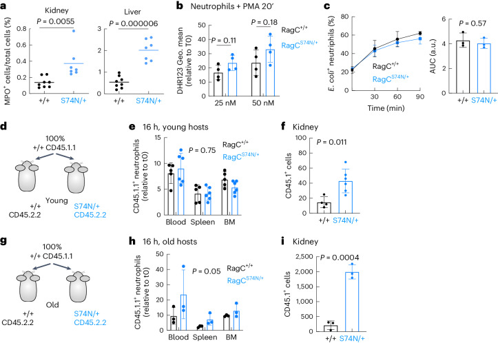
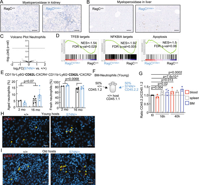
While BM-derived cells from Rragcmut/+ mice per se do not shorten longevity, chronic inflammation and inflammatory damage may be a necessary player in response to organ damage caused by increased nutrient signaling in Rragcmut/+ mice and a critical contributor to their shortened lifespan. Of note, two of the most upregulated inflammatory cytokines in kidneys from old Rragcmut/+ mice were Cxcl1 and Cxcl2 (Fig. 1l), chemokines that promote activation and extravasation of neutrophils52. Thus, to quantitatively ascertain neutrophil infiltration, we quantified myeloperoxidase (MPO)-positive cells, a marker of neutrophil inflammation, by IHC and found a significant increase in neutrophil abundance in kidney and liver from old Rragcmut/+ mice (Fig. 5a and Extended Data Fig. 5a,b). Myeloid inflammation can be detrimental for organ homeostasis53,54, so to test whether RragcS74N/+ neutrophils were cell-intrinsically dysfunctional and potential drivers of the health decline of RragcS74N/+ mice, we purified BM-derived neutrophils from Rragc+/+ and RragcS74N/+ mice and tested their cell-intrinsic ability to respond to activating stimuli in vitro. Treatment of purified neutrophils with the activating agent phorbol 12-myristate 13-acetate (PMA) resulted in similar activation and production of reactive oxygen species as indicated by the levels of the dihydrorhodamine (DHR) 123 probe (Fig. 5b) and a similar extent of activation-induced phagocytosis of Escherichia coli (Fig. 5c). Transcriptomic profiling of neutrophils purified from BM of five Rragc+/+ and five RragcS74N/+ young mice, yielded minimal transcriptomic changes (Extended Data Fig. 5c), with zero DEGs and with only few significant enrichment of signatures, which included TFEB targets, NFKBIA targets and apoptosis in Rragc+/+ neutrophils (Extended Data Fig. 5d and Supplementary Table 1).
Fig. 5. Old RragcS74N/+ organs promote neutrophil infiltration.
a, Quantification of IHC staining for myeloperoxidase in kidneys (left) and livers (right) collected from 18-mo-old Rragc+/+ (n = 8) and RragcS74N/+ (n = 7) male mice. b, Reactive oxygen species production by neutrophils isolated from Rragc+/+ (n = 4) and RragcS74N/+ (n = 4) BM as assessed by flow cytometry with DHR 123 after a 20-min treatment with PMA at the indicated concentrations. c, Kinetics of phagocytosis of GFP+ E. coli expressed as a percentage of GFP+ cells by neutrophils isolated from Rragc+/+ (n = 3) and RragcS74N/+ (n = 3) BM. Data are presented as mean ± s.d. d, Experimental setup of the neutrophil adoptive transplant experiment. BM-derived neutrophils were isolated from 2–3-mo-old Rragc+/+ mice (CD45.1.1) and transplanted to 3-mo-old Rragc+/+ (CD45.2.2) and to 3-mo-old RragcS74N/+ (CD45.2.2) male hosts. e, Quantification of wt CD45.1.1 neutrophils in the indicated tissues from CD45.2.2 Rragc+/+ (n = 5) and RragcS74N/+ (n = 6) male hosts 16 h after the adoptive transplant. f, Quantification of CD45.1.1 wt neutrophils by IF in kidneys from CD45.2.2 Rragc+/+ (n = 4) and RragcS74N/+ (n = 6) host males. g, Experimental setup of the neutrophil adoptive transplant experiment. BM-derived neutrophils were isolated from 3-mo-old Rragc+/+ mice (CD45.1.1) and transplanted to 12–14-mo-old Rragc+/+ (CD45.2.2) and RragcS74N/+ (CD45.2.2) male hosts. h, Quantification of transferred wt CD45.1.1 neutrophils in the indicated tissues from CD45.2.2 Rragc+/+ (n = 3) and RragcS74N/+ (n = 3) male hosts 16 h after adoptive transplant. i, Quantification of transferred wt CD45.1.1 neutrophils by IF in kidneys from CD45.2.2 Rragc+/+ (n = 3) and RragcS74N/+ (n = 3) host males. Statistical significance was assessed by two-tailed Student’s t-test (a–c,f,i) and two-way ANOVA (e,h). Data in b,c,e,f,h,i, are presented as mean ± s.d.
Extended Data Fig. 5. Old RragcS74N/+ organs promote neutrophil infiltration.
a. Representative IHC for myeloperoxidase (MPO) from kidneys harvested from 18-month-old Rragc+/+ and RragcS74N/+ male mice. Bars represents 100 μm. b. Representative IHC for myeloperoxidase (MPO) from livers harvested from 18-month-old Rragc+/+ and RragcS74N/+ male mice. Bars represents 100 μm. c. Volcano plot from RNA-seq performed in 6-8-week-old RragcS74N/+ (n = 4) versus 6-8-week-old Rragc+/+ (n = 5) neutrophils isolated from BM. Differential expression was performed with DESeq, using a 0.05 FDR. d. Gene set enrichment analysis (GSEA) related to TFEB target genes, NFKBIA target genes and genes involved in apoptosis in 6-8-week-old RragcS74N/+ (n = 4) versus 6-8-week-old Rragc+/+ (n = 5) neutrophils isolated from BM. e. Quantification of aged neutrophils (CD11b+/Ly6G+/CD62L-/CXCR4+, left) and fresh neutrophils (CD11b+/Ly6G+/CD62L+/CXCR2+, right) by flow cytometry in 2-month-old Rragc+/+ (n = 2) and RragcS74N/+ (n = 3) male mice and in 16-month-old Rragc+/+ (n = 5) and RragcS74N/+ (n = 5) male mice. Statistical significance was calculated by two-way ANOVA. Data are presented as mean values ± SD. f. Experimental setup of the neutrophil adoptive transplant experiment. BM-derived neutrophils were isolated from 10-week-old Rragc+/+ mice (CD45.1.2) and 10-week-old RragcS74N/+ (CD45.2.2) and transplanted at 1:1 ratio to 16-week-old Rragc+/+ (CD45.1.1) hosts. g. Ratio of CD45.2.2/CD45.1.1 transplanted neutrophils in blood, spleen and BM 16 h (n = 3 biological replicates) and 40 h (n = 3 biological replicates) after adoptive transfer as described in f. Data are presented as mean values ± SD. Statistical significance was calculated by two-tailed Student’s t-test. h. Representative images of IF depicting CD45.1 positive cells in kidneys from young male hosts. Bars represent 20 μm. i. Representative images of IF depicting CD45.1 positive cells in kidneys from old male hosts. Bars represent 20 μm.
Neutrophils have a rapid turnaround time of approximately 1 day, and fresh and aged neutrophils can be distinguished by differential expression of cell-surface markers55. We wondered whether neutrophils were retained for longer periods of time and, thus, aged neutrophils would be increased in RragcS74N/+ mice; however, aged neutrophils (CD11b+ Ly6G+, CD62L− and CXCR4+) were equally increased in Rragc+/+ and Rragcmut/+ old mice and accordingly, fresh neutrophils were reduced in old mice of both genotypes (Extended Data Fig. 5e). Taken together, the transcriptomic, activation and cell-surface analyses in neutrophils are consistent with negligible intrinsic molecular and functional differences in neutrophils from Rragc+/+ and RragcS74N/+ mice.
We next reasoned that parenchymal damage and chemoattractant signals from organs from RragcS74N/+ mice could trigger neutrophil inflammation. To conclusively distinguish between neutrophil-intrinsic and non-intrinsic neutrophil responses in RragcS74N/+ mice, we conducted a series of in vivo experiments with adoptive transfer of Rragc+/+ and RragcS74N/+ neutrophils isolated from BM (experimental cartoon in Extended Data Fig. 5f). Competitive adoptive transfer of Rragc+/+; CD45.1.2 and RragcS74N/+; CD45.2.2 in wt CD45.1.1 hosts resulted in a minimal, but significant increase in RragcS74N/+; CD45.2.2 compared to transferred Rragc+/+; CD45.1.2 neutrophils (Extended Data Fig. 5g). We next interrogated whether wt neutrophils become differentially activated in Rragc+/+ versus RragcS74N/+ hosts, so we transferred wt neutrophils (CD45.1.1) in young and old Rragc+/+ and RragcS74N/+ CD45.2.2 hosts to assess differential behavior in differential environments (hosts of different Rragc genotype). When young wt neutrophils were transferred to young Rragc+/+ and Rragcmut/+ hosts (Fig. 5d), the relative abundance in the blood, spleen and BM 16 h post-injection was similar (Fig. 5e), but the extravasation of wt neutrophils to kidneys of young RragcS74N/+ mice was significantly increased compared to extravasation of wt neutrophils in wt hosts (Fig. 5f and Extended Data Fig. 5h). Notably, the differential behavior of wt neutrophils was massively increased when transferred to old Rragc+/+ and RragcS74N/+ recipients (Fig. 5g). The wt neutrophils in RragcS74N/+ hosts were more abundant both in circulation and in the BM, spleen and kidney of old RragcS74N/+ hosts (Fig. 5h,i and Extended Data Fig. 5i). Such differential persistence and extravasation of wt neutrophils in old RragcS74N/+ hosts is consistent with damage signals stemming from old Rragcmut/+ organs that anomalously attract and activate myeloid cells. In turn, aberrantly behaving myeloid cells, through increased and sustained immune infiltration of Rragcmut/+ mice, may further contribute to perpetuate and worsen damage, senescence, SASP and further reinforce the chemoattractant signals for myeloid cells.
Pro-inflammatory signals precede inflammation in young RragcS74N/+ organs
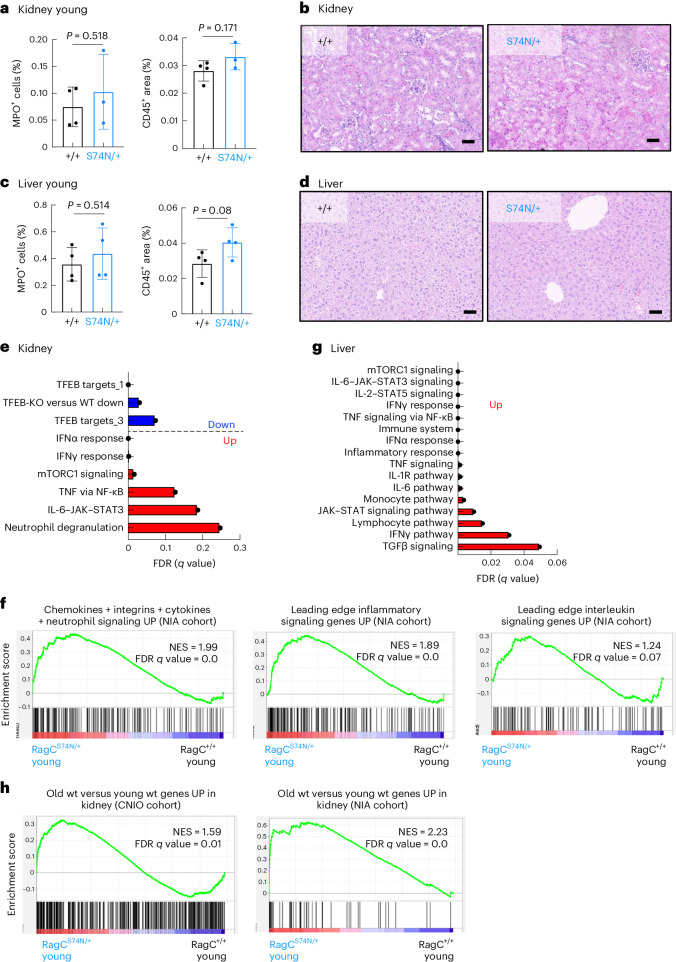
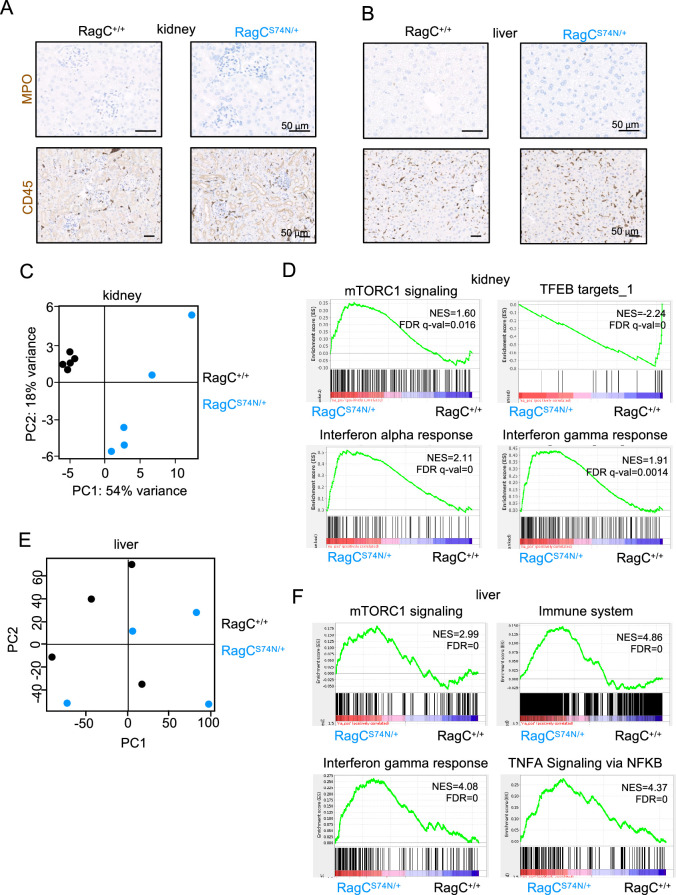
To support the notion that myeloid cells inflict additional damage to organs in Rragcmut/+ mice in response to aberrant signals from peripheral organs from Rragcmut/+ mice, we conducted bulk RNA-seq from kidneys and livers from 2–4-month-old Rragc+/+ and RragcS74N/+ mice, age in which myeloid inflammation and organ damage is not yet evident (Fig. 6a–d and Extended Data Fig. 6a,b). PCA clusters samples by genotype (Extended Data Fig. 6c), indicating that molecular differences are already evident at an early age and may underlie the extravasation of wt neutrophils observed in Fig. 5f,i. Moreover, in contrast to the absence of DEGs in purified neutrophils from Rragc+/+ versus RragcS74N/+ young mice, 689 genes from the kidney were differentially expressed in young RragcS74N/+ kidneys (335 upregulated and 354 downregulated; Supplementary Table 1). Among the top depleted signatures were Tfeb targets28,50 and mTORC1 activity and inflammation were among the top-enriched ones (Fig. 6e and Extended Data Fig. 6d). The changes in inflammatory genes and in chemokines and integrins identified in old RragcS74N/+ kidneys from the NIA cohort (Supplementary Table 2) are already present in young RragcS74N/+ organs, suggesting that these are early events during the aging process (Fig. 6f). Inflammatory signatures enriched in bulk RNA-seq were also evident in young livers (Fig. 6g, Extended Data Fig. 6e,f and Supplementary Table 1). Finally, the DEG genes between old and young wt mice from the CNIO and NIA cohorts (Supplementary Table 2) from Fig. 3 were found to be significantly enriched in young RragcS74N/+ versus young Rragc+/+ mice (Fig. 6h), supporting again the molecular overlap of the early changes seen in young RragcS74N/+ with those occurring in physiological aging.
Fig. 6. Pro-inflammatory signals precede inflammation in young RragcS74N/+ organs.
a, Quantification of IHC staining for myeloperoxidase (left) and CD45 (right) in kidneys collected from 2–4-mo-old Rragc+/+ (n = 4) and RragcS74N/+ (n = 4) male mice. b, Representative H&E pictures from the same mice as in a, showing lack of inflammatory foci in kidney. Scale bars, 50 μm. c, Quantification of IHC staining for myeloperoxidase (left) and CD45 (right) in livers collected from 2–4-mo-old Rragc+/+ (n = 4) and RragcS74N/+ (n = 4) male mice. d, Representative H&E pictures in the same mice as in a, showing lack of inflammatory foci in liver. Scale bars, 50 μm. e, Graphical representation of the FDRs from the indicated Hallmark, REACTOME, WikiPathways and curated gene sets enriched (red) and downregulated (blue) in kidneys from 3-mo-old RragcS74N/+ (n = 5) versus Rragc+/+ (n = 5) mice. f, GSEA related to chemokines, integrins, cytokines, inflammatory pathways and IL signaling genes upregulated in old kidneys of NIA cohort in kidneys from 3-mo-old RragcS74N/+ (n = 5) versus Rragc+/+ (n = 5) mice. See Supplementary Table 2 for further details of the gene sets. g, Graphical representation of the FDRs from the indicated KEGG, Hallmark, REACTOME and Biocarta gene sets enriched in livers from 2–3.5-mo-old RragcS74N/+ (n = 4) versus Rragc+/+ (n = 4) mice. h, GSEA related to upregulated genes in old kidneys from CNIO (left) NIA cohort (right) in kidneys from 3-mo-old RragcS74N/+ (n = 5) versus Rragc+/+ (n = 5) mice. See Supplementary Table 2 for further details of the gene sets. Statistical significance was assessed by two-tailed Student’s t-test (a,c). Data (a,c) are presented as mean ± s.d.
Extended Data Fig. 6. Pro-inflammatory signals precede inflammation in young RragcS74N/+ organs.
a. Representative images from anti-MPO and anti-CD45 immunostainings of kidneys harvested from 8-15-week-old Rragc+/+ and RragcS74N/+ male mice. Bars represent 50 μm b. Representative images from anti-MPO and anti-CD45 immunostainings of livers harvested from 8-15-week-old Rragc+/+and RragcS74N/+ male mice. Bars represent 50 μm. c. Principal component analysis (PCA) from the RNA-seq performed from kidney samples from 3-month-old Rragc+/+ (n = 5) and RragcS74N/+ (n = 5) mice. d. Gene set enrichment analysis (GSEA) related to inflammatory signatures, TFEB target genes, and mTORC1 signaling associated genes in kidneys from 3-month-old RragcS74N/+ (n = 5) versus Rragc+/+ (n = 5) mice. e. Principal component analysis (PCA) from the RNA-seq performed from liver samples from 3-month-old Rragc+/+ (n = 4) and RragcS74N/+ (n = 4) mice. f. Gene set enrichment analysis (GSEA) related to inflammatory signatures and mTORC1 signaling associated genes in livers from 2–3.5-month-old RragcS74N/+ (n = 4) versus Rragc+/+ (n = 4) mice.
Myeloid cell depletion extends survival in RragcS74N/+ mice
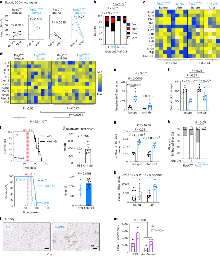
Excessive myeloid inflammation in response to organ damage can be pathogenic and inflict additional damage paracrinally, so even if RragcS74N/+ myeloid cells were not intrinsically abnormal, they could elicit further organ deterioration if activated by parenchymal signals. To determine whether secondary myeloid inflammation contributes to the health decline of Rragcmut/+ mice, we acutely depleted neutrophils and monocytes by intraperitoneal injection of a blocking antibody for Gr1 (ref. 56), in 24-month-old mice. Three anti-Gr1 injections within 1 week (Extended Data Fig. 7a) were sufficient to eliminate circulating neutrophils in both Rragc+/+ and RragcS74N/+ mice (Fig. 7a), while also partially correcting the skewed PBMC populations from old RragcS74N/+ mice (Fig. 7b). Moreover, acute treatment reduced inflammatory cytokines in blood from RragcS74N/+ mice (Fig. 7c), without correcting aging features such as SA-β-gal activity (Extended Data Fig. 7b) and dermal thickness (Extended Data Fig. 7c). A 3-week depletion of Gr1+ cells resulted in sustained reduction of neutrophils (Extended Data Fig. 7d,e), partial correction of inflammatory markers (Extended Data Fig. 7f,g) and the downregulation of the expression of inflammatory genes in livers from Rragcmut/+ mice (Fig. 7d). Notably, at this 3-week time point, a significant amelioration of several of the markers of aging were detected in Rragcmut/+ mice: significant decrease in SA-β-gal activity in the kidney (Fig. 7e and Extended Data Fig. 7h, which is also evident in Rragc+/+ mice), expansion of the dermal thickness and decrease in dermal inflammation (Fig. 7f,g) and a reduction of liver infiltration (Fig. 7h) in anti-Gr1 treated, but not isotype-antibody treated, old RragcS74N/+ mice.
Extended Data Fig. 7. Myeloid cell depletion corrects markers of inflammaging and extends survival in RragcS74N/+ mice.
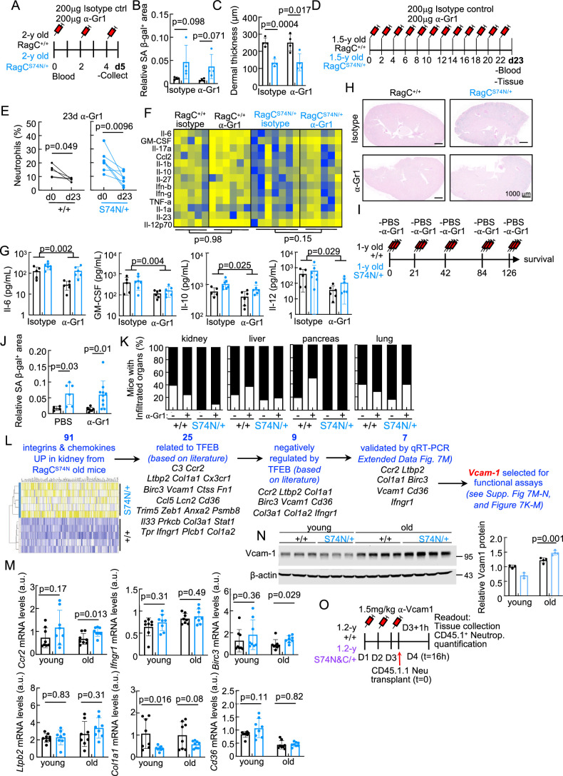
a. Experimental setup for α-Gr1 acute treatment. Rragc+/+ and RragcS74N/+ male mice were treated with 200μg of α-Gr1 or isotype at day 0, 2 and 4. At 5d, mice were euthanized and blood and tissues were collected. b. Quantification of SA-β-gal+ area within the kidney area of 2 y old isotype-treated Rragc+/+ (n = 5), anti-Gr1 treated Rragc+/+ (n = 7), isotype-treated RragcS74N/+ (n = 6), anti-Gr1-treated RragcS74N/+ (n = 7) 2 y old male mice. Data are presented as mean values ± SD. c. Dermal thickness measured in back skin of isotype-treated Rragc+/+ (n = 5), anti-Gr1-treated Rragc+/+ (n = 7), isotype-treated RragcS74N/+ (n = 6), anti-Gr1-treated RragcS74N/+ (n = 7) 2-year-old male mice. Data are presented as mean values ± SD. d. Experimental setup for three-week anti-Gr1 treatment. One-and-a-half-year-old male mice were treated with 200μg of anti-Gr1 or isotype control every other day for 23d, and mice were sacrificed and blood and tissues were processed. e. Percentage of neutrophils measured in blood before and after a 23-day treatment of anti-Gr1 treated Rragc+/+ (n = 5) and anti-Gr1-treated RragcS74N/+ (n = 7) 18-m-old male mice. Statistical significance was calculated by one-tailed Student’s t-test. f. Quantification of inflammatory cytokines in sera from isotype-treated Rragc+/+ (n = 5), anti-Gr1-treated Rragc+/+ (n = 6), isotype-treated RragcS74N/+ (n = 7), anti-Gr1-treated RragcS74N/+ (n = 6) 18-month-old male mice after a 23-day treatment. g. Concentration of the indicated cytokines in sera from isotype-treated Rragc+/+ (n = 5), anti-Gr1 treated Rragc+/+ (n = 6), isotype-treated RragcS74N/+ (n = 7), anti-Gr1 treated RragcS74N/+ (n = 6) 1.5-year-old male mice. Data are presented as mean values ± SD. h. Representative pictures of SA β-gal stains from isotype-and anti-Gr1-treated mice. i. Experimental setup for extended anti-Gr1 survival experiment. One-year-old Rragc+/+ and RragcS74N/+ male mice were treated either with PBS or anti-Gr1 every other day for two weeks, followed by a resting period of 3 weeks. This regimen was repeated five times so mice received either PBS or anti-Gr1 antibody intermittently during approximately 6 months. j. Quantification of SA-β-gal+ area within the kidney area of mice under the regime described in j. PBS-treated Rragc+/+ (n = 4), anti-Gr1-treated Rragc+/+ (n = 7), PBS-treated RragcS74N/+ (n = 6), anti-Gr1 treated RragcS74N/+ (n = 10) males. Kidneys were harvested at humane end point. Data are presented as mean values ± SD. k. Incidence of infiltrated inflammatory cells in the indicated tissues from PBS-treated Rragc+/+ (n = 5), anti-Gr1-treated Rragc+/+ (n = 7), PBS-treated RragcS74N/+ (n = 6) and anti-Gr1-treated RragcS74N/+ (n = 10) mice. Statistical significance was calculated by two-sided Fisher’s exact test. l. Heatmap of integrins and chemokines upregulated in kidney from Rragc+/+ (n = 5) and RragcS74N/+ (n = 5) mice. Out of 91 genes, 25 genes related to TFEB were selected based on literature and 9 were negatively regulated by TFEB (based on literature); 7 out of 9 were validated by qRT–PCR and finally, Vcam1 was selected for functional assays. m. qRT–PCR analysis of integrins and chemokines negatively regulated by TFEB in kidneys of young (4-month-old) and old (18-month-old) Rragc+/+ (young, n = 8; old, n = 8) and RragcS74N/+ (young, n = 8; old, n = 8) male mice. Data are presented as mean values ± SD. n. Immunoblot and quantification for Vcam1 from 4-month-old Rragc+/+ (n = 3 biological independent samples) and RragcS74N/+ (n = 3 biological independent samples) and 18-month-old Rragc+/+ (n = 4 biological independent samples) and RragcS74N/+ (n = 4 biological independent samples) male mice. Data are presented as mean values ± SD. o. Experimental setup for α-Vcam1 treatment and neutrophil adoptive transplant. Mice were treated with PBS or 1.5 mg/kg of α-Vcam1 three consecutive days and one hour after the last dose, CD45.1.1 BM-isolated neutrophils were transplanted to 16-24-month-old Rragc+/+ (CD45.2.2) and RragcS74N/+ (CD45.2.2) male and female hosts. Statistical significance in a, b, j, m, and n was calculated by two-tailed Student’s t-test. Statistical significance in f and g was calculated by two-way ANOVA.
Fig. 7. Myeloid cell depletion corrects markers of inflammaging and extends survival in RragcS74N/+ mice.
a, Quantification of neutrophils in peripheral blood before and after a 5-day treatment with anti-Gr1 or isotype control antibody from isotype-treated Rragc+/+ (n = 4), anti-Gr1-treated Rragc+/+ (n = 4), isotype-treated RragcS74N/+ (n = 4) and anti-Gr1-treated RragcS74N/+ (n = 3) 24-mo-old male mice. b, WBC count in isotype-treated Rragc+/+ (n = 5) and RragcS74N/+ (n = 7), anti-Gr1-treated Rragc+/+ (n = 6) and RragcS74N/+ (n = 7) 24-mo-old male mice. c, Quantification of cytokines in sera from isotype-treated RragcS74N/+ (n = 4) and anti-Gr1-treated RragcS74N/+ (n = 4) 24-mo-old male mice in a 5-day treatment measured by Legendplex assay. d, qRT–PCR analysis of SASP genes in the kidneys from isotype-treated Rragc+/+ (n = 5) and RragcS74N/+ (n = 7), anti-Gr1-treated Rragc+/+ (n = 6) and RragcS74N/+ (n = 7) 18-mo-old male mice. e, Quantification of SA-β-gal+ area within the kidney area of isotype-treated Rragc+/+ (n = 5) and RragcS74N/+ (n = 7), anti-Gr1-treated Rragc+/+ (n = 6) and RragcS74N/+ (n = 7) of 18-mo-old mice. f, Dermal thickness in back skin of the same mice as in e. g, Quantification of IHC of CD45+ cells in skin from the same mice as in f. h, Incidence of infiltrated inflammatory cells in livers from mice in e. i, Kaplan–Meier survival curves of control (n = 5) and anti-Gr1-treated (n = 9) Rragc+/+ mice (left) and controls (n = 6) and anti-Gr1-treated (n = 10) RragcS74N/+ mice (right). j, Survival since the first dose of anti-Gr1 of the mice in k. k, qRT–PCR analysis of Vcam1 in kidneys of young (4-mo-old) and old (18-mo-old) Rragc+/+ (young, n = 8; old, n = 8) and RragcS74N/+ (young, n = 8; old, n = 8) male mice. l, Representative IHC staining of Vcam1 in kidney from 18-mo-old Rragc+/+ and RragcS74N/+ mice. m, Quantification of transferred wt CD45.1.1 neutrophils by IF in kidneys from CD45.2.2 host males treated with PBS (Rragc+/+ (n = 3); RragcS74mut/+ (n = 3)) or anti-Vcam1 (Rragc+/+ (n = 3); RragcS74mut/+ (n = 3)). Statistical significance was assessed by one-tailed Student’s t-test (a); two-tailed Student’s t-test (e–g, j,k); two-sided Fisher’s exact test (h); two-way ANOVA (b–d,m); and log-rank test (l). Data (b,e–g,j,k,m) are mean ± s.d.
We next sought to evaluate the long-term effect of controlling myeloid inflammation. This effort constituted an experimental challenge because repetitive injections of anti-Gr1 results in a feedback response that results in the maturation of neutrophils and other myeloid cells with reduced expression of the Gr1 receptor, becoming refractory to the intervention57. Thus, we designed an intermittent regime of five cycles of anti-Gr1 injections, followed by resting periods, over a total of 6 months, to enable transient but recurrent depletion of myeloid cells (Extended Data Fig. 7i). Starting at approximately the same age as that of the cohorts used for pharmacological inhibition of mTORC1 with rapamycin (Extended Data Fig. 2g,h), long-term injections of anti-Gr1 antibody significantly extended the median and maximal lifespan of RragcS74N/+ mice (Fig. 7i,j; control, 73.36 weeks; anti-Gr1, 93.14 weeks), a ~23% extension of overall survival and a remarkable ~40% extension if measured since the day of the first dose of anti-Gr1 (Fig. 7i,j). Histopathological examination and quantification of SA-β-gal activity in the kidney revealed no differences between control mice and mice treated with anti-Gr1 antibodies at a humane end point, suggesting that the suppression of myeloid inflammation delays, rather than changes, the ultimate causes of death of Rragcmut/+ (Extended Data Fig. 7j,k). Altogether, the results of different regimes of myeloid cell depletion show that controlling inflammation under pathologic nutrient signaling, even at old ages, is an efficacious intervention to extend longevity.
To identify potential myelo-attractant genes differentially expressed in organs of RragcS74N/+ mice, and to assess their functional effect on the abnormal infiltration, we first selected all integrins and chemokines significantly increased in the bulk RNA-seq analysis from kidney samples (Extended Data Fig. 7l). From those 91 DEGs, we conducted a literature search to select those reported to be controlled by TFEB, and from those 25, we selected 9 that were reported to be negatively regulated by TFEB, as its transcriptional activity is suppressed by increased RagC–mTORC1 signaling. We validated seven of them by qPCR (Extended Data Fig. 7m) and selected Vcam1 for the functional validation of its involvement in myeloid infiltration in old RragcS74N/+ mice based on reported function58,59 and preliminary analysis of RragcS74N/+ samples by western blot and IHC (Fig. 7k,l and Extended Data Fig. 7n). Thus, we repeated the wt neutrophil transfer experiments (as in Fig. 5g) with the addition of α-Vcam1-blocking antibodies and quantified neutrophil extravasation (Extended Data Fig. 7o). Supporting our hypothesis, blockade of Vcam1 inhibited wt neutrophil extravasation in old Rragc+/+ and RragcS74N/+ mice (Fig. 7m).
Collectively, our work provides strong support for a two-step pro-aging effect of increased nutrient signaling, involving parenchymal damage and a secondary myeloid inflammation that can be therapeutically mitigated even at old age.
Discussion
The connections of the mTORC1 signaling pathway and aging across eukaryotes are extensive. Pharmacological inhibition of mTOR results in extended longevity in yeast, worms, flies and mice2. Moreover, genetic approaches to study mTORC1 signaling in aging have been undertaken in yeast, worms and flies, but equivalent genetic approaches in mice have been limited by the occurrence of specific detrimental phenotypes that precluded the analysis of time-dependent health decline. Pten-deficient and Tsc1/2-deficient mice die in utero and heterozygous deletion of Pten or Tsc1 or Tsc2 result in the development of tumors following loss of heterozygosity18,20–23. While these findings were invaluable for understanding developmental biology, metabolic physiology and cancer biology, systemic and other tissue-specific models of upregulation of mTORC1 activity yielded fewer lessons for the understanding of the processes underlying aging driven by increased mTORC1 activity60,61. Partial loss-of-function approaches have been undertaken, including heterozygous mice for several components of mTORC1 (ref. 17) and a hypomorphic mTOR mutant16, which revealed that the links between mTORC1 activity and longevity exceed the systemic metabolic effects of this cascade.
Here we report a mouse model of a systemic increase in Rag GTPase–mTORC1 activity leading to a pro-aging-like phenotype. We propose that this phenotype is revealed in Rragcmut/+ mice precisely because of a mild increase in mTORC1 activity. In contrast to the tissue-specific deleterious pathologies seen in strains with stronger activation of the mTORC1 pathway, a moderate and systemic increase in nutrient signaling allows mice to survive long enough and with minimal phenotypic alterations to manifest time-dependent decline in organ function.
Notably, the transcriptomic changes occurring in Rragcmut/+ organs from young and old mice exhibit a large overlap with the transcriptomic changes that take place during physiological aging of wt mice, thus supporting the extrapolation of the conclusions obtained from the molecular and cellular analyses of Rragcmut/+ mice for ‘normal’ mammalian aging and positing that the nutrient–Rag GTPase–mTORC1 axis is involved in mammalian longevity. Moreover, based on the overlap of transcriptomic signatures from Rragcmut/+ mice and septuagenarians (Fig. 3), the involvement of this axis could be extrapolated also to human aging.
Rragcmut/+ mice show surprisingly decreased spontaneous tumor development. A simplistic yet counterintuitive interpretation of this finding would be that activating mutations in RagC have tumor suppressive functions, in spite of being bona fide oncogenic for B cells. In this regard, recent evidence points to a liability of activating mutations in the nutrient signaling cascade in conditions of limited nutrient availability in cultured cells62.
Neither B cells nor inflammation per se drive the shortened lifespan of mice with increased nutrient signaling. Instead, peripheral organ damage results in secondary myeloid inflammation. In turn, this inflammation perpetuates and worsens the peripheral organ damage, as reported in other physiological settings63. The pharmacological intervention to suppress neutrophil inflammation with an anti-Gr1 antibody (Fig. 7), together with the in vitro activation of neutrophils, strongly support the notion that myeloid cells are unlikely to be cell-autonomously activated in an aberrant manner by RagC mutations, but rather respond to signals such as Cxcl1 and Cxcl2 and Vcam1, emanating from prematurely damaged organs of Rragcmut/+ mice.
The identification of inflammation as a mediator of the shortened longevity by increased nutrient signaling evidences the irreplaceable value of mouse genetics for understanding the inter-organ communications and interactions that mediate the complexity of processes that precipitate the evolution of aging, which can be arguable not entirely captured by approaching this in other model organisms.
The largely beneficial effects of depletion of myeloid cells contrasts with the negligible effect of pharmacological inhibition of mTORC1 with rapamycin administered in the same formulation, dose, regime and starting at an age similar to that reported elsewhere14. The effect of rapamycin in extending lifespan is proportional to the duration of the dosing14,15. Hence, it is conceivable that rapamycin extended the survival of wt mice because their remaining lifespan when dosing started was long enough to allow rapamycin to exert its longevity-promoting effect, but was insufficient in the short-lived Rragcmut/+ mice. This is a valid interpretation of our data, but is at odds with the strong protective effect of the anti-Gr1 intervention when administered at the same age as of the rapamycin-treated Rragcmut/+ mice. An alternative interpretation is that organ damage by increased mTORC1 activity in Rragcmut/+ was already extensive, irreparable and not tunable by pharmacological inhibition of mTORC1 at an advanced age. Moreover, rapamycin had barely any effect in the chronic inflammation of old Rragcmut/+, so it supports the concept that inflammation secondarily occurring in response to organ damage is a relevant player in the process of nutrient signaling-driven aging. Unlike what occurs in lymphomas29, another potential explanation for negligible effect of rapamycin is related to the program executed by the RagC–mTORC1 axis during aging. Compelling new evidence64,65 states that RagC-dependent activation of mTORC1 results in preferential phosphorylation of members of the TFEB family. Indeed, our transcriptional profiles of organs from Rragcmut/+ support a strong suppression of Tfeb transcriptional activity and, notably, phosphorylation of TFEB family members by mTORC1 is particularly insensitive to the allosteric mode of action of rapamycin. Thus, if Rragcmut/+ cells have impaired Tfeb activity due to sustained phosphorylation by mTORC1, minimal effect of rapamycin would be expected. The mitigation of myeloid infiltration by the blockade of Vcam1 in our myeloid transplantation setting supports the relevance of the RagC–mTORC1–Tfeb axis, as Tfeb activity suppresses the expression of Vcam1 (ref. 66). Finally, activating mutations in RagC may also shorten longevity by yet-to-be-identified functions independent of canonical mTORC1 activity.
Our dissection of the determinants of the aging-like phenotype in mice with increased nutrient signaling reveals that chronic inflammation is actionable as a potential therapeutic control of the health decline. Many aging-related diseases have a positive correlation with BMI67, which also associates with mild chronic inflammation; conversely, dietary restriction (DR) is an extremely efficacious intervention to extend healthy aging and longevity in mammals. While DR and mTORC1 inhibition are molecularly distinguishable5,68, some degree of epistasis between DR and mTORC1 exists3, so presumably DR acts at least partially through the modulation of the Rag GTPase–mTORC1 signaling axis.
Methods
Mice
All animal procedures carried out at the CNIO were approved by the CNIO-ISCIII Ethics Committee for Research and Animal Welfare (CEIyBA) and by the Autonomous Community of Madrid (certificates PROEX 15/18, PROEX 215.17, 139.1/22 and PROEX 225.7/22). Mouse work at the NIA was approved by the Animal Care and Use Committee of the NIA in Baltimore (277-TGB-2022). IghmμMT mice46 were obtained from The Jackson Laboratories (stock 002288). Mice were housed under specific-pathogen-free conditions at 22 °C and 45–65% humidity and with 12-h dark–light cycles. Mice were fed with a standard chow diet (Harlan Teklad 2018). All mice were observed weekly by trained personnel. Upon signs of morbidity, mice were closely inspected daily until application of humane end point criteria (http://dels.nas.edu/global/ilar/Guide). For BM reconstitution experiments, host mice were sublethally irradiated with two 4.5-Gy doses within a 4-h period following by injection of BM cells. Rapamycin was supplied encapsulated in chow diet at 42 ppm (Rapamycin Holdings and Purina Lab Diet). For anti-Gr1 treatment, mice were injected intravenously with 200–400 μg of anti-Gr1 antibody (BioXcell, clone RB6-8C5, BE0075) or isotype control (BioXcell, clone LTF-2, BE0090). For anti-Vcam1 treatment, mice were injected intravenously with 1.5 mg kg−1 of anti-Vcam1 antibody (BioXcell, clone M/K-2.7, BE0027-25MG) or PBS.
Generation of RagcS74N Crispr-edited KI mice
Blastocysts were injected with the Cas9 mRNA, a single-guide RNA targeting the Rragc locus and a single-stranded DNA oligonucleotide containing the desired mutation flanked by 40–60 bases homologous to the sequence adjoining the DNA double-strand break, as carried out previously29,69. Following clone selection, genotyping was performed by PCR and restriction fragment length polymorphisms or Sanger sequencing. We utilized a gRNA with the following sequence (in minus orientation): CGGCAAATCCTCCATCCAGA. The repair oligonucleotide contained the AAC mutations plus five additional silent mutations (CGGCAAAAACAGTATCCAAAAA).
Metabolic parameters
Blood was collected from the submandibular vein. Hemoglobin and hematocrit were determined by a blood cell counter (CVM LaserCell) and blood urea nitrogen, creatinine and amylase were measured using VetScan rotors (Abaxis, 500-0038-25).
Behavioral tests
For the tightrope assay, mice were placed on a bar of a circular section (60 cm long and 1.5 cm in diameter) and the test was considered successful when a mouse remained on the bar for 60 s in at least one of five consecutive trials70. The rotarod tests were performed in a Panlab LE 8200 using acceleration from 4–40 rpm in a period of 120 s. The time to fall was recorded and an average of three trials was used71. Mice were previously trained for three consecutive days.
Measurement of blood pressure
Blood pressure was measured using a noninvasive tail-cuff method with the CODA system (CODA-HT2, Kent Scientific Corporation)72. Mice were habituated for three consecutive days.
Detection of cytokines in serum
Sera from mice were collected and analyzed with a cytokine assay LEGENDplex Mouse Inflammation Panel (13-plex) (BioLegend, 740446). Data were collected by flow cytometry (BD FACSCanto II) and cytokines were quantified with a LEGENDplex Data Analysis Software Suite.
SA-β-gal stain in tissues
The SA-β-gal stain was performed using a Senescence β-Galactosidase Staining kit (Cell Signaling Technology, 9860). Kidneys were fixed in 1× Fixative solution overnight at room temperature (RT) and then incubated for 30 h at 37 °C. Tissues were washed twice with 50% ethanol and twice with 70% ethanol. Kidneys were embedded in paraffin blocks and nuclear fast red stain was performed.
Isolation of neutrophils for transplants
Neutrophils were purified from CD45.1.1 mouse BM using a 65% Percoll gradient (GE HealthCare, 17-0891-01) and red blood cells were lysed using Erythrocyte Lysis Buffer (QIAGEN, 79217). Then, 2–5 × 10⁶ neutrophils were transferred to recipient mice bled 2 min after transplant to determine the input number of CD45.1.1 neutrophils.
Signaling in MEFs
Subconfluent cell cultures were rinsed and placed in RPMI without amino acids (US Biological, R8999-04A), supplemented with 10% dialyzed FBS and all 20 amino acids for 1 h. After that, cells were rinsed three times and then placed in RPMI without amino acids supplemented with 10% dialyzed FBS.
Immunofluorescence in MEFs
MEFs were seeded on 12-mm-diameter coverslip in a 12-well plate (150,000 cells per well), one day before the experiment. After 1 h of the indicated treatment, cells were fixed with 8% paraformaldehyde in PBS (EMB, 15710) for 10 min at RT. Cells were permeabilized with 0.05% Triton X-100 (Sigma, T9284) in PBS for 5 min at RT, followed by three washes with PBT (PBS + Tween 0.1% (Sigma, P7949)). After blocking in 5% goat serum (Sigma, G9023) in PBT for 1 h, slides were incubated with TFEB antibody (1:100 dilution, Bethyl Laboratories, A303-673A) for 1 h at RT, washed 3× with PBT and incubated with goat anti-rabbit IgG (H+L) Cross-Adsorbed Secondary Antibody, Alexa Fluor 488 (1:300 dilution, Life Technologies, A11008) for 1 h at RT. Coverslips were then washed three times and incubated with 4,6-diamidino-2-phenylindole (DAPI; Sigma, D9542) for 5 min. After washing three times with PBT, coverslips were mounted with Fluoromount-G (Bionova Cientifica, 0100-01).
Staining and flow cytometry analysis
Mononuclear cells were isolated from mouse blood, spleen or bone marrow (BM). Cells were separated by crushing the spleens through a 70-μm mesh (Corning) in ice-cold PBS + 0.1% BSA + 3 mM EDTA, and red blood cells were lysed using Erythrocyte Lysis Buffer. Cells from blood were isolated after red blood cells lysis and BM cells were isolated by flushing cells from the tibia and femur and collecting them in ice-cold PBS + 0.1% BSA + 3 mM EDTA. Cell staining was performed on ice in PBS + 0.1% BSA + 3 mM EDTA after a step of incubation with Fc-block Reagent (anti-CD16/CD32, BD Pharmigen, 553142). Macrophages were identified as F4/80+/SSChi. NK cells were identified as F4/80−B220−/CD3−/NK.1.1+. Dendritic cells were identified as F4/80−B220−/CD3−/NK.1.1−/MHCII+/CD11c+. Neutrophils were identified as F4/80−/B220−/CD3−/NK.1.1−/MHCII−/CD11c−/CD11b+/Ly6G+. Monocytes were identified as F4/80−/B220−/CD3−/NK.1.1-/MHCII−/CD11c−/CD11b+/Ly6G−/Ly6Cl°w or high. Eosinophils were identified as F/480−/B220−/CD3−/NK.1.1-/MHCII−/CD11c−/CD11b+/Ly6G−/Ly6Cmed. Aged neutrophils55 were identified as CD11b+/Ly6G+/CD62L−/CXCR4+. Fresh neutrophils were identified as CD11b+/Ly6G+/CD62L+/CXCR2+. Flow cytometry analyses included BD LSRFortessa or BD FACSCanto II cell analyzers, running BD FACSDiva software (BD Biosciences). FlowJo software (v.9.8.1 and v.10; TreeStar) was used for data analyses and plot rendering.
Assessment of neutrophil function
Reactive oxygen species production
Neutrophils were purified from BM using a 65% Percoll gradient (GE HealthCare, 17-0891-01). Then, 5 × 10⁵ neutrophils were plated and incubated with 2.5 μg ml−1 DHR 123 (Molecular Probes, D23806) and 25 or 50 nM PMA (Sigma-Aldrich, P1585) for 20 min at 37 °C and 5% CO2. After incubation, cells were washed with ice-cold PBS + 0.1% BSA + 3 mM EDTA, stained for cell-surface markers and analyzed by flow cytometry73.
Phagocytosis of E.coliGFP
Neutrophils were purified from BM using a 65% Percoll gradient. Then, 5 × 10⁵ neutrophils were incubated with GFP-expressing E. coli in a ratio of 1:100 for 30–90 min at 37 °C. Cells were collected and stained for cell surface markers and analyzed by flow cytometry.
Immunoblotting
Cells were rinsed once with ice-cold PBS and lysed in ice-cold lysis buffer (50 mM HEPES, pH 7.4, 40 mM NaCl, 2 mM EDTA, 1.5 mM sodium orthovanadate, 50 mM NaF, 10 mM pyrophosphate, 10 mM glycerophosphate, 1% Triton X-100 and one tablet of EDTA-free complete protease inhibitors (Roche) per 25 ml). Small pieces of frozen tissues were homogenized using a FastPrep-24 5G tissue homogenizer. Lysates were cleared by centrifugation at maximum speed for 10 min. Protein extracts were denatured by the addition of sample buffer, resolved by SDS–PAGE and analyzed by immunoblotting. Data collection was performed using Odyssey Infrared Imaging System (Application Software v.3.0.30. LI-COR Biosciences, NDP.view2 software). In addition, analyses were performed according to standard procedures using FIJI ImageJ v.1.53.
Subcellular protein fractionation in tissues
Tissues were extracted with a Subcellular Protein Fractionation kit for tissues (Thermo Scientific, 87790), adjusting the volumes to the sample mass. After protein extraction, the cytosolic and nuclear fractions were quantified using BCA (Thermo Scientific, 23222). Then, 30 µg cytoplasmic and 30 µg nuclear fraction from the kidney and 25 µg cytoplasmic and 8 µg nuclear fraction from the heart were used for immunoblotting.
RNA extraction, cDNA synthesis and qRT–PCR
Total RNA was extracted from tissues with TRItidy (Panreac Applichem, A4051,0200) and using an RNeasy kit from QIAGEN (74106). RNA was extracted from cells with a TRItidy and Picopure RNA Isolation kit (Arcturus, Applied Biosystems, 12204-01). RNA was treated with DNase (QIAGEN, 79254) before cDNA synthesis. To perform cDNA synthesis, 1 μg RNA was retrotranscribed using SuperScript IV VILO Master Mix (Invitrogen, 11756500). Quantitative real-time PCR was performed using GoTaq qPCR Master Mix (Promega, 6001) in a QuantStudio 6 Flex Real-Time thermocycler (Applied Biosystems). Data were analyzed by the change-in-threshold (2−ΔCT) method, using β-actin as the housekeeping reference gene. Primers were designed using Primer3 Software (http://bioinfo.ut.ee/primer3-0.4.0/).
Gene expression profiling of neutrophils, liver and kidney from CNIO cohorts
Neutrophils (CD45+, CD11b+ and Ly6G+) were sorted from RragcS74N/+ and Rragc+/+ BM cells in a BD FACSAria Ilu (Becton Dickinson) and InFlux (Cytopeia-Becton Dickinson) cell sorters. For the liver and kidney RNA-seq, total RNA samples (300–500 ng) were processed into cDNA sequencing libraries with the QuantSeq 3′ mRNA-Seq Library Prep kit (FWD) for Illumina (Lexogen, 015). For neutrophil preparations (RNA range 16–180 ng), a unique molecular identifier second-strand synthesis module (Lexogen, 081) was used. Library generation was initiated by reverse transcription with oligodT priming and a second-strand synthesis was performed from random primers. Primers from both steps contained Illumina-compatible sequences. Libraries were completed by PCR and sequenced on an Illumina NextSeq 550 System (with v.2.5 reagent kits).
The resulting reads were analyzed with the nextpresso74 pipeline: sequencing quality was checked with FastQC v.0.11.7 (https://www.bioinformatics.babraham.ac.uk/projects/fastqc/); reads were aligned to the mouse genome (GRCm39 for neutrophils and young and old kidney RNA-seq and GRCm38 in the case of young liver RNA-seq) with TopHat2 (ref. 75) using Bowtie76 and SAMtools77, allowing three mismatches and 20 multi hits. The GENCODE vM25 gene annotation for GRCm38 was used; read counts were obtained with HTSeq78 v.0.6.1 (–stranded = yes), using the mouse gene annotation from GENCODE (gencode.vM26.GRCm39.Ensembl103 for neutrophil and young and old kidney RNA-seq, gencode.vM20.GRCm38.Ensembl95 for young liver RNA-seq). Differential expression was performed with DESeq79, using a 0.05 false discovery rate (FDR). Gene Set Enrichment Analysis (GSEA) Preranked80 was used to perform gene set enrichment analysis for several gene signatures on a preranked gene list, setting 1,000 gene set permutations. Only those gene sets with significant enrichment levels (FDR q value < 0.25) were considered.
Gene expression profiling of kidneys from NIA cohort
Four-month-old and 24–26-month-old C57LB/6J mice were killed in the morning under fed conditions and kidneys were snap frozen in liquid N2 and stored at −80 °C. For RNA isolation, 50 mg kidney tissue was weighed, resuspended in cold TRIzol Reagent (Invitrogen Life Technologies) and homogenized in a TissueLyser (QIAGEN). Downstream steps for RNA isolation were carried out automatically on a QIACube by using the RNAeasy Plus kit (QIAGEN).
For kidney RNA-seq, total RNA samples (750 ng) were processed into cDNA sequencing libraries with the SMARTer Stranded Total RNA Sample Prep kit - HI Mammalian kit and unique dual-indexed PCR primers for amplification were used (Takara Bio). Paired-end sequencing was performed for 103 cycles in an Illumina Novaseq 6000 sequencer and real-time analysis generated the base-call files, which were de-multiplexed and converted to standard FASTQ files using the bcl2fastq program (v.2.20.0.422). The quality of reads was first assessed using fastqc v.0.11.8 (https://www.bioinformatics.babraham.ac.uk/projects/fastqc/). Next, reads were trimmed using bbduk (part of the bbtools package; https://jgi.doe.gov/data-and-tools/software-tools/bbtools/). Reads were then aligned to the mm10 reference genome and ENSEMBL annotation v.89 using STAR v.2.7.8a in two-pass mode81. The BAM files generated by STAR were sorted and indexed using SAMtools v.1.9 (ref. 77). To generate a count matrix, featureCounts v.2.0.1 (ref. 82) was utilized. An overall quality control assessment was then performed using multiqc v.1.9 (ref. 83). To perform differential gene expression analysis, the genes in the count matrix were filtered by requiring at least ten samples to have a count greater than 10. Finally, DESeq2 v.1.40.2 (ref. 79) was used for analysis. The apeglm method84 was used for visualization and ranking. Genes with an adjusted P value <0.05 were considered significant.
Histological and immunohistochemical analyses of mouse tissues
Tissue samples were fixed in 10% neutral buffered formalin, paraffin-embedded, cut at 3 μm, mounted in superfrost plus slides and dried overnight. Consecutive rehydrated sections were stained with hematoxylin and eosin (H&E) and IHC reactions were performed in an automated platform (Ventana Discovery XT). Antigen retrieval was first performed with the appropriate pH buffer (CC1m, Ventana, Roche) and endogenous peroxidase was blocked (peroxide hydrogen at 3%). Slides were then incubated with rat monoclonal anti-p21 (clone 291HUGO; 1:10 dilution; CNIO Monoclonal Antibodies Core Unit, also Abcam, 107099), rabbit polyclonal MPO (1:1,250 dilution, DAKO, A0398), rabbit polyclonal CD45 (clone D3F8Q, 1:200 dilution, Cell Signaling Technology, 70257), rabbit monoclonal Vcam1 (clone D2T4N, 1:200 dilution, Cell Signaling Technology, 32653) and TFEB (1:1,500 dilution, Bethyl, A303-673A). After addition of the primary antibody, slides were incubated with the visualization systems (Omni Map anti-rabbit, Ventana, Roche) conjugated with horseradish peroxidase. IHC reactions were developed using 3, 30-diaminobenzidine tetrahydrochloride (DAB) (Chromo Map DAB, Ventana, Roche; DAB Dako) counterstained with Carazzi’s hematoxylin. For p21 and MPO quantification, whole slides were acquired with a slide scanner (AxioScan Z1, Zeiss). Different images from different slides were chosen for quantification program training (Zen Blue software package, Zeiss) and an appropriate script for p21 and MPO quantification was created: positivity was evaluated in one phase (phase 1, positive cells) and compared to tissue area (phase 2, tissue area). After training and script optimization, the quantification program was run and results were exported as Excel files with scoring data for each TIFF file. For TFEB quantification, whole slides were acquired with a slide scanner (AxioScan Z1, Zeiss). For each mouse, whole kidney sections were selected and analyzed using Qupath software85. The deep-learning-based segmentation method StarDist86 was used to perform cell segmentation and to classify the cells as positive or negative for TFEB staining, followed by TFEB subcellular location assessment.
CD45.1.1 immunofluorescence
Mouse kidneys were collected and fixed in periodate-lysine-paraformaldehyde overnight at 4 °C. Samples were washed twice and dehydrated successively in 10%, 20% and 30% sucrose for 2 h each at 4 °C with shaking. Tissues were frozen in OCT Compound Cryostat Embedding Medium (Tissue Tek, 4583) using dry ice and cut into 10-µm sections in Superfrost Plus Adhesion Microscope Slides (Epredia, J1810AMNZ) using a cryostat (Leica CM1950, Leica Biosystems). For immunofluorescence (IF) staining, sections were permeabilized in TRIS 0.1 M in PBS + 1% Triton X-100 for 1 h at RT and blocked with anti-mouse CD16/CD32 (1:100 dilution, Fc-Block, BD Pharmigen, 553142) for 1 h at RT. To identify transferred neutrophils, tissues were stained with AF647-labeled anti-CD45.1 antibody (1:100 dilution, BioLegend, 110720) in TRIS 0.1 M in PBS + 1% BSA + 0.3% Triton X-100 overnight at 4 °C. DAPI (5 μg ml−1; Sigma-Aldrich, D9542) was used for cell nuclei staining. Sections were mounted with ProLong(R) Gold Antifade Reagent (Cell Signaling Technology, 9071S) and analyzed with a confocal microscope (LSM 900, Zeiss Microscopy). Neutrophil quantification was performed manually using ZEN lite software v.3.6 (Zeiss Microscopy).
Statistics and reproducibility
The n, indicating the total number of animals per group, as well as the definition of center, dispersion and precision measures are indicated in each figure and figure legend. Unless otherwise stated, a two-tailed Student’s t-test or chi-squared test was performed as described in the figures. Error bars represent s.d. Survival in mouse experiments was represented with Kaplan–Meier curves and significance was estimated with a log-rank test. Statistical analyses were performed with Prism v.9 software (GraphPad). For the heatmap of integrins and chemokines in old kidneys, expression patterns were displayed using the pheatmap package (RRID SCR_016418) in R software (https://cran.rproject.org/web/packages/pheatmap/index.html), with expression data from RNA-seq, normalized by z-score. Hierarchical clustering was performed using Euclidean correlation distance. No statistical methods were used to predetermine sample sizes. Data distribution was assumed to be normal but this was not formally tested. Mice were randomly assigned to different treatments and conditions. Mouse studies were performed in a blinded fashion; mice had numbered IDs but no information on genotypes was available in the cage cards. Genotypes were verified at the analysis step. The genotype of the mouse was not disclosed to the pathologist. During survival experiments, some mice needed to be killed due to animal discomfort not related to aging pathologies (dermatitis and a corneal ulcer) according to the guidelines for Humane Endpoints for Animals used in Biomedical Research and were excluded from the study. Experiments in Fig. 1a were performed three times. The experiment in Extended Data Fig. 1e was performed once using four independent replicates. The experiment in Extended Data Fig. 2f was performed once using 3–4 independent replicates per genotype and treatment. The experiment in Fig. 1b and Extended Data Fig. 1g was performed once in each tissue using three independent biological replicates per genotype. The experiments in Figs. 1c–n, 2c,f,g, 3b–e, 4c–n, 5a–c, 6a,c,e–h, 7a–k,m and Extended Data Figs. 1f, j–r, 2a–e,g–k, 3c, 4b–g, 5c,e,g, 6c–f, 7b–k,m were performed once. The experiment in Extended Data Fig. 1i was performed once using four independent replicates per genotype. The experiments in Fig. 1n and Extended Data Fig. 7n were performed once with 3–4 independent replicates for genotype and age. The experiments in Fig. 2a,b were performed four times. The experiment in Figs. 4b and 5e–i and Extended Data Fig. 4a were performed twice.
Ethics approval on human samples
The ethics approval certificate for the analysis of data from previous work51 was issued on 5 February 2010 by the Ethics Committee of the Hospital Universitario de la Ribera de Alzira.
Reporting summary
Further information on research design is available in the Nature Portfolio Reporting Summary linked to this article.
Supplementary information
Supplementary Information on gating strategy for FACS.
Lists of DEGs, GSEA, signatures, primers and antibodies.
Source data
Excel tabs with source data for Fig. 1.
Excel tabs with source data for Fig. 2.
Excel tabs with source data for Fig. 3.
Excel tabs with source data for Fig. 4.
Excel tabs with source data for Fig. 5.
Excel tabs with source data for Fig. 6.
Excel tabs with Source data for Fig. 7.
Excel tabs with source data for Extended Data Fig. 1.
Excel tabs with source data for Extended Data Fig. 2.
Excel tabs with source data for Extended Data Fig. 3.
Excel tabs with source data for Extended Data Fig. 4.
Excel tabs with source data for Extended Data Fig. 5.
Excel tabs with source data for Extended Data Fig. 7.
Acknowledgements
We thank D.M. Sabatini, R. Jaenisch, S. Markoulaki and the Whitehead Institute for Biomedical Research CRISPR facility for zygote injections, the CNIO Flow Cytometry, Histopathology, Animal Facility and Genomics Core Units, the Computational Biology and Genomic Core at the NIA and the Advanced Light Microscopy Facility at the CBM for technical support. We also thank D. Sancho for providing fluorescent E. coli and A. Hidalgo for help with the analysis of the impact of myeloid cells. Research was supported by the RETOS projects Programme of Spanish Ministry of Science, Innovation and Universities, Spanish State Research Agency, co-funded by the European Regional Development Fund (grants PID2019-104012RB-I00 and PID2022-136413OB-I00), EU-H2020 Programme (ERC-2014-STG-638891), a Ramon y Cajal Award from MICIU/AEI (RYC-2013-13546), Spanish Association Against Cancer Research Scientific Foundation Laboratory Grant (LABAE16001EFEY/AECC) Beca de Investigación en Oncología Olivia Roddom, FERO Grant for Research in Oncology and La Caixa Banking Foundation (LCF/PR/HR21/0046). Miguel Servet Fellowship and grant award (MS16/00112 and CP16/00112) and project PI18/00816 within the Health Strategic Action from the ISCIII (to A.O.-M.), both co-funded by the European Regional Development Fund. A.O.M. is a recipient of a Ramon y Cajal Award from MICINN/AEI (RYC-2019-027280-I). This research was supported in part by the Intramural Research Program at the NIA, National Institutes of Health. C.L.F. is supported by a PIE-CSIC (20212AT020) grant. Y.V.G. is a beneficiary of a CNIO Friends contract funded by the Domingo Martinez Foundation. C.B. and D.M. acknowledge grant PID2020-113839RB-I00 funded by MCIN/AEI/10.13039/501100011033 and grant PID2019-108973RB-C22 funded by MCIN/AEI/10.13039/501100011033, respectively. A.B.P.-G., L.d.P.-R., N.D.-S. and E.S.G. are recipients of Ayudas de Contratos Predoctorales para la Formación de Doctores from MICIU/AEI (BES-2017-081381, PRE-2019-090891, BES-2016-077410 and PRE2020-095127), E.F.F. is a PIPF-2022/SAL-GL-24382 fellow from the Autonomous Community of Madrid and A.A.C. is a fellow from the La Caixa Foundation (ID 100010434 LCF/BQ/DR19/11740022). A.E. is an EMBO Young Investigator.
Extended data
Author contributions
A.O.-M. performed most experiments, contributed to experimental design, data analysis, paper writing and funding. C.L.-F., A.S., N.D.-S., L.d.P.R., A.B.P.-G., E.F.-F., Y.V.-G., E.S.G. and A.A.-C. provided help with experimentation. M.C.-R. and N.L.P. performed RNA-seq of old and young wt kidneys at NIA. P.G.-G. performed IHC and helped with the quantification and data analysis. O.G.-C. performed bioinformatic analysis of RNA-seq. E.C. performed and diagnosed histology and pathology. D.M. and C.B. provided transcriptomics data from young and septuagenarian individuals and contributed to its analysis. M.C.-A. provided critical expertise and help with neutrophil experimentation. R.d.C. provided guidance and experimental resources. A.E. conceived and supervised the study, analyzed the data, wrote the paper and secured funding. All authors read and commented on the paper and figures.
Peer review
Peer review information
Nature Aging thanks Jesus Gil and the other, anonymous, reviewer(s) for their contribution to the peer review of this work.
Data availability
Sequence data that support the findings of this study have been deposited in the Gene EXpression Omnibus under accession codes GSE255148, GSE255864, GSE221283, GSE221284, GSE221285 and GSE221286. The human transcriptomic data from the SCSG study are deposited in ArrayExpress under identifier E-MTAB-9988. Other data supporting the findings of this study are available in Source Data. All other data supporting the findings of this study are available as source data and from the corresponding author upon request.
Competing interests
The authors declare no competing interests.
Footnotes
Publisher’s note Springer Nature remains neutral with regard to jurisdictional claims in published maps and institutional affiliations.
Contributor Information
Ana Ortega-Molina, Email: aortega@cbm.csic.es.
Alejo Efeyan, Email: aefeyan@cnio.es.
Extended data
is available for this paper at 10.1038/s43587-024-00635-x.
Supplementary information
The online version contains supplementary material available at 10.1038/s43587-024-00635-x.
References
- 1.Suzman, R. & Beard, J. Global Health and Aging (World Health Organization, 2011).
- 2.López-Otín, C., Blasco, M. A., Partridge, L., Serrano, M. & Kroemer, G. The hallmarks of aging. Cell10.1016/j.cell.2013.05.039 (2013). [DOI] [PMC free article] [PubMed]
- 3.Singh, P. P., Demmitt, B. A., Nath, R. D. & Brunet, A. The genetics of aging: a vertebrate perspective. Cell10.1016/j.cell.2019.02.038 (2019). [DOI] [PMC free article] [PubMed]
- 4.Saxton, R. A. & Sabatini, D. M. mTOR signaling in growth, metabolism, and disease. Cell10.1016/j.cell.2017.02.004 (2017). [DOI] [PMC free article] [PubMed]
- 5.Green, C. L., Lamming, D. W. & Fontana, L. Molecular mechanisms of dietary restriction promoting health and longevity. Nat. Rev. Mol. Cell Biol.10.1038/s41580-021-00411-4 (2022). [DOI] [PMC free article] [PubMed]
- 6.Valvezan, A. J. & Manning, B. D. Molecular logic of mTORC1 signalling as a metabolic rheostat. Nat. Metab.1, 321–333 (2019). 10.1038/s42255-019-0038-7 [DOI] [PubMed] [Google Scholar]
- 7.Efeyan, A., Comb, W. C. & Sabatini, D. M. Nutrient-sensing mechanisms and pathways. Nature517, 302–310 (2015). 10.1038/nature14190 [DOI] [PMC free article] [PubMed] [Google Scholar]
- 8.Shimobayashi, M. & Hall, M. N. Multiple amino acid sensing inputs to mTORC1. Cell Res.10.1038/cr.2015.146 (2016). [DOI] [PMC free article] [PubMed]
- 9.Kaeberlein, M. et al. Cell biology: regulation of yeast replicative life span by TOR and Sch9 response to nutrients. Science10.1126/science.1115535 (2005). 10.1126/science.1115535 [DOI] [PubMed] [Google Scholar]
- 10.Long, X. et al. TOR deficiency in C. elegans causes developmental arrest and intestinal atrophy by inhibition of mRNA translation. Curr. Biol.10.1016/S0960-9822(02)01091-6 (2002). 10.1016/S0960-9822(02)01091-6 [DOI] [PubMed] [Google Scholar]
- 11.Vellai, T. et al. Influence of TOR kinase on lifespan in C. elegans. Nature10.1038/426620a (2003). 10.1038/426620a [DOI] [PubMed] [Google Scholar]
- 12.Kapahi, P. et al. Regulation of lifespan in Drosophila by modulation of genes in the TOR signaling pathway. Curr. Biol.10.1016/j.cub.2004.03.059 (2004). 10.1016/j.cub.2004.03.059 [DOI] [PMC free article] [PubMed] [Google Scholar]
- 13.Zid, B. M. et al. 4E-BP extends lifespan upon dietary restriction by enhancing mitochondrial activity in Drosophila. Cell139, 149–160 (2009). 10.1016/j.cell.2009.07.034 [DOI] [PMC free article] [PubMed] [Google Scholar]
- 14.Harrison, D. E. et al. Rapamycin fed late in life extends lifespan in genetically heterogeneous mice. Nature460, 392–395 (2009). 10.1038/nature08221 [DOI] [PMC free article] [PubMed] [Google Scholar]
- 15.Miller, R. A. et al. Rapamycin-mediated lifespan increase in mice is dose and sex dependent and metabolically distinct from dietary restriction. Aging Cell10.1111/acel.12194 (2014). 10.1111/acel.12194 [DOI] [PMC free article] [PubMed] [Google Scholar]
- 16.Wu, J. J. et al. Increased mammalian lifespan and a segmental and tissue-specific slowing of aging after genetic reduction of mTOR expression. Cell Rep.10.1016/j.celrep.2013.07.030 (2013). 10.1016/j.celrep.2013.07.030 [DOI] [PMC free article] [PubMed] [Google Scholar]
- 17.Lamming, D. W. et al. Rapamycin-induced insulin resistance is mediated by mTORC2 loss and uncoupled from longevity. Science335, 1638–1643 (2012). 10.1126/science.1215135 [DOI] [PMC free article] [PubMed] [Google Scholar]
- 18.Onda, H., Lueck, A., Marks, P. W., Warren, H. B. & Kwiatkowski, D. J. Tsc2(+/−) mice develop tumors in multiple sites that express gelsolin and are influenced by genetic background. J. Clin. Invest.104, 687–695 (1999). 10.1172/JCI7319 [DOI] [PMC free article] [PubMed] [Google Scholar]
- 19.Kwiatkowski, D. J. et al. A mouse model of TSC1 reveals sex-dependent lethality from liver hemangiomas, and up-regulation of p70S6 kinase activity in Tsc1 null cells. Hum. Mol. Genet.11, 525–534 (2002). 10.1093/hmg/11.5.525 [DOI] [PubMed] [Google Scholar]
- 20.Liang, N. et al. Regulation of YAP by mTOR and autophagy reveals a therapeutic target of tuberous sclerosis complex. J. Exp. Med.10.1084/jem.20140341 (2014). 10.1084/jem.20140341 [DOI] [PMC free article] [PubMed] [Google Scholar]
- 21.Suzuki, A. et al. High cancer susceptibility and embryonic lethality associated with mutation of the PTEN tumor suppressor gene in mice. Curr. Biol.10.1016/S0960-9822(07)00488-5 (1998). 10.1016/S0960-9822(07)00488-5 [DOI] [PubMed] [Google Scholar]
- 22.Stambolic, V. et al. Negative regulation of PKB/Akt-dependent cell survival by the tumor suppressor PTEN. Cell10.1016/S0092-8674(00)81780-8 (1998). 10.1016/S0092-8674(00)81780-8 [DOI] [PubMed] [Google Scholar]
- 23.Di Cristofano, A., Pesce, B., Cordon-Cardo, C. & Pandolfi, P. P. Pten is essential for embryonic development and tumour suppression. Nat. Genet.10.1038/1235 (1998). 10.1038/1235 [DOI] [PubMed] [Google Scholar]
- 24.Mavrakis, K. J. et al. Tumorigenic activity and therapeutic inhibition of Rheb GTPase. Genes Dev. 10.1101/gad.1690808 (2008). 10.1101/gad.1690808 [DOI] [PMC free article] [PubMed] [Google Scholar]
- 25.Nardella, C. et al. Aberrant Rheb-mediated mTORC1 activation and Pten haploinsufficiency are cooperative oncogenic events. Genes Dev.10.1101/gad.1699608 (2008). 10.1101/gad.1699608 [DOI] [PMC free article] [PubMed] [Google Scholar]
- 26.Efeyan, A. et al. Regulation of mTORC1 by the Rag GTPases is necessary for neonatal autophagy and survival. Nature493, 679–683 (2013). 10.1038/nature11745 [DOI] [PMC free article] [PubMed] [Google Scholar]
- 27.Plata-Gómez, A. B. et al. Hepatic nutrient and hormone signaling to mTORC1 instructs the postnatal metabolic zonation of the liver. Nat. Commun.15, 1878 (2024). 10.1038/s41467-024-46032-1 [DOI] [PMC free article] [PubMed] [Google Scholar]
- 28.de la Calle Arregui, C. et al. Limited survival and impaired hepatic fasting metabolism in mice with constitutive Rag GTPase signaling. Nat. Commun.10.1038/s41467-021-23857-8 (2021). 10.1038/s41467-021-23857-8 [DOI] [PMC free article] [PubMed] [Google Scholar]
- 29.Ortega-Molina, A. et al. Oncogenic Rag GTPase signalling enhances B cell activation and drives follicular lymphoma sensitive to pharmacological inhibition of mTOR. Nat Metab.10.1038/s42255-019-0098-8 (2019). 10.1038/s42255-019-0098-8 [DOI] [PMC free article] [PubMed] [Google Scholar]
- 30.Okosun, J. et al. Recurrent mTORC1-activating RRAGC mutations in follicular lymphoma. Nat. Genet.10.1038/ng.3473 (2015). 10.1038/ng.3473 [DOI] [PMC free article] [PubMed] [Google Scholar]
- 31.Green, M. R. et al. Mutations in early follicular lymphoma progenitors are associated with suppressed antigen presentation. Proc. Natl Acad. Sci. USA112, E1116–E1125 (2015). 10.1073/pnas.1501199112 [DOI] [PMC free article] [PubMed] [Google Scholar]
- 32.Ying, Z. X. et al. Recurrent mutations in the MTOR regulator RRAGC in follicular lymphoma. Clin. Cancer Res.10.1158/1078-0432.CCR-16-0609 (2016). 10.1158/1078-0432.CCR-16-0609 [DOI] [PMC free article] [PubMed] [Google Scholar]
- 33.Tsun, Z. Y. et al. The folliculin tumor suppressor is a GAP for the RagC/D GTPases that signal amino acid levels to mTORC1. Mol. Cell52, 495–505 (2013). 10.1016/j.molcel.2013.09.016 [DOI] [PMC free article] [PubMed] [Google Scholar]
- 34.Shen, K., Choe, A. & Sabatini, D. M. Intersubunit crosstalk in the Rag GTPase heterodimer enables mTORC1 to respond rapidly to amino acid availability. Mol. Cell10.1016/j.molcel.2017.09.026 (2017). [DOI] [PMC free article] [PubMed]
- 35.Egle, A., Harris, A. W., Bath, M. L., O’Reilly, L. & Cory, S. VavP-Bcl2 transgenic mice develop follicular lymphoma preceded by germinal center hyperplasia. Blood103, 2276–2283 (2004). 10.1182/blood-2003-07-2469 [DOI] [PubMed] [Google Scholar]
- 36.Bellantuono, I. et al. A toolbox for the longitudinal assessment of healthspan in aging mice. Nat. Protoc.10.1038/s41596-019-0256-1 (2020). 10.1038/s41596-019-0256-1 [DOI] [PMC free article] [PubMed] [Google Scholar]
- 37.Campisi, J. et al. From discoveries in ageing research to therapeutics for healthy ageing. Nature10.1038/s41586-019-1365-2 (2019). [DOI] [PMC free article] [PubMed]
- 38.Kim, M., Moon, H. B. & Spangrude, G. J. Major age-related changes of mouse hematopoietic stem/progenitor cells. Ann. NY Acad. Sci.10.1111/j.1749-6632.2003.tb03247.x (2003). [DOI] [PubMed]
- 39.Sudo, K., Ema, H., Morita, Y. & Nakauchi, H. Age-associated characteristics of murine hematopoietic stem cells. J. Exp. Med.10.1084/jem.192.9.1273 (2000). [DOI] [PMC free article] [PubMed]
- 40.Lasry, A. & Ben-Neriah, Y. Senescence-associated inflammatory responses: aging and cancer perspectives. Trends Immunol.10.1016/j.it.2015.02.009 (2015). [DOI] [PubMed]
- 41.De Cecco, M. et al. L1 drives IFN in senescent cells and promotes age-associated inflammation. Nature10.1038/s41586-018-0784-9 (2019). 10.1038/s41586-018-0784-9 [DOI] [PMC free article] [PubMed] [Google Scholar]
- 42.Sancak, Y. et al. The Rag GTPases bind raptor and mediate amino acid signaling to mTORC1. Science320, 1496–1501 (2008). 10.1126/science.1157535 [DOI] [PMC free article] [PubMed] [Google Scholar]
- 43.Kim, E., Goraksha-Hicks, P., Li, L., Neufeld, T. P. & Guan, K. L. Regulation of TORC1 by Rag GTPases in nutrient response. Nat. Cell Biol.10, 935–945 (2008). 10.1038/ncb1753 [DOI] [PMC free article] [PubMed] [Google Scholar]
- 44.Kang, S. A. et al. mTORC1 phosphorylation sites encode their sensitivity to starvation and rapamycin. Science341, 1236566 (2013). 10.1126/science.1236566 [DOI] [PMC free article] [PubMed] [Google Scholar]
- 45.Ballabio, A. & Bonifacino, J. S. Lysosomes as dynamic regulators of cell and organismal homeostasis. Nat. Rev. Mol. Cell Biol.10.1038/s41580-019-0185-4 (2020). [DOI] [PubMed]
- 46.Kitamura, D., Roes, J., Kuhn, R. & Rajewsky, K. A B cell-deficient mouse by targeted disruption of the membrane exon of the immunoglobulin mu chain gene. Nature350, 423–426 (1991). 10.1038/350423a0 [DOI] [PubMed] [Google Scholar]
- 47.Heintz, C. et al. Splicing factor 1 modulates dietary restriction and TORC1 pathway longevity in C. elegans. Nature541, 102–106 (2017). 10.1038/nature20789 [DOI] [PMC free article] [PubMed] [Google Scholar]
- 48.Cummings, N. E. & Lamming, D. W. Regulation of metabolic health and aging by nutrient-sensitive signaling pathways. Mol. Cell. Endocrinol.10.1016/j.mce.2016.11.014 (2017). 10.1016/j.mce.2016.11.014 [DOI] [PMC free article] [PubMed] [Google Scholar]
- 49.Palmieri, M. et al. Characterization of the CLEAR network reveals an integrated control of cellular clearance pathways. Hum. Mol. Genet.20, 3852–3866 (2011). 10.1093/hmg/ddr306 [DOI] [PubMed] [Google Scholar]
- 50.Sardiello, M. et al. A gene network regulating lysosomal biogenesis and function. Science325, 473–477 (2009). 10.1126/science.1174447 [DOI] [PubMed] [Google Scholar]
- 51.Borras, C. et al. Human exceptional longevity: Transcriptome from centenarians is distinct from septuagenarians and reveals a role of Bcl-xL in successful aging. Aging8, 3185–3208 (2016). 10.18632/aging.101078 [DOI] [PMC free article] [PubMed] [Google Scholar]
- 52.Moser, B., Clark-Lewis, I., Zwahlen, R. & Baggiolini, M. Neutrophil-activating properties of the melanoma growth-stimulatory activity. J. Exp. Med.10.1084/jem.171.5.1797 (1990). 10.1084/jem.171.5.1797 [DOI] [PMC free article] [PubMed] [Google Scholar]
- 53.Yousefzadeh, M. J. et al. An aged immune system drives senescence and ageing of solid organs. Nature10.1038/s41586-021-03547-7 (2021). 10.1038/s41586-021-03547-7 [DOI] [PMC free article] [PubMed] [Google Scholar]
- 54.Lagnado, A. et al. Neutrophils induce paracrine telomere dysfunction and senescence in ROS‐dependent manner. EMBO J.10.15252/embj.2020106048 (2021). 10.15252/embj.2020106048 [DOI] [PMC free article] [PubMed] [Google Scholar]
- 55.Casanova-Acebes, M. et al. XRhythmic modulation of the hematopoietic niche through neutrophil clearance. Cell10.1016/j.cell.2013.04.040 (2013). 10.1016/j.cell.2013.04.040 [DOI] [PMC free article] [PubMed] [Google Scholar]
- 56.Fleming, T. J., Fleming, M. L. & Malek, T. R. Selective expression of Ly-6G on myeloid lineage cells in mouse bone marrow. RB6-8C5 mAb to granulocyte-differentiation antigen (Gr-1) detects members of the Ly-6 family.J. Immunol.151, 2399–2408 (1993). 10.4049/jimmunol.151.5.2399 [DOI] [PubMed] [Google Scholar]
- 57.Moses, K. et al. Survival of residual neutrophils and accelerated myelopoiesis limit the efficacy of antibody-mediated depletion of Ly-6G + cells in tumor-bearing mice. J. Leukoc. Biol.10.1189/jlb.1hi0715-289r (2016). 10.1189/jlb.1hi0715-289r [DOI] [PubMed] [Google Scholar]
- 58.Kapitsinou, P. P. et al. Endothelial HIF-2 mediates protection and recovery from ischemic kidney injury. J. Clin. Invest.124, 2396–2409 (2014). 10.1172/JCI69073 [DOI] [PMC free article] [PubMed] [Google Scholar]
- 59.Scheiermann, C. et al. Adrenergic nerves govern circadian leukocyte recruitment to tissues. Immunity37, 290–301 (2012). 10.1016/j.immuni.2012.05.021 [DOI] [PMC free article] [PubMed] [Google Scholar]
- 60.Castets, P. et al. Sustained activation of mTORC1 in skeletal muscle inhibits constitutive and starvation-induced autophagy and causes a severe, late-onset myopathy. Cell Metab.17, 731–744 (2013). 10.1016/j.cmet.2013.03.015 [DOI] [PubMed] [Google Scholar]
- 61.Sengupta, S., Peterson, T. R., Laplante, M., Oh, S. & Sabatini, D. M. mTORC1 controls fasting-induced ketogenesis and its modulation by ageing. Nature468, 1100–1104 (2010). 10.1038/nature09584 [DOI] [PubMed] [Google Scholar]
- 62.Bielska, A. A. et al. Activating mTOR mutations are detrimental in nutrient-poor conditions. Cancer Res.82, 3263–3274 (2022). 10.1158/0008-5472.CAN-22-0121 [DOI] [PMC free article] [PubMed] [Google Scholar]
- 63.Barkaway, A. et al. Age-related changes in the local milieu of inflamed tissues cause aberrant neutrophil trafficking and subsequent remote organ damage. Immunity10.1016/j.immuni.2021.04.025 (2021). 10.1016/j.immuni.2021.04.025 [DOI] [PMC free article] [PubMed] [Google Scholar]
- 64.Napolitano, G. et al. A substrate-specific mTORC1 pathway underlies Birt–Hogg–Dubé syndrome. Nature10.1038/s41586-020-2444-0 (2020). 10.1038/s41586-020-2444-0 [DOI] [PMC free article] [PubMed] [Google Scholar]
- 65.Wada, S. et al. The tumor suppressor FLCN mediates an alternate mTOR pathway to regulate browning of adipose tissue. Genes Dev. 10.1101/gad.287953.116 (2016). 10.1101/gad.287953.116 [DOI] [PMC free article] [PubMed] [Google Scholar]
- 66.Lu, H. et al. TFEB inhibits endothelial cell inflammation and reduces atherosclerosis. Sci. Signal10, eaah4214 (2017). 10.1126/scisignal.aah4214 [DOI] [PubMed] [Google Scholar]
- 67.Di Angelantonio, E. et al. Body-mass index and all-cause mortality: individual-participant-data meta-analysis of 239 prospective studies in four continents. Lancet10.1016/S0140-6736(16)30175-1 (2016). 10.1016/S0140-6736(16)30175-1 [DOI] [PMC free article] [PubMed] [Google Scholar]
- 68.Ham, D. J. et al. Distinct and additive effects of calorie restriction and rapamycin in aging skeletal muscle. Nat. Commun.13, 2025 (2022). 10.1038/s41467-022-29714-6 [DOI] [PMC free article] [PubMed] [Google Scholar]
- 69.Ortega-Molina, A. et al. Inhibition of Rag GTPase signaling in mice suppresses B cell responses and lymphomagenesis with minimal detrimental trade-offs. Cell Rep. 10.1016/j.celrep.2021.109372 (2021). 10.1016/j.celrep.2021.109372 [DOI] [PMC free article] [PubMed] [Google Scholar]
- 70.Ortega-Molina, A. et al. Pten positively regulates brown adipose function, energy expenditure, and longevity. Cell Metab.15, 382–394 (2012). 10.1016/j.cmet.2012.02.001 [DOI] [PubMed] [Google Scholar]
- 71.Whittemore, K. et al. Telomerase gene therapy ameliorates the effects of neurodegeneration associated to short telomeres in mice. Aging10.18632/aging.101982 (2019). 10.18632/aging.101982 [DOI] [PMC free article] [PubMed] [Google Scholar]
- 72.Wang, Y., Thatcher, S. E. & Cassis, L. A. Measuring blood pressure using a noninvasive tail cuff method in mice. Meth. Mol. Biol.10.1007/978-1-4939-7030-8_6 (2017). [DOI] [PubMed]
- 73.Evrard, M. et al. Developmental analysis of bone marrow neutrophils reveals populations specialized in expansion, trafficking, and effector functions. Immunity10.1016/j.immuni.2018.02.002 (2018). 10.1016/j.immuni.2018.02.002 [DOI] [PubMed] [Google Scholar]
- 74.Graña, O., Rubio-Camarillo, M., Fdez-Riverola, F., Pisano, D. G. & Glez-Peña, D. Nextpresso: next generation sequencing expression analysis pipeline. Curr. Bioinform.10.2174/1574893612666170810153850 (2017). 10.2174/1574893612666170810153850 [DOI] [Google Scholar]
- 75.Trapnell, C. et al. Differential gene and transcript expression analysis of RNA-seq experiments with TopHat and Cufflinks. Nat. Protoc.10.1038/nprot.2012.016 (2012). 10.1038/nprot.2012.016 [DOI] [PMC free article] [PubMed] [Google Scholar]
- 76.Langmead, B., Trapnell, C., Pop, M. & Salzberg, S. Ultrafast and memory-efficient alignment of short DNA sequences to the human genome. Genome Biol. 10.1186/gb-2009-10-3-r25 (2009). 10.1186/gb-2009-10-3-r25 [DOI] [PMC free article] [PubMed] [Google Scholar]
- 77.Li, H. et al. The Sequence Alignment/Map format and SAMtools. Bioinformatics10.1093/bioinformatics/btp352 (2009). 10.1093/bioinformatics/btp352 [DOI] [PMC free article] [PubMed] [Google Scholar]
- 78.Anders, S., Theodor Pyl, P. & Huber, W. HTSeq—a Python framework to work with high-throughput sequencing data. Bioinformatics10.1093/bioinformatics/btu638 (2015). [DOI] [PMC free article] [PubMed]
- 79.Love, M. I., Huber, W. & Anders, S. Moderated estimation of fold change and dispersion for RNA-seq data with DESeq2. Genome Biol.10.1186/s13059-014-0550-8 (2014). 10.1186/s13059-014-0550-8 [DOI] [PMC free article] [PubMed] [Google Scholar]
- 80.Subramanian, A. et al. Gene set enrichment analysis: A knowledge-based approach for interpreting genome-wide expression profiles. Proc. Natl Acad. Sci. USA10.1073/pnas.0506580102 (2005). 10.1073/pnas.0506580102 [DOI] [PMC free article] [PubMed] [Google Scholar]
- 81.Dobin, A. et al. STAR: ultrafast universal RNA-seq aligner. Bioinformatics29, 15–21 (2013). 10.1093/bioinformatics/bts635 [DOI] [PMC free article] [PubMed] [Google Scholar]
- 82.Liao, Y., Smyth, G. K. & Shi, W. FeatureCounts: an efficient general purpose program for assigning sequence reads to genomic features. Bioinformatics30, 923–930 (2014). 10.1093/bioinformatics/btt656 [DOI] [PubMed] [Google Scholar]
- 83.Ewels, P., Magnusson, M., Lundin, S. & Käller, M. MultiQC: summarize analysis results for multiple tools and samples in a single report. Bioinformatics32, 3047–3048 (2016). 10.1093/bioinformatics/btw354 [DOI] [PMC free article] [PubMed] [Google Scholar]
- 84.Zhu, A., Ibrahim, J. G. & Love, M. I. Heavy-tailed prior distributions for sequence count data: removing the noise and preserving large differences. Bioinformatics35, 2084–2092 (2019). 10.1093/bioinformatics/bty895 [DOI] [PMC free article] [PubMed] [Google Scholar]
- 85.Bankhead, P. et al. QuPath: open source software for digital pathology image analysis. Sci. Rep.7, 16878 (2017). 10.1038/s41598-017-17204-5 [DOI] [PMC free article] [PubMed] [Google Scholar]
- 86.Schmidt, U., Weigert, M., Broaddus, C. & Myers, G. in Medical Image Computing and Computer Assisted Intervention – MICCAI 2018.Lecture Notes in Computer Science Vol. 11071 (Springer, 2018).
Associated Data
This section collects any data citations, data availability statements, or supplementary materials included in this article.
Supplementary Materials
Supplementary Information on gating strategy for FACS.
Lists of DEGs, GSEA, signatures, primers and antibodies.
Excel tabs with source data for Fig. 1.
Excel tabs with source data for Fig. 2.
Excel tabs with source data for Fig. 3.
Excel tabs with source data for Fig. 4.
Excel tabs with source data for Fig. 5.
Excel tabs with source data for Fig. 6.
Excel tabs with Source data for Fig. 7.
Excel tabs with source data for Extended Data Fig. 1.
Excel tabs with source data for Extended Data Fig. 2.
Excel tabs with source data for Extended Data Fig. 3.
Excel tabs with source data for Extended Data Fig. 4.
Excel tabs with source data for Extended Data Fig. 5.
Excel tabs with source data for Extended Data Fig. 7.
Data Availability Statement
Sequence data that support the findings of this study have been deposited in the Gene EXpression Omnibus under accession codes GSE255148, GSE255864, GSE221283, GSE221284, GSE221285 and GSE221286. The human transcriptomic data from the SCSG study are deposited in ArrayExpress under identifier E-MTAB-9988. Other data supporting the findings of this study are available in Source Data. All other data supporting the findings of this study are available as source data and from the corresponding author upon request.


