Abstract
Early trophoblast differentiation is crucial for embryo implantation, placentation and fetal development. Dynamic changes in DNA methylation occur during preimplantation development and are critical for cell fate determination. However, the underlying regulatory mechanism remains unclear. Recently, we derived morula-like expanded potential stem cells from human preimplantation embryos (hEPSC-em), providing a valuable tool for studying early trophoblast differentiation. Data analysis on published datasets showed differential expressions of DNA methylation enzymes during early trophoblast differentiation in human embryos and hEPSC-em derived trophoblastic spheroids. We demonstrated downregulation of DNA methyltransferase 3 members (DNMT3s) and upregulation of ten-eleven translocation methylcytosine dioxygenases (TETs) during trophoblast differentiation. While DNMT inhibitor promoted trophoblast differentiation, TET inhibitor hindered the process and reduced implantation potential of trophoblastic spheroids. Further integrative analysis identified that glutamyl aminopeptidase (ENPEP), a trophectoderm progenitor marker, was hypomethylated and highly expressed in trophoblast lineages. Concordantly, progressive loss of DNA methylation in ENPEP promoter and increased ENPEP expression were detected in trophoblast differentiation. Knockout of ENPEP in hEPSC-em compromised trophoblast differentiation potency, reduced adhesion and invasion of trophoblastic spheroids, and impeded trophoblastic stem cell (TSC) derivation. Importantly, TET2 was involved in the loss of DNA methylation and activation of ENPEP expression during trophoblast differentiation. TET2-null hEPSC-em failed to produce TSC properly. Collectively, our results illustrated the crucial roles of ENPEP and TET2 in trophoblast fate commitments and the unprecedented TET2-mediated loss of DNA methylation in ENPEP promoter.
Supplementary Information
The online version contains supplementary material available at 10.1007/s00018-024-05306-z.
Keywords: Early trophoblast development, Human expanded potential stem cells, DNA methylation, Ten-eleven translocation methylcytosine dioxygenase 2, Glutamyl aminopeptidase
Introduction
The first lineage segregation of human preimplantation embryos involves the formation of trophectoderm (TE) and inner cell mass (ICM) [1]. While the ICM further develops into the embryo proper, TE differentiates into all the lineages of the placenta, including cytotrophoblasts (CTB), syncytiotrophoblasts (STB) and extra-villous trophoblasts (EVT). The early trophoblasts are essential for a successful pregnancy by controlling the implantation, hormone production and immune protection of the fetus [2]. The mechanisms regulating the first lineage segregation are well characterized in mouse [3, 4]. Although TE initiation is generally conserved among different species including human, cow and mouse [5, 6], studies in human embryos demonstrated fundamental differences in spatial and temporal expressions of TE and ICM determinants between humans and mice [7, 8], highlighting the importance of investigation on human TE specification [9].
Substantial epigenetic reprogramming occurs after fertilization, leading to the formation of a totipotent zygote and subsequent cell fate determination of the embryonic or extra-embryonic tissues [10, 11]. TE determinants accompanied by epigenetic modifications ensure a stable inheritance of cell fate [12] and are crucial for successful embryo implantation and placental development [13]. DNA methylation is one of the critical epigenetic modifications during embryonic development. It involves the covalent addition of methyl group to the fifth position of cytosine (5mC) by DNA methyltransferases (DNMTs) [14] and active oxidization of 5mC into 5-hydroxymethylcytosine (5hmC), 5-formylcytosine (5fC), and 5-carboxylcytosine (5caC) by ten-eleven translocation methylcytosine dioxygenases (TETs) [15]. It is known that loss of DNA methylation can permit changes in cell fate commitment during early embryogenesis. A remarkable reduction of DNA methylation was observed from the 8-cell to blastocyst stage in human embryos, which is accompanied by upregulations of DNMT3A, DNMT3B, TET1 and TET2 but downregulation of DNMT1 [16, 17]. Although a global increase in DNA methylation was observed after implantation, TE exhibited a comparatively lower level of DNA methylation as compared to the epiblast [17]. However, the expression patterns of enzymes mediating DNA methylation during TE development and their roles in early trophoblast differentiation are not fully understood. Previous study demonstrated that the loss of DNA methylation prompted trophoblast differentiation from mouse Dnmt1-KO embryonic stem cells (ESCs) and DNA demethylation at Elf5 promoter and induction of its expression reinforced trophoblast fate commitment [18]. Later, researchers discovered 10 genes as the potential epigenetic gatekeepers of the trophoblast identity through screening for hypermethylated promoters between mouse ESCs and trophoblastic stem cells (TSCs) [19]. To date, only hypomethylated ELF5 has been confirmed in human trophoblast lineages [20]. On the other hand, the initiation of ELF5 during early trophoblast differentiation occurs before its promoter demethylation in humans [21]. It is unclear if the activation of TE-specific genes after loss of DNA methylation is essential for normal human trophoblast development.
The availability of human embryos for scientific research is limited due to ethical concerns. We have established TE-like spheroids from human embryonic stem cells (hESC) [22]. Upon 48 h of differentiation, trophoblastic spheroids at 48 h exhibit a blastocoel-like structure and share similar transcriptomic characteristics with TE of early human blastocysts. Upon further differentiation to polar-TE-like cells, trophoblastic spheroids at 72 h can specifically adhere to the receptive endometrial epithelial cells (EEC) [22, 23] and invade the EEC and endometrial stromal cells [22]. Our clinical trial demonstrated that its attachment rate onto EEC can predict the cumulative livebirth of women aged ≥ 35 [24]. Recently, we have derived expanded potential stem cells from human preimplantation embryos (hEPSC-em) donated for research use. Transcriptomic analysis showed that hEPSC-em resembled morula-stage human blastomeres [25]. When compared to the hESC-derived trophoblastic spheroids, hEPSC-em lines exhibited higher efficiency in trophoblast differentiation with increased expressions of trophoblast markers [25]. Besides, hEPSC-em can differentiate efficiently to TSC [25] as a valuable tool for investigation of early trophoblast development and function [26].
In this study, we aimed to delineate the roles of DNA methylation and to identify TE-specific marker activated by loss of DNA methylation during early trophoblast differentiation. With the use of the morula-like hEPSC-em cells, we demonstrated that inhibition of TETs impeded human trophoblast differentiation. Specifically, we report that glutamyl aminopeptidase (ENPEP) was epigenetically activated by TET2-mediated loss of DNA methylation. Both TET2 and ENPEP played crucial roles in human trophoblast fate commitment.
Materials and methods
hEPSC-em culture
The hEPSC-em and hEPSC-em-GATA3mCherry reporter cell lines were cultured and maintained as previously reported [25]. Briefly, the cells were cultured on STO feeder cells mitotically inactivated by mitomycin-C (Thermo Fisher Scientific, USA) or gamma irradiation (Gammacell® 3000 Elan, Nordion, Canada). hEPSC-em culture medium was prepared as follows: Dulbecco’s Modified Eagle’s Medium/Nutrient Mixture F-12 Ham (DMEM/F12), 1% l-glutamine, 1% penicillin–streptomycin, 1% NEAA, 0.1 mM 2-mercaptoethanol, 0.5% N-2 supplement, 1% B-27 supplement (Thermo Fisher Scientific), 65 µg/mL l-ascorbic acid 2-phosphate (Sigma-Aldrich, USA), 2.5 µM XAV939 (Sigma-Aldrich), 0.15 µM A419259 trihydrochloride (Tocris Bioscience, UK), 1.0 µM CHIR 99021 (Stemgent, USA), 0.25 µM SB 590885 (Tocris Bioscience) and 10 ng/mL recombinant human leukemia inhibitory factor (LIF, Peprotech, USA). The medium was changed daily, and the cells were regularly passaged every 3–4 days using 0.05% trypsin. On the day of seeding, 20% Knock-out serum replacement (KOSR, Thermo Fisher Scientific) and 10 μm Y-27632 (STEMCELL Technologies, USA) were supplemented to the hEPSC-em culture medium.
BAP-induced trophoblast differentiation from hEPSC-em
The hEPSC-em were differentiated into trophoblast cells using a 2-dimensional (2D) or 3D model according to published protocols [22, 27]. The trophoblast differentiation medium consisted of mouse embryonic fibroblast conditioned medium (MEF-CM): Knock-out DMEM, 15% KOSR, 1% NEAA, 1% penicillin–streptomycin, 1% l-Glutamine, and 0.1 mM 2-mercaptoethanol. The BAP supplementation referred to 10 ng/mL BMP4 (R&D system, USA), 1 µM A83-01 (STEMGENT, USA) and 0.1 µM PD 173074 (Sigma-Aldrich). hEPSC-em were digested into single cells with 0.05% trypsin. For 2D differentiation, the cells were seeded at 0.25 × 10⁵ cells/cm2 on pre-coated matrigel (Corning) plates in MEF-CM medium supplemented with 10 µM of Y27632. From the second day onwards, the cultured medium was switched to MEF-CM medium containing BAP, and it was changed every day thereafter. For 3D trophoblastic spheroid differentiation, the cells were seeded into AggreWell 400 plate (STEMCELL Technologies) pretreated with Anti-Adherence Rinsing Solution at a density of 150 cells/microwell in MEF-CM medium supplemented with 10 µM of Y27632. The cells were aggregated for 24 h and then transferred into ultra-low adherence 6-well plates (Corning) in MEF-CM medium supplemented with BAP. The medium was changed daily.
Derivation of TSC lines from hEPSC-em
TSC-em lines were derived from hEPSC-em as described [26] with minor modifications. Briefly, hEPSC-em were digested into single cells and seeded onto geltrex (Corning) coated plate at a density of 0.5 × 10⁴ cells/cm2. The cells were passaged after reaching 70%–80% confluency with Tryple Express (Thermo Fisher Scientific). TSC-like colonies appeared after 3–5 passages and stable TSC lines were formed after 8–10 passages. The hTSC culture medium was prepared as described [26]: DMEM/F12 was supplemented with 1% l-glutamine, 0.5% penicillin–streptomycin, 0.3% Bovine serum albumin (BSA) (FUJIFILM Wako, Japan), 0.2% Fetal bovine serum (FBS), 1% ITS-X supplement (Thermo Fisher Scientific), 0.1 mM 2-mercaptoethanol, 50 µg/mL l-ascorbic acid 2-phosphate, 50 ng/mL recombinant human EGF (R&D system), 2 µM CHIR-99021, 0.5 µM A83-01, 1 µM SB 431542, 0.8 mM VPA (FUJIFILM Wako) and 5 µM Y-27632.
STB and EVT differentiations from TSC-em were conducted according to a previous protocol [26] with minor modifications. The basal medium for STB and EVT differentiations was prepared as follows: DMEM/F12, 0.1 mM 2-mercaptoethanol, 0.5% penicillin–streptomycin, 0.3% BSA, 1% ITS-X supplement and 4% KOSR. TSC-em were digested into single cells and seeded at a density of 0.2 × 10⁵ cells/cm2 onto matrigel (Corning) coated plates. During the first 3 days of EVT differentiation, the cells were cultured with basal medium supplemented with 7.5 μM A83-01, 100 ng/ml NRG1 (R&D System), 2.5 μM Y27632 and 2% Matrigel (Corning). On day 3, the medium was refreshed using basal medium with 7.5 μM A83-01, 2.5 μM Y27632 and 0.5% Matrigel. For the STB differentiation, cells were cultured in basal medium supplemented with 2.5 μM Y27632 and 2 μM Forskolin (FUJIFILM Wako) and the medium was changed every three days. On day 6, the EVT and STB cells were collected for gene expression analysis.
Trophoblastic spheroid attachment assay and spreading area calculation
The attachment of hEPSC-em-derived trophoblastic spheroids onto Ishikawa cells, a receptive endometrial epithelial cell derived from human endometrial adenocarcinoma [28] which commonly used for early implantation study [22, 29], was conducted according to the published protocols [22, 23]. The attachment rates were calculated as the number of firmly attached trophoblastic spheroids out of the total number of seeded trophoblastic spheroids. The attached trophoblastic spheroids were co-cultured with Ishikawa cells overnight. The spreading area of trophoblastic spheroids into Ishikawa cell was calculated by the software ImageJ (National Institutes of Health, USA). Briefly, the outline of each spread trophoblastic spheroid was manually drawn [22] and the area was then analysed by the “Measure” function of ImageJ.
5-AzaC and DMOG treatment to BAP-induced trophoblast differentiation
5-Azacytidine (5-AzaC, Sigma-Aldrich, USA) stock solution at a concentration of 500 µM in DMEM/F12 supplemented with 10% KOSR was prepared. A 5 mM dimethyloxallyl glycine (DMOG, MCE, USA) stock solution in 11.42 mL Milli Q water was also prepared. Different concentrations of 5-Aza (0.1, 0.2, 0.5, and 1.0 µM) and DMOG (10, 20, 40, and 80 µM) were used for treatments during BAP-induced trophoblast differentiation from our established hEPSC-em-GATA3mCherry reporter cell line [25]. The GATA3mCherry fluorescent signal was detected by Infinite 200 PRO microplate reader (Tecan, Switzerland) at a wavelength of 580 nm.
Gene knockout in hEPSC-em using CRISPR-Cas9 approach
Two gRNAs targeting exon 3 (3′-TGGAGAAAGACGTAACTTCG-5′) and exon 6 (3′-CGGAGCTTACCGAGACGCTG-5′) of the TET2 locus, and two gRNAs targeting exon 2 (gRNA1: 3′-AAGAATACGGAGCACTTTCA-5′) and exon 10 (gRNA2: 3′-GTGAAAGAAGTAATGGACAC-5′) of the ENPEP locus were inserted into the pKLV2-U6-gRNA-PGK-puro vectors (Addgene #67974) for deletion of the respective genes. Cas9 vector (6 μg of pKLV2-EF1a-Cas9Bsd; Addgene #68343) and 3 μg of gRNA vectors were electroporated into hEPSC-em using a Neon transfection system (Thermo Fisher Scientific) with 1 pulse of 1400 V for 20 ms. The electroporated cells were selected with 10 µg/mL blasticidin and 2 µg/mL puromycin. To confirm the correct deletions, the regions covering the gRNA cutting sites were PCR amplified for Sanger sequencing at the Centre for PanorOmic Sciences (CPOS), the University of Hong Kong. The Sanger sequencing results were visualized by the SnapGene Viewer (Dotmatics, https://www.snapgene.com/). Deletion of ENPEP and TET2 proteins were validated using Western Blotting or immunocytochemistry. The PCR primers and antibodies used are listed in Supplementary Tables S1 and S2, respectively.
Bisulfite sequencing
Genomic DNA was bisulfite converted using the EpiTect Plus DNA Bisulfite Kit (QIAGEN, German) following manufacturer’s instruction. The converted DNA was subjected to PCR amplification using the Meth-Primer of ENPEP designed by an online platform [30]. The CpG islands in the promoter of ENPEP were identified. The primer sequences used were: Forward primer (F1): TTTAGGTTGAGTGGTAAAGGTTGAG; Reverse primer (R1): TACAAAAAAATTATCACAACTCCCC. The PCR products were purified using the GeneJET PCR Purification Kit (Thermo Fisher Scientific) and ligated into the pGEM®-T Easy vector (Promega). Blue/White colony screening for successful integration was conducted using the X-Gal/IPGT LB-agar plates. The white bacteria colonies were picked and expanded in LB-Broth. Plasmid DNA was extracted using the QIAprep® Spin Miniprep Kit (QIAGEN, German). The DNA was Sanger sequenced using the T7 primer (5′ TAATACGACTCACTATAGGG 3′) and SP6 primer (5′ ATTTAGGTGACACTATAG 3′) by CPOS. The unmethylated CpG sites (CG) in the original DNA were converted into UG after sodium bisulfite treatment and the methylated CpG sites (mCG) were unmodified. The proportion of methylated cytosines was defined as the number of methylated CpG sites out of the total CpG sites.
Real-time quantitative polymerase chain reaction (RT-qPCR)
Total RNA was isolated using the mirVana PARIS RNA kit (Thermo Fisher Scientific) and was reverted to complementary DNA (cDNA) by the PrimeScript Reverse Transcription Kit (Takara, Japan). RT-qPCR was performed using the TaqMan Gene Expression Assay in an Applied Biosystems QuantStudio™ 5 Real-Time PCR System (Thermo Fisher Scientific). The mRNA levels were quantified using the 2−ΔΔCT method and normalized with the endogenous 18S ribosomal RNA. The probes used are listed in Supplementary Table S3.
Chromatin immunoprecipitation (ChIP)-qPCR
hEPSC-em, trophoblastic spheroids at 24 h and trophoblastic spheroids at 48 h were collected for ChIP using the SimpleChIP Enzymatic Chromatin IP Kit (Cell Signaling Technology, USA). To digest the nucleus DNA to a length of 150–900 bp, 0.5 µL of Micrococcal Nuclease followed by sonication (Soniprep 150, MSE) was adopted. For each IP sample, 5 µg of digested, cross-linked chromatin was incubated with 2 µg of antibody overnight. Normal rabbit IgG was set as negative control and the rabbit anti-Histone H3 antibody was used as positive control. The DNA fragments bound with target proteins were pulled down by the Protein G Magnetic Beads. After cross-linking reversal and DNA purification, the DNA was subjected to RT-qPCR. The DNA level relative to the Input was calculated using the formula: 2% × 2(Ct value of 2%Input Sample – Ct value of IP Sample). The antibodies and primers used are listed in Supplementary Tables S2 and S4, respectively.
Immunofluorescence staining
Cells were fixed in 4% paraformaldehyde and permeabilized with 0.1% Triton X-100 (Sigma-Aldrich). After blocking with 10% normal goat serum, the cells were incubated with primary antibodies at 4 °C overnight. In the next day, the cells were incubated with fluorescence-conjugated secondary antibodies (Thermo Fisher Scientific). The nuclei were stained with Hoechst 33258 (Thermo Fisher Scientific). Images were captured using the confocal microscope (LSM 980, Carl Zeiss AG) at the CPOS. The antibodies used in this study are listed in Supplementary Table S2.
Detection of hCG in conditioned medium
Spent culture media were collected and the hCG level in the media was detected by the ARCHITECT total β-hCG reagent kit (Abbott, USA) according to the manufacturer’s instruction.
Cell viability assay (XTT)
The XTT assay was performed using the Cell Proliferation Kit II (Sigma-Aldrich) according to the manufacturer’s instructions. Briefly, the culture medium was replaced with phenol red free DMEM/F12 medium and XTT reagents were then added to each well. After incubation at 37 ℃ for 4 h, the absorbance was detected by an Infinite 200 PRO microplate reader at a wavelength of 450 nm.
Bioinformatics analysis
The published RNA sequencing datasets used in this study included pre-implantation [31] and post-implantation (GSE136447) [32] human embryos profiled by single-cell RNA sequencing. The violin plots were plotted with the R package ggplot2 using log2(TPM) or log2(count) values calculated for target genes across different samples. The published reduced representation bisulphite sequencing (RRBS) datasets analyzed in this study were obtained from GSE49828 [16] and analyzed with the R package MethylKit [33]. The hypo- and hyper-methylated CpGs were defined as methylation differences greater than 25% with p < 0.01. Promoters were defined as regions 1 kb upstream and downstream of transcriptional start sites (TSS). The differentially methylated CpGs within the promoter region were then mapped to the human hg38 genome using the R package genomation and annotated in Ensembl. The Ensembl ID was then converted to Gene symbols using the R package biomaRt. An online platform (https://bioinformatics.psb.ugent.be/webtools/Venn/) was used to generate the Venn diagram.
Statistics
Data were analyzed and plotted using Prism 9 (Graphpad). The normal distribution of data was assessed using normality test of Shapiro–Wilk (W) and Kolmogorov–Smirnov (Distance). The p values were calculated using unpaired t-test if data were normally distributed. Otherwise, Mann–Whitney U test was performed. The threshold for statistical significantly difference was p < 0.05.
Results
Differential expression patterns of enzymes mediating DNA methylation during early trophoblast development
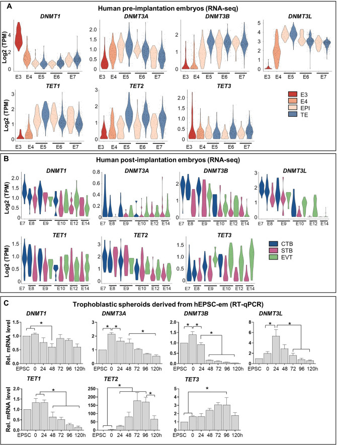
The differential expression patterns of DNMTs and TETs were analysed using published datasets of pre- and post-implantation embryos [31, 32]. It was found that DNMT3A, DNMT3B, DNMT3L, TET1 and TET2 were induced substantially from embryonic day 3 (E3) to day 5 (E5). While DNMT1 was decreased substantially, constant expression of TET3 was observed within the same period (Fig. 1A). Notably, TET2 was the only gene showing higher expression level in TE than in EPI from E5 to E7 (Fig. 1A). We further analysed the dataset of post-implantation embryos from E7 to E14 [32], downregulations of DNMTs and TETs were observed during differentiation of CTB into STB and EVT (Fig. 1B). The mRNA expression levels of these two family members were studied in our trophoblastic spheroid differentiation model, in which early TE-like and trophoblast-like signatures were induced upon induced differentiation for 48 h and 96 h, respectively [23, 25]. As expected, quantitative PCR analysis demonstrated similar expression patterns of these enzymes during trophoblastic differentiation from hEPSC-em (Fig. 1C); the initially induced DNMT3 members and TET1 were gradually down-regulated upon further trophoblast differentiation. Similarly, down-regulation of DNMT1 was observed at the first 48 h of differentiation. Although both TET2 and TET3 expressions were induced from 0 to 96 h, significant induction of TET2 occurred from 24 h post-differentiation whereas significant augmentation of TET3 was only observed at 96 h (Fig. 1C). These results suggested that the downregulation of DNMTs and upregulation of TETs might play critical roles in early trophoblast development.
Fig. 1.
Expression patterns of DNMTs and TETs in early human embryos and hEPSC-em derived trophoblastic spheroids. A, B The gene expression patterns of DNMTs (DNMT1, DNMT3A, DNMT3B and DNMT3L) and TETs (TET1, TET2 and TET3) in TE and EPI of human preimplantation embryos from embryonic day 3 (E3) to E7 (A) and human post-implantation embryos from E7 to E14 (B). C The relative mRNA levels of DNMTs and TETs during trophoblastic spheroid differentiation from hEPSC-em at 0 h to 120 h. Data were presented as mean ± SEM, n = 3. t-test was performed. *p < 0.05
Effects of DNMTs and TETs inhibitors on trophoblast differentiation
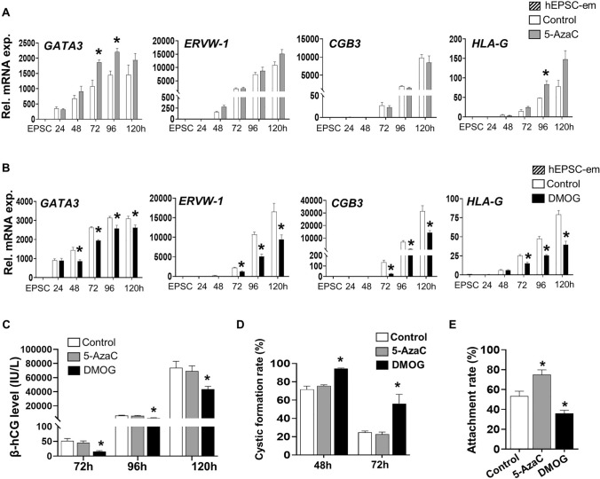
We utilized 5-AzaC (DNMTs inhibitor) [34] and DMOG (TETs inhibitor) [35] that altered global DNA methylome for studying the effects of methylation on trophoblast differentiation. The time- and dose-dependent effects of both chemicals on BAP-induced trophoblast differentiation were first investigated using our hEPSC-em-GATA3mCherry reporter cell line [25]. The treatments only covered the first 48 h of trophoblastic spheroid formation when differentiation of the TE lineage was initiated [23]. The different doses of 5-AzaC (0.1, 0.2, 0.5, and 1.0 μM) and DMOG (10, 20, 40, and 80 μM) used did not affect cell viability of the differentiating cells (Fig. S1A) within a period of 120 h. While treatments with 0.5 μM and 1.0 μM of 5-AzaC at 0–24 h significantly increased the GATA3mCherry signal at 120 h, treatments with 40 μM and 80 μM of DMOG at 24–48 h significantly reduced the GATA3mCherry signal (Fig. S1B). We further analysed the gene expressions of different trophoblast markers in the 0.5 μM 5-AzaC (0–24 h) and 40 μM DMOG (24-48 h) treated cells. While 5-AzaC had no effect on the expressions of pluripotent marker OCT4, it significantly increased the expressions of markers of early trophoblast (KRT7 and GATA3) and EVT (HLA-G) (Fig. 2A, Fig. S1C). In contrast, treatment of DMOG significantly decreased the expressions of GATA3, ERVW-1, CGB3 and HLA-G at 72–120 h after BAP treatment and delayed the reductions of KRT7 at 96 h and 120 h (Figs. 2B, S1D), when the early trophoblast marker KRT7 was reported to decrease at these two time points [25]. Concordantly, DMOG significantly lowered the levels of β-hCG secretions while no significant effect was observed in the 5-AzaC treated group (Fig. 2C). The effects of 5-AzaC and DMOG on trophoblastic spheroid formation and attachment onto receptive EEC (Ishikawa cells) were investigated using our published protocol [22]. Upon 48 h of trophoblast differentiation, around 70% of trophoblastic spheroids at 48 h formed cystic structures in the control and the 5-AzaC treated groups, and most of the cystic structures collapsed at 72 h post-differentiation. However, treatment with DMOG significantly delayed the collapse of the cystic structures, leading to significantly higher cystic formation rates at 48 h and 72 h (Figs. 2D, S1E). The attachment rate of 5-AzaC treated trophoblastic spheroids at 72 h was significantly higher than the control group, while DMOG significantly reduced the attachment rate of trophoblastic spheroids onto the Ishikawa cells (Fig. 2E). These results suggested that loss of DNA methylation is a critical process for proper trophoblast differentiation.
Fig. 2.
Effects of 5-AzaC and DMOG on trophoblast differentiation and trophoblastic spheroid formation. A, B The relative mRNA expression of early trophoblast marker GATA3, STB markers (ERVW-1 and CGB3) and EVT marker (HLA-G) after treatments with 0.5 μM of 5-AzaC (0–24 h) (A) or 40 μM of DMOG (24-48 h) (B) during BAP-induced trophoblast differentiation from hEPSC-em-GATA3mCherry reporter line (n = 3). C The β-hCG levels in the conditioned media collected from 72 to 120 h after treatments with 0.5 μM of 5-AzaC at 0-24 h or 40 μM of DMOG at 24-48 h during BAP-induced trophoblast differentiation from hEPSC-em-GATA3mCherry reporter line (n = 3). D The percentages of cystic structure formed in control, 5-AzaC or DMOG treated trophoblastic spheroids at 48 h and 72 h (n = 4–5). E The 1-h attachment rates of control, 5-AzaC- and DMOG-treated trophoblastic spheroids at 72 h on receptive Ishikawa cells (n = 4). Data were presented as mean ± SEM. *p < 0.05 compared to control group
ENPEP is a TE marker regulated by DNA methylation
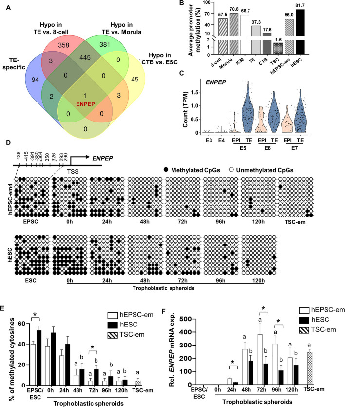
To identify TE determinants that were regulated by DNA methylation, we compared four sets of genes in the published datasets: hypomethylated genes in TE relative to 8-cell embryos and to morulae [16], hypomethylated genes in CTB relative to hESC [26], and the top 100 TE specific genes in preimplantation embryos [31]. It was found that glutamyl aminopeptidase (ENPEP), a trophoblast progenitor marker [36], was commonly found in these 4 gene sets (Fig. 3A). The average DNA methylation levels in the promoter of ENPEP in different cell types in the published datasets [16, 26] were analyzed. The ENPEP promoter displayed high levels of DNA methylation at 8-cell (57.5%), morula (70.8%) and ICM of blastocyst (66.7%) stages. Its level in TE (37.3%) was comparably lower (Fig. 3B). Much lower methylation levels were observed in CTB (17.6%) and TSC-derived CTB (1.6%) when compared to hESC (81.7%) and hEPSC-em (56.0%) (Fig. 3B). Concomitantly, analysis on single-cell RNA sequencing data of preimplantation embryos [31] demonstrated a profound induction of ENPEP at E5 TE as compared to embryos at E3 (8-cell) and E4 (morula), and its levels were manifestly higher in TE when compared to EPI from E5 to E7 (Fig. 3C).
Fig. 3.
Identification of ENPEP as a candidate TE-marker regulated by DNA methylation. A)Venn diagram showing the clustering of hypomethylated genes in TE (verse 8-cell and morula) and CTB (verse hESC) with the top 100 TE-specific genes. B The average DNA methylation levels of ENPEP promoter in pre-implantation embryos (8-cell embryos, morula stage embryos, ICM, TE) [16], CTB [26], TSC derived from CTB [26] and hESC [26] obtained from published datasets and hEPSC-em (this study). C Violin plot showing the expression patterns of ENPEP in published dataset of human preimplantation embryos [31]. D, E Representative bisulfite sequencing results (D) and the average percentages of methylated cytosines (E) of ENPEP promoter in hEPSC-em- and hESC-derived trophoblastic spheroids at different time points, and in TSC-em. Each horizontal line represented the sequencing result of one clone. Filled circles and open circles represented the methylated and unmethylated CpG dinucleotide, respectively. F The relative mRNA expression of ENPEP in hEPSC-em- and hESC-derived trophoblastic spheroids and in TSC-em was determined by RT-qPCR. Data were presented as mean ± SEM, n = 4. t-test was performed for gene expression analysis. Mann–Whitney U test was performed for DNA methylation analysis. ap < 0.05 when compared to hEPSC-em; bp < 0.05 when compared to hESC; *p < 0.05 pairwise comparison between trophoblastic spheroids derived from hEPSC-em and hESC at the same time points
Using the online platform MethPrimer, one CpG island was identified in the ENPEP promoter (Fig. S2). Bisulfite sequencing was used to investigate the differential DNA methylation levels in the ENPEP promoter during trophoblast differentiation. The promoters were differentially methylated between hEPSC-em and hESC, with average percentages of methylated cytosine of 40% ± 2.89% and 53.33% ± 4.08%, respectively (Fig. 3D and E). In hEPSC-em- and hESC-derived trophoblastic spheroids, the DNA methylation levels of the ENPEP promoter decreased significantly at 48 h and maintained at low levels thereafter (Fig. 3D and E). Concordantly, pronounced inductions of ENPEP mRNA expression was observed from 48 h onwards (Fig. 3F). Notably, hEPSC-em derived trophoblastic spheroids exhibited a faster loss of DNA methylation in the ENPEP promoter and higher gene expression when compared with the hESC-derived trophoblastic spheroids, reflecting better trophoblast differentiation potency of hEPSC-em as reported [25]. Moreover, ENPEP was hypomethylated and highly expressed in TSC-em (Fig. 3D–F). Immunofluorescence staining confirmed the protein expressions of ENPEP in hEPSC-em-derived trophoblastic spheroids and TSC-em (Fig. S3).
ENPEP is essential in trophoblast cell fate commitment
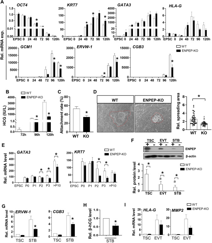
ENPEP is a trophoblast progenitor marker [36], but its role during trophoblast differentiation is unknown. We knockout ENPEP (ENPEP-KO) in the hEPSC-em line using the CRISPR-Cas9 approach (Fig. S4A). The deletions of ENPEP in hEPSC-em and its derived trophoblasts were confirmed by Sanger sequencing (Fig. S4B), quantitative PCR (Fig. S4C) and western blotting analyses (Fig. S4D). The gene expressions during trophoblast differentiation were compared between the ENPEP wildtype (WT) and ENPEP-KO hEPSC-em lines. The results showed that ENPEP deletion delayed the downregulation of OCT4 at 48 h and early trophoblast marker KRT7 at 120 h in trophoblastic spheroids, reduced the expressions of STB markers (GCM1, ERVW-1, and CGB3) but had no effect on the expression of EVT marker HLA-G (Fig. 4A). In agreement, β-hCG secretion was significantly lower in the ENPEP-KO trophoblastic spheroids (Fig. 4B). Moreover, the attachment rates of ENPEP-KO trophoblastic spheroids onto EEC were significantly lower than the WT group (Fig. 4C). After co-cultured with EEC for 24 h, the average spreading area of trophoblastic spheroids was significantly smaller in the ENPEP-KO group (Fig. 4D).
Fig. 4.
Deletion of ENPEP impaired trophoblast differentiation from hEPSC-em. A The relative mRNA expression of pluripotent marker (OCT4), early trophoblast markers (KRT7 and GATA3), EVT marker (HLA-G) and STB markers (GCM1, ERVW-1 and CGB3) as determined by RT-qPCR in trophoblastic spheroids generated from WT and ENPEP-KO hEPSC-em. (n = 3–4). B The β-hCG secretion levels in the conditioned media of trophoblastic spheroids from 72 to 120 h (n = 3). C The attachment rates of WT and ENPEP-KO hEPSC-em derived trophoblastic spheroids at 72 h after 1 h of coculture with receptive Ishikawa cells (n = 5). D Representative pictures of trophoblastic spheroids cocultured with Ishikawa cells for 24 h (left) and the spreading area of trophoblastic spheroids. E The relative mRNA expression of TSC markers (GATA3 and KRT7) during TSC derivation from WT and KO hEPSC-em at passage 0–3 and passage 10–12 (n = 4). F Western blotting analysis of ENPEP protein levels in TSC, EVT and STB differentiated from TSC (n = 4). G, H The relative mRNA expressions of STB markers (ERVW-1 and CGB3) as determined by RT-qPCR (n = 4–5) (G) and the β-hCG secretion levels in the spent culture media of differentiated STB from WT and KO TSC-em as detected by enzyme immunoassay (n = 6) (H). I The relative mRNA expressions of EVT markers (HLA-G and MMP2). Data were presented as mean ± SEM. t-test was performed for gene and protein expression analysis. Mann–Whitney U test was used for the analysis of attachment rates and β-hCG levels. ap < 0.05 when compared to WT hEPSC-em or WT TSC-em; *p < 0.05 pairwise comparison between WT and ENPEP-KO group at the same time points
TSC lines were derived from the WT and the ENPEP-KO hEPSC-em lines. Downregulation of OCT4 and upregulation of TSC markers (GATA3, KRT7 and TFAP2C) were observed during TSC derivation (Figs. 4E, S4E). The expression levels of TSC markers were significantly lower in the ENPEP-KO group than the WT group in the first 3 passages of TSC derivation but only the GATA3 gene expression was significantly lower in the ENPEP-KO-derived TSC at passages > 10 (Fig. 4E). In addition, TSC devoid of ENPEP exhibited lower levels of TSC-specific miR-525-3p (Fig. S4F), though HLA-A, -B and -C were undetectable in both the WT and ENPEP-KO TSC (Fig. S4G).
The two groups of TSC were induced to differentiate into EVT and STB as reported [25]. Upon inductions of differentiations into EVT and STB, the protein levels of ENPEP were significantly downregulated in the ENPEP-WT cells (Fig. 4F), consistent with the patterns observed in human post-implantation embryos [32] (Fig. S4H). The absence of ENPEP in TSC impeded the differentiation as demonstrated by significant reduction of expressions of STB markers (ERVW-1 and CGB3) (Fig. 4G) and β-hCG secretion level (Fig. 4H). Although not statistically significant, decreasing trends of EVT markers (HLA-G and MMP2) were observed in ENPEP-KO group (Fig. 4I). Taken together, deletion of ENPEP impaired early trophoblast development, in particular the STB differentiation.
TET2 mediated loss of DNA methylation and induction of ENPEP is essential for early trophoblast differentiation
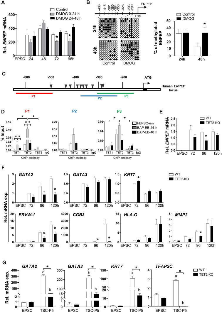
The involvements of TETs in regulating the expression and methylation of ENPEP were investigated by DMOG treatments during trophoblastic spheroid differentiation. Treatment with DMOG at 0-24 h suppressed ENPEP expressions at 72 h and 96 h (Fig. 5A) and hindered loss of DNA methylation in ENPEP promoter as compared to the control group at 48 h of differentiation (Fig. 5B), suggesting a role of TET proteins in regulating DNA methylation of ENPEP promoter. To verify the TET-mediated ENPEP expression, enrichment of TET proteins in ENPEP promoter was determined by ChIP-qPCR. Three pairs of primers were designed to cover the ENPEP promoter regions (P1, P2 and P3) near the CpG sites (Schematic Fig. 5C). The results showed high enrichment of TET2 binding but not that of TET1 and TET3 in all the promoter regions studied (Fig. 5D). Notably, the enrichments of TET2 protein at P1 and P3 were significantly higher than that of TET1 and TET3 in trophoblastic spheroids at 24 h (Fig. 5D). In addition, the binding of TET1 or TET2 at P1 was significantly higher in the trophoblastic spheroids at 24 h than undifferentiated hEPSC-em and trophoblastic spheroids at 48 h, though the enrichment was substantially higher for TET2 (Fig. 5D). Although not statistically significant, increased trends of TET2 binding at P2 and P3 of the ENPEP promoter were also observed in the trophoblastic spheroids at 24 h (Fig. 5D). These results suggested that loss of DNA methylation of ENPEP involved TET proteins, particularly TET2 during trophoblast differentiation.
Fig. 5.
TET2 mediated ENPEP expression and its role during trophoblast differentiation. A The relative mRNA expression of ENPEP after treatments with 40 μM DMOG at different time points during BAP-induced differentiation (n = 3). B Representative bisulfite sequencing results (left) and the average percentages of methylated cytosines (right) of ENPEP promoter in untreated (control) and DMOG treated groups at 24 h and 48 h during BAP-induced trophoblast differentiation. Each horizontal line representing the sequencing result of one clone. Filled circles and open circles represented the methylated and unmethylated CpG dinucleotide, respectively. C Schematic diagram showing the positions of the primer pairs (P1, P2 and P3) of human ENPEP promoter used for ChIP-qPCR analysis. CpG sites were denoted as triangles. D ChIP-qPCR results showing the relative enrichment of ENPEP promoter DNA by antibodies against TET1, TET2 and TET3 proteins. IgG of the same species were included as the controls for ChIP analysis (lower panel) (n = 3). E The relative mRNA expression of ENPEP in TET2-WT and TET2-KO hEPSC-em derived trophoblasts (n = 4). F The relative mRNA expression of early trophoblast markers (GATA2, GATA3 and KRT7), STB markers (ERVW-1 and CGB3) and EVT marker (HLA-G and MMP2) during BAP-induced trophoblast differentiation from WT and TET2-KO hEPSC-em lines (n = 5). G The relative mRNA expression of TSC markers (GATA2, GATA3, KRT7 and TFAP2C) at passage 5 during TSC derivation from WT and TET2-KO hEPSC-em. The data were presented as mean ± SEM. Mann–Whitney U test was performed for DNA methylation analysis. t-test was performed for gene expression analysis. ap < 0.05 when compared to WT hEPSC-em; bp < 0.05 when compared to TET2-KO hEPSC-em; *p < 0.05 compared between WT and TET2-KO
We next delineate the role of TET2 during early trophoblast differentiation by knocking out TET2 in hEPSC-em using the CRISPR-Cas9 approach (Fig. S5A). The absence of TET2 in hEPSC-em and its derived trophoblasts was confirmed by Sanger sequencing (Fig. S5B), quantitative PCR (Fig. S5C) and immunofluorescence staining (Fig. S5D). The gene expressions during BAP-induced trophoblast differentiation were measured in wildtype (WT) and TET knockout (TET2-KO) hEPSC-em lines. The results showed that TET2-KO significantly suppressed the induction of ENPEP at 96 h post-BAP treatment, and induced decreased trends of ENPEP expression at 72 h and 120 h (Fig. 5E). Meanwhile, TET2-KO led to pronounced repressions of STB markers (ERVW-1 and CGB3) but not EVT marker (HLA-G) (Fig. 5F). We also attempted to derive TSC from the WT and TET2-KO hEPSC-em lines. TSC colonies were established from the TET2-KO hEPSC-em at passage 5, with remarkable induction of TSC markers GATA2, GATA3, KRT7 and TFAP2C (Fig. 5G). However, no TSC-like colonies were observed in the TET2-KO group at passage 5 and the expression levels of TSC markers in the remaining cells were significantly lower than those in the WT TSC colonies (Fig. 5G). These results indicated the critical role of TET2 during early trophoblast differentiation.
Discussion
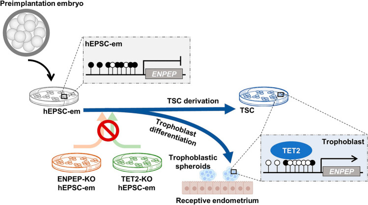
Understanding the intricate processes and deciphering the mechanisms of the first cell differentiation into distinct cell lineages are hampered by limited availability of human embryos. Here, with the use of hEPSC-em with enhanced trophoblast differentiation potential, we demonstrated that TET2-mediated ENPEP hypomethylation and gene expression were critical for early trophoblast differentiation (Fig. 6).
Fig. 6.
Schematic diagram of TET2-mediated loss of DNA methylation in ENPEP promoter and gene activation during early trophoblast differentiation. Hypermethylated ENPEP in morula-like hEPSC-em is demethylated through TET2 recruitment on ENPEP promoter during trophoblastic spheroid differentiation and TSC derivation. Deletion of ENPEP or TET2 in hEPSC-em compromises trophoblast differentiation potency and reduces adhesion and invasion of derived trophoblastic spheroids
The comparable expression patterns of DNMTs and TETs in hEPSC-em derived trophoblastic spheroids and TSC with the published transcriptomic datasets of human preimplantation [31] and post-implantation [32] embryos supported the use of the current model for the study. As speculated in a previous study [16], the reduced DNA methylation maintainer DNMT1 and increased DNA methylation erasers TET1 and TET2 might contribute to the erasure of parental epigenomes, whereas the upregulations of DNMT3s during first lineage specification might be involved in gaining de novo methylation marks in the embryos. The genome-wide DNA methylation profiles of the mid-gestation mouse embryos and placenta revealed that the majority (96.8%) of the differentially methylated regions (DMRs) are hypomethylated in the placenta, resulting in an overall low level of methylation in extraembryonic tissues [37]. Intriguingly, a higher level of TET2 was observed in TE than the EPI of E5 to E7 human blastocyst. Previous study demonstrated that DMRs are hypermethylated in mouse ESCs devoid of Tet2 when compared to both WT and Tet1-KO ESCs, which are accompanied by a larger number of downregulated genes during differentiation induced by leukemia inhibitory factor (LIF) withdrawal [38]. The significance of TET2 in trophoblast differentiation was further supported by the substantial upregulation of TET2 expressions in early TE-like trophoblastic spheroids at 48 h and pTE-like trophoblastic spheroids at 72 h.
We then utilized two inhibitors targeting the DNMTs and TETs to first confirm the roles of DNA methylation. The metabolites of 5-AzaC can be incorporated into DNA, forming covalent adducts with cellular DNMTs. This led to the degradation of the covalently trapped enzymes and subsequent rapid passive loss of methylation during DNA replication [34, 39]. On the other hand, DMOG inhibits TET enzyme activities by acting as a small-molecule inhibitor of 2OG-dependent oxygenases [35]. In this study, stimulatory effects of 5-AzaC but inhibitory effects of DMOG were observed during initiation of trophoblast differentiation. Indeed, Dnmt1 knockout induces efficient trophoblast differentiation in post-implantation epiblast-like mouse ESCs [40] originally lacking trophoblast differentiation potential [41]. On the contrary, triple knockout of Tet1-3 in mouse embryos induced hypermethylation and diminished expressions of Lefty1 and Lefty2, thereby hyperactivating Nodal signaling [42], a critical pathway required to be inactivated during trophoblast differentiation [22]. The differential time-dependent effects of 5-AzaC and DMOG on the expression of trophoblast markers might be attributed to the dissimilar expression patterns of DNMTs and TETs during early trophoblast differentiation. In particular, the positive effects of 5-AzaC on trophoblast differentiation were exclusively observed when 5-AzaC treatment covered the first 24 h of differentiation during which DNMTs were expressed at higher levels. In contrast, obvious effects of DMOG were only observed when the treatment covered 24–48 h of differentiation, which aligned with the gradual induction of TET2 from 24 to 72 h. The observation might further imply that DMOG induced TET2 inhibition played a critical role in early trophoblast differentiation.
We studied the endometrial attachment competencies of 5-AzaC- or DMOG-treated trophoblastic spheroids. Trophoblastic spheroids at 72 h with collapsed cystic structures secret HCG and are developmentally competent for attaching onto receptive endometrial epithelial cells [22–24]. HCG produced by human embryos can increase the expression of LIF, a marker of endometrial receptivity, thereby enhancing the receptivity of EECs [43]. In this connection, the reduced HCG productions, delayed collapse of cystic structures and reduced attachment rates further reinforced that DMOG treatment reduced the differentiation potential of trophoblastic spheroids into pTE-like stage. Although 5-AzaC had no effect on CGB3 expression and HCG secretion, it significantly enhanced the attachment rate of trophoblastic spheroids at 72 h. It was reported that 5′-aza-2’-deoxycytidine treatment induced the expressions of E-cadherin and γ-catenin, two adhesion molecules critical for embryo attachment [44, 45], in a trophoblast cell line (BeWo) [46]. Thus, 5-AzaC might also be involved in increasing the expressions of adhesion molecules on the trophoblastic spheroids, leading to higher EEC attachment potential. Taken together, the opposite effects of 5-AzaC and DMOG highlighted the crucial roles of DNA methylation during trophoblast differentiation.
Recent innovative techniques such as low-input chromatin analysis have emerged to broaden our accessibility of human early-stage embryos and discover the mechanisms of DNA methylation modifications on a whole-genome-wide level [16, 47, 48]. With the substantial inductions of TETs and higher impacts of DMOG during early trophoblast differentiation, we identified epigenetically regulated genes in human TE. By matching the published DNA methylomes of human embryos [16, 26] with TE-specific genes [31], ENPEP was identified as a hypomethylated gene in early TE lineages when compared to 8-cell/morula stage human embryos and epiblast-like hESC. Further validations confirmed the gradual loss of DNA methylation and induction of ENPEP expression during the first 72 h of trophoblastic spheroid differentiation. An observed decline of ENPEP gene expression at later time points when DNA methylation remained low could be due to other regulatory mechanisms on ENPEP during trophoblast subtypes establishment. For examples, the 3′UTR region of ENPEP contained several miRNA binding sites [49], which may contribute to the reductions of ENPEP at later time points when its DNA methylation level remained low. Similar to the reported findings that more rapid loss of DNA methylation in the ELF5 promoter and its higher expression in the hEPSC-em derived trophoblastic spheroids as compared to that in the primed hESC [25], the faster demethylation and higher expression of ENPEP in hEPSC-em derived trophoblastic spheroids might contribute to the expanded potential of hEPSC-em into trophoblast lineages.
ENPEP is a cell surface marker of TE progenitor cells [21, 36, 50], but its roles in trophoblast development is unknown. In human placenta, ENPEP was predominantly expressed in the apical membrane of CTB and on the surface of apical microvilli of STB to act as a component of the fetal-maternal barrier to oligopeptides [51]. Here, we demonstrated that the deletion of ENPEP impaired trophoblast differentiation. Placental development initiates in a hypoxic environment, where the induced hypoxia-inducible factors (HIFs) play an essential role in determining trophoblast fate [52, 53]. Hypoxia-induced HIF-1α expression was hindered by ENPEP deprivation in murine aortic endothelial cells [54]. We also found an upregulation of HIF-1α expression during trophoblast differentiation from hEPSC [55]. Thus, ENPEP might control trophoblast differentiation by regulating the HIF-1α pathway. The delayed reduction of OCT4 in the ENPEP-KO trophoblastic spheroids may be associated with a blunted HIF-1α signaling, as HIF-1α could suppress Oct4 expression via direct binding to the reverse hypoxia-responsive elements in the Oct4 promoter [56].
ENPEP is known as a glutamyl aminopeptidase which cleaves the N-terminal aspartic acid of angiotensin (ANG) II [57]. Human placenta can synthesize ANG II [58, 59]. Notably, ANG II decreases expression and secretion of E-cadherin in EEC [60] and downregulates the production of invasion-related proteolytic enzyme, plasmin in trophoblasts [61, 62]. The absence of ENPEP might result in accumulation of ANG II at the interface of trophoblastic spheroids and EEC, thereby reducing the E-cadherin mediated attachment process and trophoblast invasion as observed in the ENPEP-null trophoblastic spheroids. Furthermore, TSCs derived from ENPEP-KO hEPSC-em failed to differentiate into STB properly, but they could still form EVT-like cells, suggesting ENPEP might be dispensable for early EVT differentiation.
We next attempted to delineate the roles of TET proteins in epigenetic regulation of ENPEP during early trophoblast differentiation. Our ChIP-qPCR results demonstrated a prominent recruitment of TET2 protein, rather than TET1 and TET3, at the ENPEP promoter during initiation of trophoblast differentiation in trophoblastic spheroids at 24 h. The reduced TET2 bindings at the ENPEP promoter in trophoblastic spheroids at 48 h might allow exposure of newly unmethylated DNAs and facilitate their subsequent transcriptions [63, 64]. These results also explained the findings that DMOG treatment had the highest effect at 0–24 h but not 24–48 h in delaying the reduction of DNA methylation level in the ENPEP promoter and suppression of its expression. Since the transition from the morula-like stage to TE-like stage occurred from trophoblastic spheroids at 24 h to 48 h [23], the TET2-mediated loss of DNA methylation in ENPEP promoter at 24–48 h may reinforce the trophoblast fate during this process. The involvement of TET2 in regulating ENPEP was further supported by the findings that TET2 deletion significantly decreased ENPEP expression during trophoblast differentiation.
TETs are methylcytosine dioxygenases involved in successive oxidation and conversion of 5mC to 5hmC, 5fC, and 5caC [65, 66]. It was reported that TET2 preferentially bound to 5mC over 5hmC [67], which poses a challenge for bisulfite sequencing since it cannot differentiate between the two [68]. As the oxidized methylcytosines are not recognized by DNMT1 [69], the loss of methylation in ENPEP promoter might be due to TET2 initiated replication-dependent passive dilution [70] or thymine DNA glycosylase (TDG)-dependent active demethylation [65, 71]. Further examination is required to confirm if there exist other molecules affecting the methylation status of ENPEP promoter during trophoblast formation. The function of TET2 has been widely studied in hematopoiesis and immune regulation [72, 73]. Whether TET2 is critical in regulating early human trophoblast differentiation and the underlying mechanisms remain largely unknown. It was reported that Tet2 deletion impaired the stem cell state of mouse TSC, with reduced TSC marker expressions and loss of epithelial characteristics [74]. In human, downregulation of TET2 expression was detected in placenta from the preeclampsia patients and was related to the reduced trophoblast migration and invasion abilities [75]. In this study, we also found that the TET2 null hEPSC-em exhibited attenuation on trophoblast differentiation and TSC formation ability, indicating the critical roles of TET2 in governing early trophoblast differentiation. The alternative targets of TET2 or other epigenetically regulated molecules during early trophoblast differentiation warrant further investigation.
In summary, this study highlighted the crucial roles of loss of DNA methylation in prompting early trophoblast differentiation. More importantly, TET2-mediated loss of DNA methylation in ENPEP promoter played a vital role in trophoblast fate commitment.
Supplementary Information
Below is the link to the electronic supplementary material.
Acknowledgements
The authors acknowledge the assistance from Centre for PanorOmic Sciences (CPOS) at Li Ka Shing Faculty of Medicine, the University of Hong Kong.
Author contributions
Wen Huang: Conceptualization; Resources; Methodology; Investigation; Formal analysis; Validation; Visualization; Writing—original draft. Andy Chun Hang Chen: Conceptualization; Resources; Formal analysis; Investigation; Visualization; Supervision; Validation; Methodology; Writing—review and editing. Xujin Wei: Investigation; Formal analysis; Validation; Visualization. Sze Wan Fong: Resources; Methodology. William Shu Biu Yeung: Data curation; Project administration; Writing—review and editing. Yin Lau Lee: Conceptualization; Data curation; Supervision; Funding acquisition; Methodology; Project administration; Writing—review and editing.
Funding
Innovation Technology Commission (Health@InnoHK); National Key Research and Development Program of China, 2022YFC2702503; Shenzhen Science and Technology Innovation Program, KQTD20190929172749226; Sanming Project of Medicine in Shenzen Municipality, SZSM202211014; The University of Hong Kong-Shenzhen Hospital Fund for Shenzhen Key Medical Discipline, SZXK2020089.
Data availability
This study includes no data deposited in external repositories. The data that support the findings of this study are available from the corresponding author upon reasonable request.
Declarations
Conflict of interest
The authors have no relevant financial or non-financial interests to disclose.
Ethical approval
Not applicable.
Consent to participate
Not applicable.
Consent to publish
Not applicable.
Footnotes
Publisher's Note
Springer Nature remains neutral with regard to jurisdictional claims in published maps and institutional affiliations.
Wen Huang and Andy Chun Hang Chen contributed equally.
Contributor Information
William Shu Biu Yeung, Email: wsbyeung@hku.hk.
Yin Lau Lee, Email: cherielee@hku.hk.
References
- 1.Rossant J, Tam PPL. Early human embryonic development: blastocyst formation to gastrulation. Dev Cell. 2022;57(2):152–165. doi: 10.1016/j.devcel.2021.12.022. [DOI] [PubMed] [Google Scholar]
- 2.Burton GJ, Fowden AL. The placenta: a multifaceted, transient organ. Philos Trans R Soc Lond B Biol Sci. 2015;370(1663):20140066. doi: 10.1098/rstb.2014.0066. [DOI] [PMC free article] [PubMed] [Google Scholar]
- 3.Yao C, Zhang W, Shuai L. The first cell fate decision in pre-implantation mouse embryos. Cell Regen. 2019;8(2):51–57. doi: 10.1016/j.cr.2019.10.001. [DOI] [PMC free article] [PubMed] [Google Scholar]
- 4.Lanner F. Lineage specification in the early mouse embryo. Exp Cell Res. 2014;321(1):32–39. doi: 10.1016/j.yexcr.2013.12.004. [DOI] [PubMed] [Google Scholar]
- 5.Gerri C, McCarthy A, Mei Scott G, Regin M, Stamatiadis P, Brumm S, et al. A conserved role of the Hippo signalling pathway in initiation of the first lineage specification event across mammals. Development. 2023 doi: 10.1242/dev.201112. [DOI] [PMC free article] [PubMed] [Google Scholar]
- 6.Gerri C, McCarthy A, Alanis-Lobato G, Demtschenko A, Bruneau A, Loubersac S, et al. Initiation of a conserved trophectoderm program in human, cow and mouse embryos. Nature. 2020;587(7834):443–447. doi: 10.1038/s41586-020-2759-x. [DOI] [PMC free article] [PubMed] [Google Scholar]
- 7.Ortega NM, Winblad N, Plaza Reyes A, Lanner F. Functional genetics of early human development. Curr Opin Genet Dev. 2018;52:1–6. doi: 10.1016/j.gde.2018.04.005. [DOI] [PubMed] [Google Scholar]
- 8.Soncin F, Natale D, Parast MM. Signaling pathways in mouse and human trophoblast differentiation: a comparative review. Cell Mol Life Sci. 2015;72(7):1291–1302. doi: 10.1007/s00018-014-1794-x. [DOI] [PMC free article] [PubMed] [Google Scholar]
- 9.Hanna CW, Demond H, Kelsey G. Epigenetic regulation in development: is the mouse a good model for the human? Hum Reprod Update. 2018;24(5):556–576. doi: 10.1093/humupd/dmy021. [DOI] [PMC free article] [PubMed] [Google Scholar]
- 10.Chen C, Gao Y, Liu W, Gao S. Epigenetic regulation of cell fate transition: learning from early embryo development and somatic cell reprogramming†. Biol Reprod. 2022;107(1):183–195. doi: 10.1093/biolre/ioac087. [DOI] [PMC free article] [PubMed] [Google Scholar]
- 11.Xu Q, Xie W. Epigenome in early mammalian development: inheritance. Reprogramm Establ Trends Cell Biol. 2018;28(3):237–253. doi: 10.1016/j.tcb.2017.10.008. [DOI] [PubMed] [Google Scholar]
- 12.Reik W. Stability and flexibility of epigenetic gene regulation in mammalian development. Nature. 2007;447(7143):425–432. doi: 10.1038/nature05918. [DOI] [PubMed] [Google Scholar]
- 13.Kohan-Ghadr HR, Kadam L, Jain C, Armant DR, Drewlo S. Potential role of epigenetic mechanisms in regulation of trophoblast differentiation, migration, and invasion in the human placenta. Cell Adh Migr. 2016;10(1–2):126–135. doi: 10.1080/19336918.2015.1098800. [DOI] [PMC free article] [PubMed] [Google Scholar]
- 14.Moore LD, Le T, Fan G. DNA methylation and its basic function. Neuropsychopharmacology. 2013;38(1):23–38. doi: 10.1038/npp.2012.112. [DOI] [PMC free article] [PubMed] [Google Scholar]
- 15.Kohli RM, Zhang Y. TET enzymes, TDG and the dynamics of DNA demethylation. Nature. 2013;502(7472):472–479. doi: 10.1038/nature12750. [DOI] [PMC free article] [PubMed] [Google Scholar]
- 16.Guo H, Zhu P, Yan L, Li R, Hu B, Lian Y, et al. The DNA methylation landscape of human early embryos. Nature. 2014;511(7511):606–610. doi: 10.1038/nature13544. [DOI] [PubMed] [Google Scholar]
- 17.Zhou F, Wang R, Yuan P, Ren Y, Mao Y, Li R, et al. Reconstituting the transcriptome and DNA methylome landscapes of human implantation. Nature. 2019;572(7771):660–664. doi: 10.1038/s41586-019-1500-0. [DOI] [PubMed] [Google Scholar]
- 18.Ng RK, Dean W, Dawson C, Lucifero D, Madeja Z, Reik W, et al. Epigenetic restriction of embryonic cell lineage fate by methylation of Elf5. Nat Cell Biol. 2008;10(11):1280–1290. doi: 10.1038/ncb1786. [DOI] [PMC free article] [PubMed] [Google Scholar]
- 19.Cambuli F, Murray A, Dean W, Dudzinska D, Krueger F, Andrews S, et al. Epigenetic memory of the first cell fate decision prevents complete ES cell reprogramming into trophoblast. Nat Commun. 2014;5:5538. doi: 10.1038/ncomms6538. [DOI] [PMC free article] [PubMed] [Google Scholar]
- 20.Lee CQ, Gardner L, Turco M, Zhao N, Murray MJ, Coleman N, et al. What is trophoblast? A combination of criteria define human first-trimester trophoblast. Stem Cell Rep. 2016;6(2):257–272. doi: 10.1016/j.stemcr.2016.01.006. [DOI] [PMC free article] [PubMed] [Google Scholar]
- 21.Krendl C, Shaposhnikov D, Rishko V, Ori C, Ziegenhain C, Sass S, et al. GATA2/3-TFAP2A/C transcription factor network couples human pluripotent stem cell differentiation to trophectoderm with repression of pluripotency. Proc Natl Acad Sci U S A. 2017;114(45):E9579–e9588. doi: 10.1073/pnas.1708341114. [DOI] [PMC free article] [PubMed] [Google Scholar]
- 22.Lee YL, Fong SW, Chen AC, Li T, Yue C, Lee CL, et al. Establishment of a novel human embryonic stem cell-derived trophoblastic spheroid implantation model. Hum Reprod. 2015;30(11):2614–2626. doi: 10.1093/humrep/dev223. [DOI] [PubMed] [Google Scholar]
- 23.Yue C, Chen ACH, Tian S, Fong SW, Lee KC, Zhang J, et al. Human embryonic stem cell-derived blastocyst-like spheroids resemble human trophectoderm during early implantation process. Fertil Steril. 2020;114(3):653–664. doi: 10.1016/j.fertnstert.2020.01.009. [DOI] [PubMed] [Google Scholar]
- 24.Lee YL, Ruan H, Lee KC, Fong SW, Yue C, Chen ACH, et al. Attachment of a trophoblastic spheroid onto endometrial epithelial cells predicts cumulative live birth in women aged 35 and older. Fertil Steril. 2023;120(2):268–276. doi: 10.1016/j.fertnstert.2023.03.013. [DOI] [PubMed] [Google Scholar]
- 25.Chen ACH, Lee YL, Ruan H, Huang W, Fong SW, Tian S, et al. Expanded potential stem cells from human embryos have an open chromatin configuration with enhanced trophoblast differentiation ability. Adv Sci (Weinh) 2023;10(11):e2204797. doi: 10.1002/advs.202204797. [DOI] [PMC free article] [PubMed] [Google Scholar]
- 26.Okae H, Toh H, Sato T, Hiura H, Takahashi S, Shirane K, et al. Derivation of human trophoblast stem cells. Cell Stem Cell. 2018;22(1):50–63.e6. doi: 10.1016/j.stem.2017.11.004. [DOI] [PubMed] [Google Scholar]
- 27.Amita M, Adachi K, Alexenko AP, Sinha S, Schust DJ, Schulz LC, et al. Complete and unidirectional conversion of human embryonic stem cells to trophoblast by BMP4. Proc Natl Acad Sci U S A. 2013;110(13):E1212–E1221. doi: 10.1073/pnas.1303094110. [DOI] [PMC free article] [PubMed] [Google Scholar]
- 28.Nishida M. The Ishikawa cells from birth to the present. Hum Cell. 2002;15(3):104–117. doi: 10.1111/j.1749-0774.2002.tb00105.x. [DOI] [PubMed] [Google Scholar]
- 29.Liu Y, Kodithuwakku SP, Ng PY, Chai J, Ng EH, Yeung WS, et al. Excessive ovarian stimulation up-regulates the Wnt-signaling molecule DKK1 in human endometrium and may affect implantation: an in vitro co-culture study. Hum Reprod. 2010;25(2):479–490. doi: 10.1093/humrep/dep429. [DOI] [PubMed] [Google Scholar]
- 30.Li LC, Dahiya R. MethPrimer: designing primers for methylation PCRs. Bioinformatics. 2002;18(11):1427–1431. doi: 10.1093/bioinformatics/18.11.1427. [DOI] [PubMed] [Google Scholar]
- 31.Petropoulos S, Edsgärd D, Reinius B, Deng Q, Panula SP, Codeluppi S, et al. Single-cell RNA-Seq reveals lineage and X chromosome dynamics in human preimplantation embryos. Cell. 2016;165(4):1012–1026. doi: 10.1016/j.cell.2016.03.023. [DOI] [PMC free article] [PubMed] [Google Scholar]
- 32.Xiang L, Yin Y, Zheng Y, Ma Y, Li Y, Zhao Z, et al. A developmental landscape of 3D-cultured human pre-gastrulation embryos. Nature. 2020;577(7791):537–542. doi: 10.1038/s41586-019-1875-y. [DOI] [PubMed] [Google Scholar]
- 33.Akalin A, Kormaksson M, Li S, Garrett-Bakelman FE, Figueroa ME, Melnick A, et al. methylKit: a comprehensive R package for the analysis of genome-wide DNA methylation profiles. Genome Biol. 2012;13(10):R87. doi: 10.1186/gb-2012-13-10-r87. [DOI] [PMC free article] [PubMed] [Google Scholar]
- 34.Schermelleh L, Spada F, Easwaran HP, Zolghadr K, Margot JB, Cardoso MC, et al. Trapped in action: direct visualization of DNA methyltransferase activity in living cells. Nat Methods. 2005;2(10):751–756. doi: 10.1038/nmeth794. [DOI] [PubMed] [Google Scholar]
- 35.Uh K, Ryu J, Farrell K, Wax N, Lee K. TET family regulates the embryonic pluripotency of porcine preimplantation embryos by maintaining the DNA methylation level of NANOG. Epigenetics. 2020;15(11):1228–1242. doi: 10.1080/15592294.2020.1762392. [DOI] [PMC free article] [PubMed] [Google Scholar]
- 36.Io S, Kabata M, Iemura Y, Semi K, Morone N, Minagawa A, et al. Capturing human trophoblast development with naive pluripotent stem cells in vitro. Cell Stem Cell. 2021;28(6):1023–1039.e13. doi: 10.1016/j.stem.2021.03.013. [DOI] [PubMed] [Google Scholar]
- 37.Legault LM, Doiron K, Lemieux A, Caron M, Chan D, Lopes FL, et al. Developmental genome-wide DNA methylation asymmetry between mouse placenta and embryo. Epigenetics. 2020;15(8):800–815. doi: 10.1080/15592294.2020.1722922. [DOI] [PMC free article] [PubMed] [Google Scholar]
- 38.Reimer M, Jr, Pulakanti K, Shi L, Abel A, Liang M, Malarkannan S, et al. Deletion of Tet proteins results in quantitative disparities during ESC differentiation partially attributable to alterations in gene expression. BMC Dev Biol. 2019;19(1):16. doi: 10.1186/s12861-019-0196-6. [DOI] [PMC free article] [PubMed] [Google Scholar]
- 39.Mund C, Brueckner B, Lyko F. Reactivation of epigenetically silenced genes by DNA methyltransferase inhibitors: basic concepts and clinical applications. Epigenetics. 2006;1(1):7–13. doi: 10.4161/epi.1.1.2375. [DOI] [PubMed] [Google Scholar]
- 40.Sakaue M, Ohta H, Kumaki Y, Oda M, Sakaide Y, Matsuoka C, et al. DNA methylation is dispensable for the growth and survival of the extraembryonic lineages. Curr Biol. 2010;20(16):1452–1457. doi: 10.1016/j.cub.2010.06.050. [DOI] [PubMed] [Google Scholar]
- 41.Brons IG, Smithers LE, Trotter MW, Rugg-Gunn P, Sun B, de Sousa C, Lopes SM, et al. Derivation of pluripotent epiblast stem cells from mammalian embryos. Nature. 2007;448(7150):191–195. doi: 10.1038/nature05950. [DOI] [PubMed] [Google Scholar]
- 42.Dai HQ, Wang BA, Yang L, Chen JJ, Zhu GC, Sun ML, et al. TET-mediated DNA demethylation controls gastrulation by regulating Lefty-Nodal signalling. Nature. 2016;538(7626):528–532. doi: 10.1038/nature20095. [DOI] [PubMed] [Google Scholar]
- 43.Sherwin JR, Sharkey AM, Cameo P, Mavrogianis PM, Catalano RD, Edassery S, et al. Identification of novel genes regulated by chorionic gonadotropin in baboon endometrium during the window of implantation. Endocrinology. 2007;148(2):618–626. doi: 10.1210/en.2006-0832. [DOI] [PubMed] [Google Scholar]
- 44.Liu G, Zhang X, Lin H, Wang H, Li Q, Ni J, et al. Effects of E-cadherin on mouse embryo implantation and expression of matrix metalloproteinase-2 and -9. Biochem Biophys Res Commun. 2006;343(3):832–838. doi: 10.1016/j.bbrc.2006.02.189. [DOI] [PubMed] [Google Scholar]
- 45.Jha RK, Titus S, Saxena D, Kumar PG, Laloraya M. Profiling of E-cadherin, beta-catenin and Ca(2+) in embryo-uterine interactions at implantation. FEBS Lett. 2006;580(24):5653–5660. doi: 10.1016/j.febslet.2006.09.014. [DOI] [PubMed] [Google Scholar]
- 46.Rahnama F, Shafiei F, Gluckman PD, Mitchell MD, Lobie PE. Epigenetic regulation of human trophoblastic cell migration and invasion. Endocrinology. 2006;147(11):5275–5283. doi: 10.1210/en.2006-0288. [DOI] [PubMed] [Google Scholar]
- 47.Yang M, Tao X, Scott K, Zhan Y, Scott RT, Seli E. Evaluation of genome-wide DNA methylation profile of human embryos with different developmental competences. Hum Reprod. 2021;36(6):1682–1690. doi: 10.1093/humrep/deab074. [DOI] [PubMed] [Google Scholar]
- 48.Okae H, Chiba H, Hiura H, Hamada H, Sato A, Utsunomiya T, et al. Genome-wide analysis of DNA methylation dynamics during early human development. PLoS Genet. 2014;10(12):e1004868. doi: 10.1371/journal.pgen.1004868. [DOI] [PMC free article] [PubMed] [Google Scholar]
- 49.Holmes RS, Spradling-Reeves KD, Cox LA. Mammalian glutamyl aminopeptidase genes (ENPEP) and proteins: comparative studies of a major contributor to arterial hypertension. J Data Mining Genom Proteom. 2017 doi: 10.4172/2153-0602.1000211. [DOI] [PMC free article] [PubMed] [Google Scholar]
- 50.Drukker M, Tang C, Ardehali R, Rinkevich Y, Seita J, Lee AS, et al. Isolation of primitive endoderm, mesoderm, vascular endothelial and trophoblast progenitors from human pluripotent stem cells. Nat Biotechnol. 2012;30(6):531–542. doi: 10.1038/nbt.2239. [DOI] [PMC free article] [PubMed] [Google Scholar]
- 51.Nomura M, Tsukahara S, Ando H, Katsumata Y, Okada M, Itakura A, et al. Differential distribution of placental leucine aminopeptidase/oxytocinase and aminopeptidase A in human trophoblasts of normal placenta and complete hydatidiform mole. Placenta. 2002;23(8–9):631–639. doi: 10.1053/plac.2002.0861. [DOI] [PubMed] [Google Scholar]
- 52.Cowden Dahl KD, Fryer BH, Mack FA, Compernolle V, Maltepe E, Adelman DM, et al. Hypoxia-inducible factors 1alpha and 2alpha regulate trophoblast differentiation. Mol Cell Biol. 2005;25(23):10479–10491. doi: 10.1128/mcb.25.23.10479-10491.2005. [DOI] [PMC free article] [PubMed] [Google Scholar]
- 53.Zhao H, Wong RJ, Stevenson DK. The impact of hypoxia in early pregnancy on placental cells. Int J Mol Sci. 2021 doi: 10.3390/ijms22189675. [DOI] [PMC free article] [PubMed] [Google Scholar]
- 54.Kubota R, Numaguchi Y, Ishii M, Niwa M, Okumura K, Naruse K, et al. Ischemia-induced angiogenesis is impaired in aminopeptidase A deficient mice via down-regulation of HIF-1α. Biochem Biophys Res Commun. 2010;402(2):396–401. doi: 10.1016/j.bbrc.2010.10.043. [DOI] [PubMed] [Google Scholar]
- 55.Gao X, Nowak-Imialek M, Chen X, Chen D, Herrmann D, Ruan D, et al. Establishment of porcine and human expanded potential stem cells. Nat Cell Biol. 2019;21(6):687–699. doi: 10.1038/s41556-019-0333-2. [DOI] [PMC free article] [PubMed] [Google Scholar]
- 56.Lee SW, Jeong HK, Lee JY, Yang J, Lee EJ, Kim SY, et al. Hypoxic priming of mESCs accelerates vascular-lineage differentiation through HIF1-mediated inverse regulation of Oct4 and VEGF. EMBO Mol Med. 2012;4(9):924–938. doi: 10.1002/emmm.201101107. [DOI] [PMC free article] [PubMed] [Google Scholar]
- 57.Benigni A, Cassis P, Remuzzi G. Angiotensin II revisited: new roles in inflammation, immunology and aging. EMBO Mol Med. 2010;2(7):247–257. doi: 10.1002/emmm.201000080. [DOI] [PMC free article] [PubMed] [Google Scholar]
- 58.Poisner AM. The human placental renin-angiotensin system. Front Neuroendocrinol. 1998;19(3):232–252. doi: 10.1006/frne.1998.0166. [DOI] [PubMed] [Google Scholar]
- 59.Sakura H, Kobayashi H, Mizutani S, Sakura N, Hashimoto T, Kawashima Y. Kinetic properties of placental aminopeptidase A: N-terminal degradation of angiotensin II. Biochem Int. 1983;6(5):609–615. [PubMed] [Google Scholar]
- 60.Shan T, Zhang L, Zhao C, Chen W, Zhang Y, Li G. Angiotensin-(1–7) and angiotensin II induce the transdifferentiation of human endometrial epithelial cells in vitro. Mol Med Rep. 2014;9(6):2180–2186. doi: 10.3892/mmr.2014.2128. [DOI] [PMC free article] [PubMed] [Google Scholar]
- 61.Zheng Q, Yang Y, Cui X, Zhang D, Liu S, Yan Q. AP1 mediates uPA/uPAR induced FUT4 expression and trophoblast invasion. J Cell Biochem. 2018;119(8):6442–6451. doi: 10.1002/jcb.26648. [DOI] [PubMed] [Google Scholar]
- 62.Xia Y, Wen HY, Kellems RE. Angiotensin II inhibits human trophoblast invasion through AT1 receptor activation. J Biol Chem. 2002;277(27):24601–24608. doi: 10.1074/jbc.M201369200. [DOI] [PubMed] [Google Scholar]
- 63.Sapozhnikov DM, Szyf M. Unraveling the functional role of DNA demethylation at specific promoters by targeted steric blockage of DNA methyltransferase with CRISPR/dCas9. Nat Commun. 2021;12(1):5711. doi: 10.1038/s41467-021-25991-9. [DOI] [PMC free article] [PubMed] [Google Scholar]
- 64.Yin Y, Morgunova E, Jolma A, Kaasinen E, Sahu B, Khund-Sayeed S, et al. Impact of cytosine methylation on DNA binding specificities of human transcription factors. Science. 2017 doi: 10.1126/science.aaj2239. [DOI] [PMC free article] [PubMed] [Google Scholar]
- 65.He YF, Li BZ, Li Z, Liu P, Wang Y, Tang Q, et al. Tet-mediated formation of 5-carboxylcytosine and its excision by TDG in mammalian DNA. Science. 2011;333(6047):1303–1307. doi: 10.1126/science.1210944. [DOI] [PMC free article] [PubMed] [Google Scholar]
- 66.Ito S, Shen L, Dai Q, Wu SC, Collins LB, Swenberg JA, et al. Tet proteins can convert 5-methylcytosine to 5-formylcytosine and 5-carboxylcytosine. Science. 2011;333(6047):1300–1303. doi: 10.1126/science.1210597. [DOI] [PMC free article] [PubMed] [Google Scholar]
- 67.Hu L, Lu J, Cheng J, Rao Q, Li Z, Hou H, et al. Structural insight into substrate preference for TET-mediated oxidation. Nature. 2015;527(7576):118–122. doi: 10.1038/nature15713. [DOI] [PubMed] [Google Scholar]
- 68.Huang Y, Pastor WA, Shen Y, Tahiliani M, Liu DR, Rao A. The behaviour of 5-hydroxymethylcytosine in bisulfite sequencing. PLoS ONE. 2010;5(1):e8888. doi: 10.1371/journal.pone.0008888. [DOI] [PMC free article] [PubMed] [Google Scholar]
- 69.Hashimoto H, Liu Y, Upadhyay AK, Chang Y, Howerton SB, Vertino PM, et al. Recognition and potential mechanisms for replication and erasure of cytosine hydroxymethylation. Nucleic Acids Res. 2012;40(11):4841–4849. doi: 10.1093/nar/gks155. [DOI] [PMC free article] [PubMed] [Google Scholar]
- 70.Otani J, Kimura H, Sharif J, Endo TA, Mishima Y, Kawakami T, et al. Cell cycle-dependent turnover of 5-hydroxymethyl cytosine in mouse embryonic stem cells. PLoS ONE. 2013;8(12):e82961. doi: 10.1371/journal.pone.0082961. [DOI] [PMC free article] [PubMed] [Google Scholar]
- 71.Maiti A, Drohat AC. Thymine DNA glycosylase can rapidly excise 5-formylcytosine and 5-carboxylcytosine: potential implications for active demethylation of CpG sites. J Biol Chem. 2011;286(41):35334–35338. doi: 10.1074/jbc.C111.284620. [DOI] [PMC free article] [PubMed] [Google Scholar]
- 72.Feng Y, Li X, Cassady K, Zou Z, Zhang X. TET2 function in hematopoietic malignancies, immune regulation, and DNA repair. Front Oncol. 2019;9:210. doi: 10.3389/fonc.2019.00210. [DOI] [PMC free article] [PubMed] [Google Scholar]
- 73.Cong B, Zhang Q, Cao X. The function and regulation of TET2 in innate immunity and inflammation. Protein Cell. 2021;12(3):165–173. doi: 10.1007/s13238-020-00796-6. [DOI] [PMC free article] [PubMed] [Google Scholar]
- 74.Chrysanthou S, Senner CE, Woods L, Fineberg E, Okkenhaug H, Burge S, et al. A critical role of TET1/2 proteins in cell-cycle progression of trophoblast stem cells. Stem Cell Rep. 2018;10(4):1355–1368. doi: 10.1016/j.stemcr.2018.02.014. [DOI] [PMC free article] [PubMed] [Google Scholar]
- 75.Li X, Wu C, Shen Y, Wang K, Tang L, Zhou M, et al. Ten-eleven translocation 2 demethylates the MMP9 promoter, and its down-regulation in preeclampsia impairs trophoblast migration and invasion. J Biol Chem. 2018;293(26):10059–10070. doi: 10.1074/jbc.RA117.001265. [DOI] [PMC free article] [PubMed] [Google Scholar]
Associated Data
This section collects any data citations, data availability statements, or supplementary materials included in this article.
Supplementary Materials
Data Availability Statement
This study includes no data deposited in external repositories. The data that support the findings of this study are available from the corresponding author upon reasonable request.


