Abstract
A 7-year-old Korean shorthair cat was admitted to our hospital with chronic constipation. Abdominal ultrasonography incidentally revealed a focal asymmetric gastric mass. The mass was submucosal and hypoechoic without loss of wall layering. Histopathological examination revealed a gastric submucosal lipoma (GSL). Although there have been reports of gastric submucosal fat infiltration in cats, there have been no reports regarding GSL. To our knowledge, this is the first report describing the ultrasonographic characteristics of GSL in a cat. Gastric submucosal lipoma should be considered as a differential diagnosis when a focal hypoechoic submucosal mass without loss of wall layering in the stomach is observed on ultrasound images.
Key clinical message:
This case report describes the ultrasonographic characteristics of GSL in a cat and aims to provide useful information for the diagnosis of lipoma occurring in the feline gastrointestinal tract. The ultrasonographic features and histological results we describe should be helpful in diagnosing submucosal lipoma in cats with similar conditions.
RÉSUMÉ
Caractéristiques échographiques d’un lipome sous-muqueux gastrique chez un chat: une étude de cas
Un chat coréen à poil court âgé de 7 ans a été admis à notre hôpital pour constipation chronique. L’échographie abdominale a révélé de manière fortuite une masse gastrique focale asymétrique. La masse était dans la sousmuqueuse et hypoéchogène sans perte de stratification murale. L’examen histopathologique a révélé un lipome sous-mucosal gastrique (GSL). Bien qu’il y ait eu des rapports d’infiltration de graisse dans la sous-muqueuse gastrique chez le chat, aucun rapport n’a été signalé concernant le GSL. À notre connaissance, il s’agit du premier rapport décrivant les caractéristiques échographiques du GSL chez un chat. Le lipome sous-muqueux gastrique doit être envisagé comme diagnostic différentiel lorsqu’une masse sous-muqueuse hypoéchogène focale sans perte de stratification de la paroi de l’estomac est observée sur les images échographiques.
Message clinique clé:
Ce rapport de cas décrit les caractéristiques échographiques du GSL chez un chat et vise à fournir des informations utiles pour le diagnostic des lipomes survenant dans le tractus gastro-intestinal félin. Les caractéristiques échographiques et les résultats histologiques que nous décrivons devraient être utiles pour diagnostiquer le lipome sous-muqueux chez les chats présentant des conditions similaires.
(Traduit par Dr Serge Messier)
Lipomas are benign soft-tissue tumors derived from mature adipocytes, and can develop in almost all organs. When referring to studies in human medicine, gastrointestinal tract (GIT) lipomas are rare (1). Most GIT lipomas are asymptomatic (1,2); moreover, < 50% of patients with intestinal lipomas become symptomatic, which can be attributed to intussusception, obstruction, or hemorrhage (3). Gastric lipomas are very rare, accounting for only 5% of all GIT lipomas and < 1 to 3% of all gastric tumors (1,2,4,5). Most gastric lipomas are detected incidentally; however, relatively large tumors can be symptomatic (2). Because fewer cases of GIT lipomas have been reported in veterinary compared to human medicine, relatively little is known about their distribution and characteristics in animals.
This article describes a feline case of gastric submucosal lipoma (GSL), which was incidentally identified on ultrasonography. Although there have been reports in human medicine describing the computed tomographic (CT) features of GSL, studies in veterinary medicine describing the ultrasonographic features of GSL have been rare. The ultrasonographic features of this case, with lipoma confirmed through biopsy, should be helpful in the diagnosis and treatment of cats with similar diseases.
CASE DESCRIPTION
A 7-year-old castrated male Korean shorthair cat weighing 8.2 kg was referred to our hospital primarily for chronic constipation. According to the referring veterinarian, the cat had recurrent constipation that was unresponsive to lactulose enemas. Physical examination revealed palpable fecal impaction along the length of the distended colon. The complete blood (cell) count was unremarkable. Abdominal radiography revealed massive fecal retention from the colon to the rectum, without pelvic narrowing.
Abdominal ultrasonography (Aplio i700; Canon Medical System, Otawara, Japan) did not reveal any specific findings related to the clinical signs in the colon and other intestinal segments. However, there was an obvious focal thickening of the upper gastric fundus/body. The lesion appeared as a heterogenous submucosal mass, isoechoic to mildly hyperechoic compared to the mucosa; moreover, there was no loss of wall layering (Figure 1). The lesion was 4 to 5 cm long and the maximum cross-sectional thickness was 9 mm. Based on ultrasonographic findings, the differential diagnoses primarily included inflammatory bowel disease, such as eosinophilic or lymphocytic-plasmocytic gastritis; and neoplastic lesions, most likely lymphoma or mast cell tumors. The formation of parasitic nodules was considered less likely since there have been no reported cases in South Korea.
FIGURE 1.
Abdominal ultrasound images of gastric submucosal lipoma in a 7-year-old Korean shorthair cat. On sagittal (A) and transverse (B) ultrasonography, there was a homogeneous, hypoechoic, submucosal mass (asterisk) that was isoechoic and mildly hyperechoic compared to the mucosa. The mass was located at the center of the hyperechoic submucosa and appeared clearly distinct from the submucosa; further, there was no loss of wall layering. The lesion was 4 to 5 cm long, the maximum cross-sectional gastric wall thickness was 9 mm, and the mass thickness was ~7 mm.
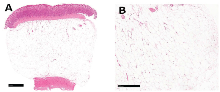
After discussion with the owner, the decision was to perform a subtotal colectomy and biopsy of the gastric mass for the purpose of treating frequently recurrent constipation and diagnosing the gastric mass. During exploratory laparotomy, a generally dilated and atonic colon was mobilized; further, a subtotal colectomy was completed with colocolic anastomosis. Simultaneously, a well-localized, solid mass discovered during gastrectomy was completely resected and subjected to histological examination. Histopathological examination confirmed the diagnosis of GSL (Figure 2). The cat’s postoperative course was uneventful and he was discharged after 1 wk.
FIGURE 2.
Histological assessment of a gastric submucosal lipoma in a 7-year-old Korean shorthair cat. A — Note the benign and well-circumscribed adipose tissue in the submucosa. The adipose tissue appeared to make a mass effect grossly as well as expansile lobules of adipose histologically consistent with a lipoma. Hematoxylin and eosin stain, 1.5× magnification (scale bar: 1 mm). B — Mature lobules of fat did not exhibit anisocytosis or anisokaryosis. Hematoxylin and eosin stain, 4× magnification (scale bar: 500 μm).
DISCUSSION
Gastrointestinal tract lipomas are benign gastric intestinal tumors of mesenchymal origin. In humans, they are relatively rare, accounting for only 2.6% of nonmalignant GIT tumors (6). Compared to the human medical literature, the veterinary literature on GSL and its incidence is limited. In cats, there have been reports of fatty infiltration of the gastric submucosa (7), but there have been no reports of GSL. The apparent rarity of these tumors may be attributed to the lack of related clinical signs, perhaps leading to underdiagnosis or incorrect diagnoses. Most lipomas are solitary; however, multiple lipomas can occur anywhere in the GIT (8). There are 3 pathological types of gastrointestinal lipoma: i) intramuscular, ii) subserosal, and iii) submucosal. Tumors arise from the submucosa in 90% of cases and from the subserosal or intramuscular layers in the remaining 10% of cases (9).
Submucosal lipomas are typically sessile or pedunculated (10). In humans, they have slow growth and an uneventful clinical course; further, they are usually discovered incidentally (11). Symptoms such as abdominal pain, diarrhea, constipation, and bleeding tend to occur when the lesion is ≥ 2 cm (12). Lesions < 1 cm rarely cause symptoms, whereas 75% of lesions > 4 cm are more likely to cause symptoms (10). Unlike the results based on human studies (10,12), there is no information on occurrence of clinical signs according to the size of GSL in veterinary cases. Therefore, treatment should be based on clinical signs. For symptomatic GSL, surgical resection may be considered. Treatment is usually definitive since these lesions do not recur frequently. In humans, malignant transformation of lipomas has not been reported anywhere in the body and post-resection recurrence is not expected (13,14).
Gastric submucosal lipoma can be diagnosed through histological examination with the help of imaging. There have been reports, mainly from human medicine, on endoscopic findings, CT findings, and sonographic findings for submucosal lipoma occurring in the GIT (1,4,5,11). In CT scans, gastrointestinal lipomas appear as well-circumscribed, round, homogeneous masses with fat attenuation numbers (−40 to −120 Hounsfield units) in humans (2,15). The CT characteristics of lipoma reported in veterinary medicine were similar to those in humans (16,17). In this case, CT imaging could have been helpful in diagnosing lipoma based on the CT characteristics of lipoma described in previous studies, but we did not obtain CT scans for this cat due to the lack of clinical symptoms and the cost burden. Considering the lack of veterinary reports on the ultrasonographic characteristics of GSL occurring within the GIT, the ultrasonographic features and histological results we describe here should be helpful in diagnosing submucosal lipoma in cats with similar conditions.
In conclusion, this report describes the imaging features of GSL in a cat, which was identified incidentally. On ultrasonography, the GSL lesion had segmental submucosal wall thickening without the loss of wall layering. Further studies are warranted to determine the ultrasonographic characteristics of GSL and the clinical relevance of GSL in cats. CVJ
Footnotes
Copyright is held by the Canadian Veterinary Medical Association. Individuals interested in obtaining reproductions of this article or permission to use this material elsewhere should contact Permissions.
REFERENCES
- 1.Taylor AJ, Stewart ET, Dodds WJ. Gastrointestinal lipomas: A radiologic and pathologic review. AJR Am J Roentgenol. 1990;155:1205–1210. doi: 10.2214/ajr.155.6.2122666. [DOI] [PubMed] [Google Scholar]
- 2.Jeong IH, Maeng YH. Gastric lipomatosis. J Gastric Cancer. 2010;10:254–258. doi: 10.5230/jgc.2010.10.4.254. [DOI] [PMC free article] [PubMed] [Google Scholar]
- 3.Triantopoulou C, Vassilaki A, Filippou D, Velonakis S, Dervenis C, Koulentianos E. Adult ileocolic intussusception secondary to a submucosal cecal lipoma. Abdom Imaging. 2004;29:426–428. doi: 10.1007/s00261-003-0137-4. [DOI] [PubMed] [Google Scholar]
- 4.Ferrozzi F, Tognini G, Bova D, Pavone P. Lipomatous tumors of the stomach: CT findings and differential diagnosis. J Comput Assist Tomogr. 2000;24:854–858. doi: 10.1097/00004728-200011000-00006. [DOI] [PubMed] [Google Scholar]
- 5.Thompson WM. Imaging and findings of lipomas of the gastrointestinal tract. Am J Roentgenol. 2005;184:1163–1171. doi: 10.2214/ajr.184.4.01841163. [DOI] [PubMed] [Google Scholar]
- 6.Ackerman NB, Chughtai SQ. Symptomatic lipomas of the gastrointestinal tract. Surg Gynecol Obstet. 1975;141:565–568. [PubMed] [Google Scholar]
- 7.Heing HG, Teoh WT, Sheikh Omar AR. Gastric submucosal fat in cats. Anat Histol Embryol. 2008;37:362–365. doi: 10.1111/j.1439-0264.2008.00857.x. [DOI] [PubMed] [Google Scholar]
- 8.Deeths TM, Madden PN, Dodds WJ. Multiple lipomas of the stomach and duodenum. Am J Dig Dis. 1975;20:771–774. doi: 10.1007/BF01070835. [DOI] [PubMed] [Google Scholar]
- 9.Charalambous G, Katergiannakis V, Manouras A. Jejunojejunal lipoma causing intussusception. Case Rep Gastroenterol. 2012;6:684–688. doi: 10.1159/000345379. [DOI] [PMC free article] [PubMed] [Google Scholar]
- 10.Janevska V, Spasevska L, Dukova B, Janevski V. Intestinal submucosal lipomas. Mac J Med Sci. 2012;5:49–54. [Google Scholar]
- 11.Turi S, Röckelein G, Dobroschke J, Wiedmann KH. Lipoma of the small bowel [article in German] Z Gastroenterol. 2004;42:147–151. doi: 10.1055/s-2004-812837. [DOI] [PubMed] [Google Scholar]
- 12.McGrath FP, Moote DJ, Langer JC, Orr W, Somers S. Duodenojejunal intussusception secondary to a duodenal lipoma presenting in a young boy. Pediatr Radiol. 1991;21:590–591. doi: 10.1007/BF02012606. [DOI] [PubMed] [Google Scholar]
- 13.Case records of the Massachusetts General Hospital. Weekly clinicopathological exercises. Case 24-1997. A six-year-old boy with bouts of abdominal pain, vomiting, and a left-sided abdominal mass. N Engl J Med. 1997;337:329–336. doi: 10.1056/NEJM199707313370508. [DOI] [PubMed] [Google Scholar]
- 14.Eisen LK, Cunningham JD, Aufses AH. Intussusception in adults: Institutional review. J Am Coll Surg. 1999;188:390–395. doi: 10.1016/s1072-7515(98)00331-7. [DOI] [PubMed] [Google Scholar]
- 15.Chekan EG, Westcott C, Low VHS, Ludwig KA. Small bowel intussusception and laparoscopy. Surg Laparosc Endosc. 1998;8:324–326. [PubMed] [Google Scholar]
- 16.Spoldi E, Schwarz T, Sabattini S, Vignoli M, Cancedda S, Rossi F. Comparisons among computed tomographic features of adipose masses in dogs and cats. Vet Radiol Ultras. 2017;58:29–37. doi: 10.1111/vru.12445. [DOI] [PubMed] [Google Scholar]
- 17.McEntee MC, Thrall DE. Computed tomographic imaging of infiltrative lipoma in 22 dogs. Vet Radiol Ultras. 2001;42:221–225. doi: 10.1111/j.1740-8261.2001.tb00928.x. [DOI] [PubMed] [Google Scholar]


