

Abstract
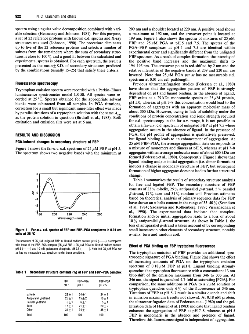
C.d. and fluorescence spectroscopy have been used to investigate the effect of ligand binding on the structure and stability of folate-binding protein (FBP) from cow's whey. The c.d. spectrum of unligated FBP predicts the following secondary structure: 22% helix, 25% antiparallel beta-strand, 5% parallel beta-strand, 17% turn and 31% random-coil structure. Folate binding to FBP results in significant changes in the c.d. spectrum. Analysis of the spectrum shows a 10% decrease in antiparallel beta-strand as a result of ligand binding. Folate binding also leads to strong quenching of FBP tryptophan fluorescence. The magnitude of the quench is proportional to ligand binding. The guanidinium chloride-induced unfolding of FBP is shown to be a multistate process. Detection by c.d. and fluorescence spectroscopy lead to non-identical transitions. Modelling studies are consistent with the existence of a stable folding intermediate. Ligand binding to FBP increases the apparent folding stability of the molecule. Simultaneous detection by c.d. and fluorescence indicate that the apparent increased folding stability is derived from ligand-induced aggregation of FBP.
Full text
PDF


Selected References
These references are in PubMed. This may not be the complete list of references from this article.
- Anderson R. G., Kamen B. A., Rothberg K. G., Lacey S. W. Potocytosis: sequestration and transport of small molecules by caveolae. Science. 1992 Jan 24;255(5043):410–411. doi: 10.1126/science.1310359. [DOI] [PubMed] [Google Scholar]
- Antony A. C. The biological chemistry of folate receptors. Blood. 1992 Jun 1;79(11):2807–2820. [PubMed] [Google Scholar]
- Aune K. C., Tanford C. Thermodynamics of the denaturation of lysozyme by guanidine hydrochloride. II. Dependence on denaturant concentration at 25 degrees. Biochemistry. 1969 Nov;8(11):4586–4590. doi: 10.1021/bi00839a053. [DOI] [PubMed] [Google Scholar]
- Bhandari S. D., Joshi S. K., McMartin K. E. Folate binding and transport by rat kidney brush-border membrane vesicles. Biochim Biophys Acta. 1988 Jan 22;937(2):211–218. doi: 10.1016/0005-2736(88)90243-x. [DOI] [PubMed] [Google Scholar]
- Birdsall B., King R. W., Wheeler M. R., Lewis C. A., Jr, Goode S. R., Dunlap R. B., Roberts G. C. Correction for light absorption in fluorescence studies of protein-ligand interactions. Anal Biochem. 1983 Jul 15;132(2):353–361. doi: 10.1016/0003-2697(83)90020-9. [DOI] [PubMed] [Google Scholar]
- Elwood P. C., Kane M. A., Portillo R. M., Kolhouse J. F. The isolation, characterization, and comparison of the membrane-associated and soluble folate-binding proteins from human KB cells. J Biol Chem. 1986 Nov 25;261(33):15416–15423. [PubMed] [Google Scholar]
- Elwood P. C. Molecular cloning and characterization of the human folate-binding protein cDNA from placenta and malignant tissue culture (KB) cells. J Biol Chem. 1989 Sep 5;264(25):14893–14901. [PubMed] [Google Scholar]
- Hansen S. I., Holm J., Lyngbye J., Pedersen T. G., Svendsen I. Dependence of aggregation and ligand affinity on the concentration of the folate-binding protein from cow's milk. Arch Biochem Biophys. 1983 Oct 15;226(2):636–642. doi: 10.1016/0003-9861(83)90333-8. [DOI] [PubMed] [Google Scholar]
- Hennessey J. P., Jr, Johnson W. C., Jr Information content in the circular dichroism of proteins. Biochemistry. 1981 Mar 3;20(5):1085–1094. doi: 10.1021/bi00508a007. [DOI] [PubMed] [Google Scholar]
- Johnson W. C., Jr Protein secondary structure and circular dichroism: a practical guide. Proteins. 1990;7(3):205–214. doi: 10.1002/prot.340070302. [DOI] [PubMed] [Google Scholar]
- Lee H. C., Shoda R., Krall J. A., Foster J. D., Selhub J., Rosenberry T. L. Folate binding protein from kidney brush border membranes contains components characteristic of a glycoinositol phospholipid anchor. Biochemistry. 1992 Mar 31;31(12):3236–3243. doi: 10.1021/bi00127a027. [DOI] [PubMed] [Google Scholar]
- Luhrs C. A., Pitiranggon P., da Costa M., Rothenberg S. P., Slomiany B. L., Brink L., Tous G. I., Stein S. Purified membrane and soluble folate binding proteins from cultured KB cells have similar amino acid compositions and molecular weights but differ in fatty acid acylation. Proc Natl Acad Sci U S A. 1987 Sep;84(18):6546–6549. doi: 10.1073/pnas.84.18.6546. [DOI] [PMC free article] [PubMed] [Google Scholar]
- Pace C. N. Determination and analysis of urea and guanidine hydrochloride denaturation curves. Methods Enzymol. 1986;131:266–280. doi: 10.1016/0076-6879(86)31045-0. [DOI] [PubMed] [Google Scholar]
- Perkins J. P., Bertino J. R. Dihydrofolate reductase from the L1210R murine lymphoma. Fluorometric measurements of the interaction of the enzyme with coenzymes, substrates, and inhibitors. Biochemistry. 1966 Mar;5(3):1005–1012. doi: 10.1021/bi00867a028. [DOI] [PubMed] [Google Scholar]
- Sadasivan E., Rothenberg S. P. The complete amino acid sequence of a human folate binding protein from KB cells determined from the cDNA. J Biol Chem. 1989 Apr 5;264(10):5806–5811. [PubMed] [Google Scholar]
- Tanford C. Protein denaturation. C. Theoretical models for the mechanism of denaturation. Adv Protein Chem. 1970;24:1–95. [PubMed] [Google Scholar]
- Viswanadhan V. N., Weinstein J. N., Elwood P. C. Secondary structure of the human membrane-associated folate binding protein using a joint prediction approach. J Biomol Struct Dyn. 1990 Feb;7(4):985–1001. doi: 10.1080/07391102.1990.10508537. [DOI] [PubMed] [Google Scholar]