

Abstract
Background:
Unique among all amino acids, Ser is encoded by two sets of codons, TCN and AGY (N=any nucleotide, Y=pyrimidine), that cannot interconvert through single nucleotide substitutions. Both codons are documented at the essential residues S195 and S214 within the active site of serine proteases. However, it is not known how the codons interconverted during evolution because replacement of S195 or S214 by other amino acids typically results in loss of activity.
Objective:
Characterize the prevalence of codon switching among essential and non-essential Ser residues in coagulation and fibrinolytic proteases from different vertebrate lineages.
Methods:
TCN and AGY codon usage was analyzed in >550 sequences.
Results:
Evolutionary pressure to preserve the codon of S195 is absolute, with no evidence of interconversion. Pressure to preserve the codon of S214 is also strong, but an AGY↔TCN interconversion is observed in factor VII-inactive and protein C from ray-finned fish. In both cases, the interconversion occurred in genes that were rapidly evolving. In contrast, codon switching at non-essential Ser residues in the kringle domains of coagulation and fibrinolytic proteases are quite common and could be identified in half of the kringles analyzed.
Conclusion:
Codon interconversion of essential Ser residues of coagulation and fibrinolytic proteases only occurred in genes rapidly evolving and which—at least in some cases—evolved following genome duplication. Interconversion is common at non-essential Ser residues as found in kringle domains.
Keywords: Blood coagulation, codons, evolution, fibrinolysis, vitamin K-dependent clotting factors
Introduction
Serine proteases have been classified into evolutionarily unrelated clans, which differ in overall fold and order of catalytic residues in the primary sequence [1, 2]. Clan SA is the largest and includes proteases involved in digestive and degradative processes, blood clotting, fibrinolysis, immunity, and wound repair [3]. A conserved active site geometry includes residues of the catalytic triad H57, D102 and the nucleophile S195 (chymotrypsin numbering) [2, 4, 5]. A closely associated S214, known as “the fourth member” of the catalytic triad [6, 7], is also conserved and contributes a necessary polar environment around the catalytic D102 [8, 9]. Serine is unique among amino acids as it is encoded by two sets of codons, TCN and AGY (N=any nucleotide, Y=pyrimidine), that cannot interconvert through single nucleotide substitutions [10]. Because both codons are documented for S195 and S214 in proteases of clan SA [6, 10], the question arises as to how TCN and AGY interconverted during evolution. Possible mechanisms involve simultaneous double substitution of two nucleotides (e.g., TC→AG or AG→TC) or, more commonly, consecutive single substitutions through Cys:TGY (TCN↔TGY↔AGY) or Thr:ACN (TCN↔ACN↔AGY) intermediates [10–12]. Importantly, such codon switches involving consecutive single substitutions are primarily driven by positive selection where the initial deleterious mutation of Ser is followed by a compensatory mutation that reverts the Cys or Thr intermediate to Ser encoded by a different codon [12]. However, it is unclear how Cys, Thr or other intermediate residues are tolerated at position 195 or 214 given their profound deleterious effects on activity [7, 9, 13–15]. We addressed this question with the analysis of >550 sequences of coagulation and fibrinolytic proteases from all vertebrate classes (Figure S1).
Methods
Predicted sequences of the vitamin K (VK) dependent coagulation proteases prothrombin, factor (f) VII, fIX, fX and protein C (PC) and the fibrinolytic proteases plasminogen, tissue-type plasminogen activator (tPA) and urokinase plasminogen activator (uPA) were downloaded from the National Center for Biotechnology Information database (Supporting Information).
Results and Discussion
Table 1 summarizes the codon usage of S195 and S214 in all vertebrate lineages (>550 sequences) for the VK-dependent coagulation proteases prothrombin, fVII, fIX, fX and PC and for the fibrinolytic proteases plasminogen, tPA and uPA (Table 1, Figures S2–S4). Evolutionary pressure to preserve the codon of S195 is absolute, with no evidence of AGY↔TCN codon interconversion at this position among orthologous vertebrate proteases from jawless fish to mammals. S195 is encoded by TCN codons in tPA and uPA, and by AGY codons in plasminogen and all VK-dependent coagulation proteases. This residue was mutated only among proteases like fVII-inactive from ray-finned fish and protein Z that eventually lost their catalytic function and evolved into inhibitors [16]. Ray-finned fish are unique among vertebrates insofar as their entire genome was duplicated in the last common ancestor of Teleostei [17]. Most of the duplicated genes were lost during species radiation, but a conservative estimate indicates that >5% were retained in some species [18]. Teleost are the only vertebrates with three copies of the F7 gene arranged in tandem as F7-like, F7, and F7-inactive, with F7-inactive neighboring the F10 gene [19, 20]. F7-inactive is only present in Teleostei and not in basal ray-fins (e.g., bichirs, reedifish, paddlefish, and gars), implicating that this gene emerged following teleost genome duplication [17]. Among non-teleost, paddlefish and gars have a copy of F7-like, while earlier diverging ray-fins such as bichirs and reedfish do not. S195 in fVII and fVII-like is encoded by AGY, as in fVII from all vertebrates (Table 1, Figures S2, S4). All sequences of fVII-inactive carry the S195D mutation that abolishes catalytic activity [16, 19, 20].
Table 1.
Distribution of TCN and AGY codons of S195 and S214 among coagulation and fibrinolytic proteases
| ProT | PC | fVII | fVII-like | fVII-inactive | fIX | fX | tPA | uPA | Plg | |
|---|---|---|---|---|---|---|---|---|---|---|
| Jawless fish | S195:AGY S214:TCN N=3 |
N.A. | S195:AGY S214:AGY N=1 |
N.A. | N.A. | N.A. | S195:AGY S214:AGY N=1 |
N.A. | N.A. | S195:AGY S214:TCN N=1 |
| Cartilaginous fish | S195:AGY S214:TCN N=5 |
S195:AGY S214:AGY N=2 |
S195:AGY S214:AGY N=3 |
N.A. | N.A. | S195:AGY S214:AGY N=2 |
S195:AGY S214:AGY N=2 |
S195:TCN S214:AGY N=4 |
N.A. | S195:AGY S214:TCN N=8 |
| Ray-finned fish | S195:AGY S214:TCN N=30 |
S195:AGY S214:TCN N=26 |
S195:AGY S214:AGY N=31 |
S195:AGY S214:AGY N=29 |
D195:GAY S214:TCN A214:GCN G214:GGN N=27 |
S195:AGY S214:AGY N=9 |
S195:AGY S214:AGY N=10 |
S195:TCN S214:AGY N=10 |
S195:TCN S214:AGY N=25 |
S195:AGY S214:TCN N=11 |
| Amphibians | S195:AGY S214:TCN N=4 |
S195:AGY S214:AGY N=3 |
S195:AGY S214:AGY N=3 |
N.A. | N.A. | S195:AGY S214:AGY N=3 |
S195:AGY S214:AGY N=4 |
S195:TCN S214:AGY N=5 |
S195:TCN S214:AGY N=7 |
S195:AGY S214:TCN N=10 |
| Birds | S195:AGY S214:TCN N=10 |
S195:AGY S214:AGY N=10 |
S195:AGY S214:AGY N=10 |
N.A. | N.A. | S195:AGY S214:AGY N=10 |
S195:AGY S214:AGY N=10 |
S195:TCN S214:AGY N=10 |
S195:TCN S214:AGY N=15 |
S195:AGY S214:TCN N=9 |
| Reptiles | S195:AGY S214:TCN N=8 |
S195:AGY S214:AGY N=8 |
S195:AGY S214:AGY N=7 |
N.A. | N.A. | S195:AGY S214:AGY N=8 |
S195:AGY S214:AGY N=8 |
S195:TCN S214:AGY N=11 |
S195:TCN S214:AGY N=12 |
S195:AGY S214:TCN N=6 |
| Monotremes | S195:AGY S214:TCN N=2 |
S195:AGY S214:AGY N=2 |
S195:AGY S214:AGY N=1 |
N.A. | N.A. | S195:AGY S214:AGY N=2 |
S195:AGY S214:AGY N=1 |
S195:TCN S214:AGY N=2 |
S195:TCN S214:AGY N=2 |
N.A. |
| Marsupials | S195:AGY S214:TCN N=6 |
S195:AGY S214:AGY N=2 |
S195:AGY S214:AGY N=2 |
N.A. | N.A. | S195:AGY S214:AGY N=2 |
S195:AGY S214:AGY N=2 |
S195:TCN S214:AGY N=8 |
S195:TCN S214:AGY N=8 |
S195:AGY S214:TCN N=8 |
| Placentals | S195:AGY S214:TCN N=11 |
S195:AGY S214:AGY N=10 |
S195:AGY S214:AGY N=10 |
N.A. | N.A. | S195:AGY S214:AGY N=10 |
S195:AGY S214:AGY N=10 |
S195:TCN S214:AGY N=8 |
S195:TCN S214:AGY N=10 |
S195:AGY S214:TCN N=20 |
N=number of sequences, N.A.=not available, ProT=prothrombin, Plg=plasminogen
Pressure to preserve the codon at S214 was also strong. This Ser is encoded by AGY in fVII, fIX, fX, PC, tPA and uPA, but TCN encodes the same Ser in prothrombin and plasminogen. Notable exceptions are PC and fVII-inactive from ray-fins where an AGY→TCN conversion took place (Table 1, Figures 1, S3, S4). In the case of fVII-inactive, the residue at position 214 varies as Gly, Ala or Ser in different species of ray-finned fish (Table 1, Figure 1A). Interestingly, in the species of fish that express fVII-inactive with S214, this Ser residue is encoded by TCN (Table 1, Figure 1B). In both F7 and F7-like, that evolved with F7-inactive from the same ancestral F7 gene, the codon is AGY. Hence, the codon of S214 likely switched after duplication of the F7 gene in Teleostei. Mechanistically the AGY→TCN conversion might have proceeded via a double simultaneous nucleotide substitution (e.g., AG→TC) or a single nucleotide substitution through Cys/Thr intermediates. However, the occurrence of A214 and G214 in some species of ray-finned fish suggests that Ala and Gly were intermediates in a codon interconversion pathway such as Ser:AGY→Gly:GGY→Ala:GCY→Ser:TCN. The somewhat convoluted pathway would have been tolerated after duplication of the F7 gene and inactivation of the protease with the S195D mutation, as long as the nature of residue 214 did not perturb the overall folding of the protein.
Figure 1ABC. AGY→TCN codon interconversion in fVII-inactive and PC from ray-finned fish.

(A) Alignment of representative fVII sequences from several species of ray-finned fish reveals the presence of D195 in fVII-inactive (fVII-i) as opposed to S195 in fVII and fVII-like. Different species of ray-finned fish express fVII-inactive with either S214, A214 or G214 as opposed to S214 that is always found in fVII and fVII-like. Alignment of representative sequences shows evidence of an AGY→TCN interconversion at position 214 in fVII (B) and PC (C). Shown are the aligned amino acids (capital letter) of a conserved sequence in the protease domain where S214 is located (colored) together with the nucleotides (small letter) that encode specific residues.
The codon of S214 in PC is TCN in ray-finned fish but AGY in all other vertebrates and even in the cartilaginous fish that predate ray-finned fish (Table 1, Figures 1C, S3). The Ser214:TCN codon is present in both basal and teleost ray-fins, suggesting that the codon switch was not associated with the teleost genome duplication, which contrasts with F7-inactive. However, both genes where codon switch at position S214 took place display signs of rare mutations in residues important for zymogen activation and catalysis which provides evidence that the F7 and PC genes in ray-finned fish were evolving at a fairly rapid rate. In addition to the S195D mutation that abolishes catalytic activity, we identified species of fish that express fVII-inactive with mutations at residues R15, G19 and D194 that control zymogen activation and D189 that defines the primary specificity of the protease [2, 4, 5, 21]. All ray-fins express PC with unusual mutations like the presence of aromatic residues at the site of activation and the Y225L mutation in the Na+ binding site [22, 23]. Some species of ray-finned fish carry Thr or Met at positions 16 and 17 in the protease domain of PC, as well as residues at position 99 in the active site region not found in other vertebrates [22]. In the case of fVII-inactive, the introduction of such mutations were buffered by the presence of more than one F7 gene in the ancestral teleost, averting hemostatic deficiencies. However, it is less clear how mutations at such important sites were tolerated in PC, unless the ancestor of ray-fins had more than one copy of the PC gene which was eventually lost. Some basal ray-fins like paddlefish have 2 copies of PC (S214:TCN present in both) arranged in tandem which border the sirtilin gene [24], but no additional copy of PC is present in bichirs and reedfish which are the earliest diverging ray-fins, implicating that the localized PC duplication might be unique to paddlefish. Overall, codon switching at S214 coincided with acquisition of novel mutations at functionally important positions, suggesting that codon interconversion may occur in rapidly evolving genes that, at least in some cases, are known to be duplicated. It remains to be seen whether codon switching at conserved Ser in other proteases coincides with accumulation of mutations at functionally important residues and whether these genes have ever been duplicated.
Interestingly, although the selective pressure to avert AGY↔TCN interconversion at conserved Ser is exceptionally strong, synonymous mutations at the third nucleotide in Ser codons are quite prevalent (Figures S2–4). Synonymous mutations at the third nucleotide position result in AGC↔AGT interconversions and TCG↔TCC↔TCA↔TCT interconversions, and their high prevalence contrasts with the scarcity of AGY↔TCN interconversions. The low prevalence of AGY↔TCN switches at conserved Ser likely has to do with the requirement for such interconversions to take place through a mechanism that would produce Thr/Cys intermediates [12], which would profoundly compromise catalysis [7, 9, 13–15]. That such intermediates are not tolerated is evident from severe bleeding phenotypes observed in hemophilia B patients carrying mutations at S195 and S214 in F9 [25]. Mutations at S195 and S214 have also been reported in fX- [26] and fVII-deficiencies [27], respectively.
We also examined the prevalence of codon switches at moderately conserved Ser located in the kringle domains of the fibrinolytic proteases and prothrombin. Kringle domains are comprised of 80 amino acids and the fold is stabilized by three disulfides with 1–6, 2–3, and 3–5 connectivity [28]. The number of kringles varies from one in uPA, two in prothrombin and tPA, and five in plasminogen. Kringle domains contain a CXXWXS motif located after the second Cys, with Cys and Trp perfectly conserved but Ser less so (Table 2, Figure 2). This enables a test of codon usage and switching of a Ser that may not play an essential role in stabilizing the fold. Codon switching at the Ser of the CXXWXS motif was detected in half of the kringles analyzed in this study and specifically in kringle 1 of tPA, kringles 1 and 3 of plasminogen and both kringles of prothrombin (Table 2, Figure 2). The observation confirms that codon interconversion is more prevalent among non-essential Ser residues. A pathway of interconversion could be established for kringle 3 of plasminogen and kringle 1 of tPA (Table 2, Figure 2). The Ser of the CXXWXS motif in kringle 3 of plasminogen is encoded by TCN in most vertebrates including fish, amphibians, birds and some reptiles, but is AGY in most marsupials that also carry Thr at this position in some cases. Hence, the TCN codon present in more ancient vertebrates switched to AGY via a Thr intermediate during the radiation of marsupials. Kringle 1 of tPA offers another example of such switch. Fish and amphibians use TCN for the Ser of the CXXWXS motif but birds and mammals use AGY. Snakes carry a Thr intermediate and most other reptiles carry Ile. The TCN codon in earlier diverging vertebrates was converted into Thr in the last common ancestor of amniotes. As amniotes began to radiate, some reptiles (e.g., snakes) retained the original Thr:ACY combination, whilst birds and mammals acquired a second mutation to generate Ser:AGY. Codon switching with no obvious intermediates is also evident in kringle 2 of prothrombin. The TCN codon found in nearly all vertebrates was converted to AGY upon the emergence of placental mammals (Table 2, Figure 2). On the other hand, AGY found in kringle 1 of prothrombin in most vertebrates switched to TCN in some species of ray-finned fish (e.g., Thunnus albacares and Thunnus maccoyii). Dichotomous distributions of TCN and AGY codons are also present in kringle 1 of plasminogen (Table 2, Figure 2). Lastly, even in kringle domains with no evidence of codon switching (e.g., uPA, kringle 2 of tPA, kringles 2, 4, and 5 of plasminogen), the Ser in the CXXWXS motif was mutated to some other residue (Table 2 and Figure 2). This implies that a position tolerant to substitutions has high prevalence of codon interconversion.
Table 2:
Distribution of codons of Ser or other residues in the last position of the CXXWXS motif of kringle domains
| Prothrombin | tPA | uPA | Plasminogen | |||||||
|---|---|---|---|---|---|---|---|---|---|---|
| K1 | K2 | K1 | K2 | K1 | K1 | K2 | K3 | K4 | K5 | |
|
Jawless fish (3,0,0,1) |
S:AGY | A:GCN 66% G:GGN 33% |
N.A. | N.A. | N.A. | Q:CAG | S:TCN | E:GAG | S:TCN | S:TCN |
|
Cartilaginous fish (5,4,0,8) |
S:AGY 60% R:AGA 40% |
S:TCN 80% L:TTG 20% |
S:TCN | S:TCN | N.A. | S:AGY 88% I:ATC 12% |
S:TCN 50% L:TTN 25% I:ATA 12% T:ACN 12% |
S:TCN 38% A:GCN 38% T:ACN 12% V:GTT 12% |
S:TCN N=8 |
S:TCN 25% A:GCN 63% E:GAA 12% |
|
Ray-finned fish (29,11,26,11) |
S:TCN 7% S:AGY 38% H, G, R |
S:TCN 83% A:GCN 10% L:TTN 7% |
S:TCN | S:TCN | S:TCN 43% P:CCN 23% L:CTN 34% |
S:TCN 9% A:GCN 91% |
S:TCN | S:TCN 45% T:ACN 27% A:GCN 18% V:GTT 10% |
S:TCN 45% A:GCN 45% T:ACN 10% |
A:GCN |
|
Amphibians (4,5,7,10) |
S:AGY | S:TCN | S:TCN | S:TCN | S:TCN | S:TCN 40% S:AGY 30% A:GCN 30% |
S:TCN 50% Q, E, L, V |
S:TCN 70% A:GCN 20% V:GTT 10% |
S:TCN | S:TCN 80% L:TTA 20% |
|
Reptiles (8,11,11,6) |
S:AGY | S:TCN | T:ACN 27% I:ATN 55% N, L |
S:TCN | S:TCN | D:GAC 67% A:GCN 33% |
S:TCN 50% A:GCN 50% |
S:TCN 33% A:GCN 67% |
S:TCN | S:TCN 84% L:TTA 16% |
|
Birds (10,10,15,9) |
S:AGY | S:TCN | S:AGY 90% N:AAN |
S:TCN | S:TCN 20% L:CTT 74% H:CAT 6% |
D:GAT | S:TCN | S:TCN 89% A:GCN 11% |
S:TCN | S:TCN |
|
Monotremes (2,2,2,0) |
S:AGY | S:TCN | S:AGY | S:TCN | S:TCN | N.A. | N.A. | N.A. | N.A. | N.A. |
|
Marsupials (6,8,8,8) |
S:AGY | S:TCN | S:TCN 12% M:ATG 50% K, L, Q |
S:TCN 88% T:ACT 12% |
S:TCN 75% L:TTA 12% A:GCN 12% |
D:GAT 88% G:GGT 12% |
S:TCN | S:AGY 50% T:ACA 25% K:AAA 25% |
S:TCN | A:GCN |
|
Placentals (11,8,10,19) |
S:AGY | S:AGY | S:AGY | S:TCN | S:TCN | S:TCN 32% A:GCN 21% D:GAC 42% N:AAC 5% |
S:TCN | A:GCN 37% E:GAG 63% |
S:TCN 79% A:GCN 21% |
A:GCN |
N.A.=Not available; K1–5=kringle 1–5; numbers in parentheses refer to the sequences analyzed for prothrombin, tPA, uPA and plasminogen
Figure 2. Codon distribution in kringle domains.
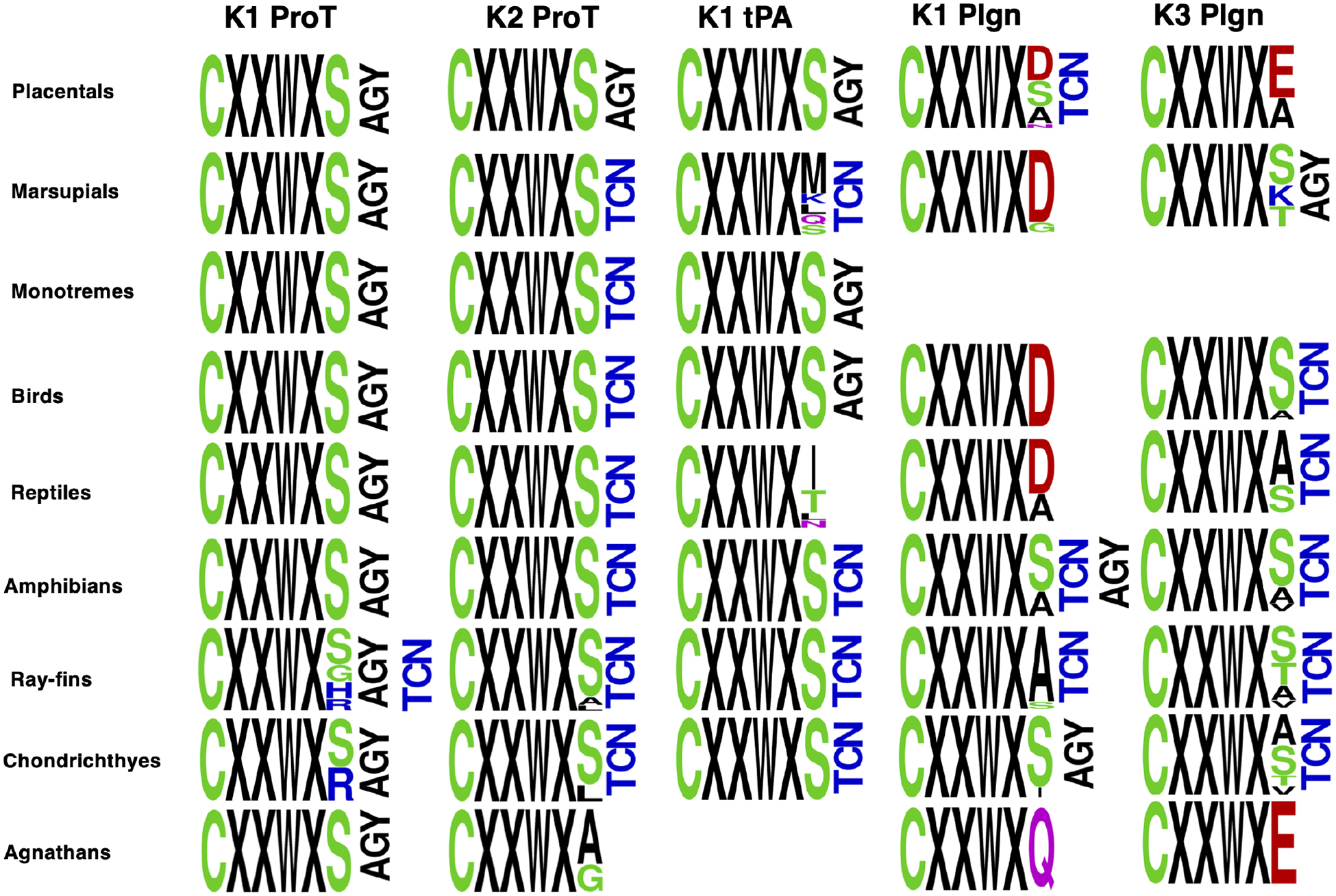
TCN and AGY codons of the Ser residue in the CXXWXS motif following the second Cys residue in kringle domains. Codons shows refer only to the last residue (Ser or other) in the motif. Abbreviations are: ProT (prothrombin), tPA (tissue-type plasminogen activator), plgn (plasminogen), K (kringle).
It was originally proposed that serine proteases carrying TCN or AGY codons for S195 evolved independently from a Cys protease ancestor [10], but this hypothesis was later challenged [29] and an alternative proposal posited that these proteases evolved from inactive proteins carrying mutations at the catalytic residues [30]. Our analysis indirectly supports this hypothesis and implies that codon interconversion at highly conserved Ser residues likely occurred during periods of evolutionary innovation when the genes were duplicated and rapidly evolving followed by selective pressure to restore Ser with a different codon [12]. Interestingly, although most TCN↔AGY interconversions are predicted to occur via Cys [10, 12], we found no evidence of such intermediate in any of the protease or kringle domains analyzed. Unpaired Cys residues may produce mispairing of disulfide bonds and this could compromise folding of protease and kringle domains stabilized by several disulfides. Indeed, all Ser residues analyzed in our study are a few residues away from a Cys engaged in critical disulfide pairing and not a candidate for codon switching through a Cys intermediate.
Supplementary Material
Acknowledgments.
We gratefully acknowledge Tracey Baird for her assistance with illustrations. This study was supported in part by the National Institutes of Health Research Grants HL049413, HL139554 and HL147821 (E.D.C).
Footnotes
Publisher's Disclaimer: This is a PDF file of an unedited manuscript that has been accepted for publication. As a service to our customers we are providing this early version of the manuscript. The manuscript will undergo copyediting, typesetting, and review of the resulting proof before it is published in its final form. Please note that during the production process errors may be discovered which could affect the content, and all legal disclaimers that apply to the journal pertain.
Competing Interests. The Authors declare no competing financial interests.
References
- 1.Barrett AJ, Rawlings ND. ‘Species’ of peptidases. Biol Chem. 2007; 388: 1151–7. [DOI] [PubMed] [Google Scholar]
- 2.Page MJ, Di Cera E. Serine peptidases: classification, structure and function. Cell Mol Life Sci. 2008; 65: 1220–36. [DOI] [PMC free article] [PubMed] [Google Scholar]
- 3.Krem MM, Di Cera E. Evolution of enzyme cascades from embryonic development to blood coagulation. Trends Biochem Sci. 2002; 27: 67–74. [DOI] [PubMed] [Google Scholar]
- 4.Perona JJ, Craik CS. Structural basis of substrate specificity in the serine proteases. Protein Sci. 1995; 4: 337–60. [DOI] [PMC free article] [PubMed] [Google Scholar]
- 5.Hedstrom L Serine protease mechanism and specificity. Chem Rev. 2002; 102: 4501–24. [DOI] [PubMed] [Google Scholar]
- 6.Krem MM, Di Cera E. Molecular markers of serine protease evolution. Embo J. 2001; 20: 3036–45. [DOI] [PMC free article] [PubMed] [Google Scholar]
- 7.Krem MM, Prasad S, Di Cera E. Ser(214) is crucial for substrate binding to serine proteases. J Biol Chem. 2002; 277: 40260–4. [DOI] [PubMed] [Google Scholar]
- 8.Perona JJ, Craik CS. Evolutionary divergence of substrate specificity within the chymotrypsin-like serine protease fold. J Biol Chem. 1997; 272: 29987–90. [DOI] [PubMed] [Google Scholar]
- 9.McGrath ME, Vasquez JR, Craik CS, Yang AS, Honig B, Fletterick RJ. Perturbing the polar environment of Asp102 in trypsin: consequences of replacing conserved Ser214. Biochemistry. 1992; 31: 3059–64. [DOI] [PubMed] [Google Scholar]
- 10.Brenner S The molecular evolution of genes and proteins: a tale of two serines. Nature. 1988; 334: 528–30. [DOI] [PubMed] [Google Scholar]
- 11.Averof M, Rokas A, Wolfe KH, Sharp PM. Evidence for a high frequency of simultaneous double-nucleotide substitutions. Science. 2000; 287: 1283–6. [DOI] [PubMed] [Google Scholar]
- 12.Rogozin IB, Belinky F, Pavlenko V, Shabalina SA, Kristensen DM, Koonin EV. Evolutionary switches between two serine codon sets are driven by selection. Proc Natl Acad Sci U S A. 2016; 113: 13109–13. [DOI] [PMC free article] [PubMed] [Google Scholar]
- 13.Baird TT Jr., Wright WD, Craik CS. Conversion of trypsin to a functional threonine protease. Protein Sci. 2006; 15: 1229–38. [DOI] [PMC free article] [PubMed] [Google Scholar]
- 14.Higaki JN, Evnin LB, Craik CS. Introduction of a cysteine protease active site into trypsin. Biochemistry. 1989; 28: 9256–63. [DOI] [PubMed] [Google Scholar]
- 15.Pelc LA, Chen Z, Gohara DW, Vogt AD, Pozzi N, Di Cera E. Why Ser and not Thr brokers catalysis in the trypsin fold. Biochemistry. 2015; 54: 1457–64. [DOI] [PMC free article] [PubMed] [Google Scholar]
- 16.Hanumanthaiah R, Day K, Jagadeeswaran P. Comprehensive analysis of blood coagulation pathways in teleostei: evolution of coagulation factor genes and identification of zebrafish factor VIIi. Blood Cells Mol Dis. 2002; 29: 57–68. [DOI] [PubMed] [Google Scholar]
- 17.Glasauer SM, Neuhauss SC. Whole-genome duplication in teleost fishes and its evolutionary consequences. Mol Genet Genomics. 2014; 289: 1045–60. [DOI] [PubMed] [Google Scholar]
- 18.Aparicio S, Chapman J, Stupka E, Putnam N, Chia JM, Dehal P, Christoffels A, Rash S, Hoon S, Smit A, Gelpke MD, Roach J, Oh T, Ho IY, Wong M, Detter C, Verhoef F, Predki P, Tay A, Lucas S, Richardson P, Smith SF, Clark MS, Edwards YJ, Doggett N, Zharkikh A, Tavtigian SV, Pruss D, Barnstead M, Evans C, Baden H, Powell J, Glusman G, Rowen L, Hood L, Tan YH, Elgar G, Hawkins T, Venkatesh B, Rokhsar D, Brenner S. Whole-genome shotgun assembly and analysis of the genome of Fugu rubripes. Science. 2002; 297: 1301–10. [DOI] [PubMed] [Google Scholar]
- 19.Davidson CJ, Hirt RP, Lal K, Snell P, Elgar G, Tuddenham EG, McVey JH. Molecular evolution of the vertebrate blood coagulation network. Thromb Haemost. 2003; 89: 420–8. [PubMed] [Google Scholar]
- 20.Jiang Y, Doolittle RF. The evolution of vertebrate blood coagulation as viewed from a comparison of puffer fish and sea squirt genomes. Proc Natl Acad Sci U S A. 2003; 100: 7527–32. [DOI] [PMC free article] [PubMed] [Google Scholar]
- 21.Huber R, Bode W. Structural basis of the activation and action of trypsin. Acc Chem Res. 1978; 11: 114–22. [Google Scholar]
- 22.Stojanovski BM, Di Cera E. Comparative sequence analysis of vitamin K-dependent coagulation factors. J Thromb Haemost. 2022; 20: 2837–49. [DOI] [PMC free article] [PubMed] [Google Scholar]
- 23.Stojanovski BM, Pelc LA, Di Cera E. Thrombin has dual trypsin-like and chymotrypsin-like specificity. J Thromb Haemost. 2023. [DOI] [PMC free article] [PubMed] [Google Scholar]
- 24.Dahms SO, Demir F, Huesgen PF, Thorn K, Brandstetter H. Sirtilins–the new old members of the vitamin K-dependent coagulation factor family. Journal of thrombosis and haemostasis. 2019; 17: 470–81. [DOI] [PMC free article] [PubMed] [Google Scholar]
- 25.Li T, Miller CH, Payne AB, Craig Hooper W. The CDC Hemophilia B mutation project mutation list: a new online resource. Molecular genetics & genomic medicine. 2013; 1: 238–45. [DOI] [PMC free article] [PubMed] [Google Scholar]
- 26.Harris VA, Lin W, Perkins SJ. Analysis of 180 genetic variants in a new interactive FX variant database reveals novel insights into FX deficiency. TH Open. 2021; 5: e557–e69. [DOI] [PMC free article] [PubMed] [Google Scholar]
- 27.Giansily-Blaizot M, Rallapalli PM, Perkins SJ, Kemball-Cook G, Hampshire DJ, Gomez K, Ludlam CA, McVey JH. The EAHAD blood coagulation factor VII variant database. Human Mutation. 2020; 41: 1209–19. [DOI] [PubMed] [Google Scholar]
- 28.Soriano-Garcia M, Padmanabhan K, de Vos AM, Tulinsky A. The Ca2+ ion and membrane binding structure of the Gla domain of Ca-prothrombin fragment 1. Biochemistry. 1992; 31: 2554–66. [DOI] [PubMed] [Google Scholar]
- 29.Koonin EV, Gorbalenya AE. Tale of two serines. Nature. 1989; 338: 467–8. [DOI] [PMC free article] [PubMed] [Google Scholar]
- 30.Rawlings ND, Barrett AJ. Families of serine peptidases. Methods Enzymol. 1994; 244: 19–61. [DOI] [PMC free article] [PubMed] [Google Scholar]
Associated Data
This section collects any data citations, data availability statements, or supplementary materials included in this article.