Abstract
Background
Echinocandins belong to the fourth generation of antifungals, and there are no systematic studies on their risk in coagulation dysfunction; this study will predict the risk of coagulation dysfunction of echinocandins using the US Food and Drug Administration Adverse Event Reporting System (FAERS) database.
Method
Data from January 2004 to March 2024 were obtained from FAERS. We examined the clinical characteristics of the coagulation dysfunction events and conducted disproportionality analysis by using reporting odds ratios (ROR) to compare echinocandins with the full database.
Results
There were 313 reports of coagulation dysfunction related to echinocandins as the primary suspect (PS) drug. The median time to incident for coagulation dysfunction was 3 (interquartile range [IQR] 1–9) days. Compared to triazoles and polyenes, echinocandins have a stronger signal (ROR 3.18, 95%CI 2.81–3.51, p < 0.01) of coagulation dysfunction. Compared to caspofungin and micafungin, anidulafungin has a stronger signal (ROR 6.84, 95%CI 4.83–9.70, p < 0.01). The strongest signal corresponding to disseminated intravascular coagulation (DIC), platelet count decreased, thrombocytopenia, gastrointestinal haemorrhage, cerebral haemorrhage, pulmonary haemorrhage and thrombotic thrombocytopenic purpura (TTP) is micafungin (ROR 27.19, 95%CI 18.49–39.98), micafungin (ROR 3.50, 95%CI 2.36–5.19), anidulafungin (ROR 9.75, 95%CI 5.22–18.19), micafungin (ROR 3.17, 95%CI 2.02–4.97), micafungin (ROR 4.95, 95%CI 2.81–8.72), caspofungin (ROR 20.76, 95%CI 11.77–36.59), micafungin (ROR 20.43, 95%CI 8.49–49.14), respectively.
Conclusions
For coagulation dysfunction, we found stronger signals for echinocandins than triazoles and polyenes, and stronger signals for anidulafungin than micafungin and caspofungin. Coagulation parameters should be closely monitored while using the respective drugs.
Supplementary Information
The online version contains supplementary material available at 10.1186/s12959-024-00641-4.
Keywords: Caspofungin, Micafungin, Anidulafungin, Coagulation dysfunction, FAERS, Pharmacovigilance
Highlights
Real world data were used to predict the risk of coagulopathy for three classes of drugs: echinocandins, triazoles, and polyenes.
Real-world data were used to compare the risk of coagulopathy between caspofungin, micafungin, and anidulafungin.
Different causes of coagulation dysfunction were analyzed by subgroups.
Supplementary Information
The online version contains supplementary material available at 10.1186/s12959-024-00641-4.
Background
Fungal infections have a high morbidity and mortality rate and pose a serious threat to human health [1]. Commonly used antifungal drugs include echinocandins, triazoles and polyenes. Echinocandins belong to the fourth generation of antifungal drugs. FDA-approved echinocandins include caspofungin, micafungin, anidulafungin, and rezafungin. These drugs have demonstrated significant antifungal efficacy through the specific inhibition of β-(1,3) -D-glucan synthesis, which forms 30–60% of the fungal cell wall [2–6]. All of them can be used as anti-infective therapy for Candida, and caspofungin is approved for the treatment of invasive aspergillosis in patients who are refractory to or intolerant of other therapies [7].
The main adverse events (AEs) of echinocandins in premarketing studies were elevated alanine aminotransferase (ALT), elevated aspartate aminotransferase (AST), fever, phlebitis, gastrointestinal reactions, headache and rash [8–11]. Meanwhile, thrombocytopenia has been reported in clinical trials of micafungin and anifungin [9, 10]. DIC has also been reported in clinical trials of rezafungin [11]. However, there are few post-marketing studies on coagulation dysfunction caused by echinocandins. This study will use real-world data from the FAERS database to explore the risk of coagulopathy with echinomycin for clinicians to reference.
Methods
Study design and data sources
The target drugs in this study include caspofungin, micafungin, and anidulafungin (rezafungin has no reported AEs related to coagulation dysfunction due to a marketing date of 2023.03.22). Control drugs included voriconazole, itraconazole, fluconazole, posaconazole, isavuconazonium, and amphotericin B. This study included all records in the FAERS database from January 2004 to March 2024 (https://fis.fda.gov/extensions/FPD-QDE-FAERS/FPD-QDE-FAERS.html). Generic and brand names were employed to define target/control drugs (supplementary Table 1). Reports in the DRUG table where role_code is “primary suspect” were included in the study.
We define coagulation dysfunction events through the preferred terms (PT) level in the Medical Dictionary for Regulatory Activities terms (MeDRA V25.1). The specific list is shown in supplementary Table 2. The primary outcome was overall coagulation-related AEs. The secondary outcomes comprised the reporting of coagulation dysfunction events with a high frequency: DIC, platelet count decreased, thrombocytopenia, gastrointestinal haemorrhage, cerebral haemorrhage, pulmonary haemorrhage, TTP Table 1 and 2
Table 1.
Clinical characteristics of patients with echinocandins-associated coagulation dysfunction events
| No. of coagulation dysfunction events in echinocandins (%) | No. of other AEs in echinocandins (%) | |
|---|---|---|
| Reports | 313 | 4395 |
| Gender | ||
| Male | 175 (55.91) | 2300 (52.33) |
| Female | 116 (37.06) | 1538 (34.99) |
| Unknown | 22 (7.03) | 557 (12.67) |
| Age | ||
| < 20 | 32 (10.22) | 442 (10.06) |
| ≥ 20 and < 40 | 32 (10.22) | 475 (10.81) |
| ≥ 40 and < 60 | 63 (20.13) | 939 (21.37) |
| ≥ 60 | 146 (46.65) | 1325 (30.15) |
| Reporter | ||
| Healthcare workers | 242 (77.32) | 3130 (71.22) |
| Non-health care workers | 64 (20.45) | 1154 (26.26) |
| Unknown | 7 (2.24) | 111 (2.53) |
| Year | ||
| 2021–2024 | 25 (7.99) | 597 (13.58) |
| 2016–2020 | 51 (16.29) | 1497 (34.06) |
| 2011–2015 | 89 (28.43) | 1359 (30.92) |
| 2004–2010 | 117 (37.38) | 941 (21.41) |
| Outcome | ||
| Death | 167 (53.35) | 1579 (35.93) |
| Life-threatening | 71 (22.68) | 365 (8.30) |
| Disability | 17 (5.43) | 51 (1.16) |
| Hospitalization | 101 (32.27) | 1128 (25.67) |
| Other serious | 190 (60.70) | 1848 (42.05) |
| Congenital Anomaly | 0 (0) | 3 (0.07) |
| Required intervention | 1 (0.32) | 12 (0.27) |
| Report countries (Top 3) | Japan 51 (16.29) | America 960 (21.84) |
| China 30 (9.58) | Japan 480 (10.92) | |
| America 15 (4.79) | France 277 (6.30) | |
| Indications (Top 3) | Prophylaxis 57 (18.21) | Fungal infection 596 (13.56) |
| Febrile neutropenia 49 (15.65) | Febrile neutropenia 329 (7.49) | |
| Fungal infection 39 (12.46) | Bronchopulmonary aspergillosis 293 (6.67) | |
| Onset time (Median [IQR] / d) | 3 (1–8) | - |
Table 2.
Quartile of coagulopathy events with different antifungal drugs
| Drugs Onset time (d) |
Micafungin | Caspofungin | Anidulafungin | Voriconazole | Itraconazole | Fluconazole | Posaconazole | Isavuconazole | Amphotericin B |
|---|---|---|---|---|---|---|---|---|---|
| Median | 4 | 2.5 | 0 | 7 | 9.5 | 6 | 7 | 5.5 | 4 |
| The first quartile | 1 | 0.25 | 0 | 2 | 2.25 | 2 | 1 | 2.25 | 1 |
| The third quartile | 9 | 7 | 10.5 | 20.75 | 40 | 13 | 27 | 10.25 | 10 |
Statistical analysis
In the disproportionality analyses, a positive signal occurs when there may be a significant correlation between the target AEs and the target drugs. In our study, ROR was used to calculate the signal strength of AEs. The ROR calculates the signal strength of a target drug by comparing the frequency of a target AE with background data. In this study, caspofungin, micafungin and anidulafungin were the target drugs and coagulopathy related events were the target AEs. A significant correlation between the target drugs and the target AEs was considered to exist when the following conditions were met: lower limit of the 95% CI is greater than 1, and number of reports is greater than 3. If one of the two conditions is not met, the correlation is considered insignificant (supplementary Table 3). Navicat Premium (16.0.1.2) and Graph Pad Prism (10.1.1) were used for data extraction and analysis. Pearson’s chi-square test was used for statistical analysis and p < 0.05 indicated that the results were statistically significant.
Robustness analysis (primary outcome)
To verify the robustness of the results, we performed four comparisons: (1) Coagulation dysfunction signal strength was calculated separately for 9 drugs (including 3 target drugs and 6 control drugs); (2) Total signal strength of coagulation dysfunction was calculated separately for echinocandins, polyenes, and triazoles; (3) Pearson’s chi-square test was used to compare other drugs with amphotericin B and anidulafungin, respectively, to determine whether there were significant differences in signal strength between them. (4) Re-calculating the ROR (95%CI) after excluding consumer source reports.
Subgroup analysis (secondary outcomes)
Also, to further explore the correlation between each drug and secondary outcomes, we did subgroup analyses for secondary outcomes. These secondary outcomes mainly included DIC, platelet count decreased, thrombocytopenia, gastrointestinal haemorrhage, cerebral haemorrhage, pulmonary haemorrhage and TTP.
Time to onset and death outcome statistics
We measured the onset time of all coagulopathy events that may have been associated with target/control drugs included in the study. The median and quartiles of onset time were used as an indicator of the latency of response to AEs. At the same time, we counted the death outcomes of the included reports.
Results
Descriptive analysis
A total of 313 reports of the target drugs were associated with coagulation dysfunction (caspofungin 128, micafungin 161, anidulafungin 24). Interestingly, although micafungin has been on the market for a shorter time than caspofungin, more coagulopathy reports have been reported than caspofungin. Anidulafungin was marketed only a year later than micafungin, but there were far fewer reports related to coagulation dysfunction. Males accounted for a higher proportion of reports associated with coagulation dysfunction than females (55.91% vs. 37.06%), and 46.65% of them were in the age group above 60 years old. Age distribution trends were consistent across the three target drugs The most reported indication was prophylaxis (18.21%), followed by febrile neutropenia (15.65%) and fungal infection (12.46%). The outcomes of death and life-threatening were reported in 167 (53.35%) and 71 (22.68%) reports respectively. Of course, the high proportion of death and life-threatening outcomes could also be due to the progression of the disease itself. Most AEs were reported from Japan (16.29%), followed by China (9.58%), and America (4.79%). The period with the highest number of reports is 2004–2010 (37.38%), followed by 2011–2015 (28.43%), 2016–2020 (16.29%) and 2021–2024 (7.99%), respectively.
Signal strength of overall coagulation dysfunction events (primary outcome)
The signal strength of each drug in terms of coagulation dysfunction is shown in Fig. 1. All three target drugs and six control drugs showed positive signals. Among the echinocandins, anidulafungin had the highest signal strength (ROR 6.84, 95%CI: 4.83–9.70), followed by micafungin (ROR 4.19, 95%CI: 3.63–4.83), and the weakest was caspofungin (ROR 3.31, 95%CI: 2.83–3.87). Among the triazoles, fluconazole had the highest signal strength (ROR 2.36, 95%CI: 2.20–2.54) and the weakest was voriconazole (ROR 1.40, 95%CI: 1.28–1.53).
Fig. 1.
Overall coagulation dysfunction signals and RORs comparing different drugs with the full database. (ROR reporting odds ratios, 95%CI 95% confidence interval)
Among the three major classes of drugs, echinocandins still had the strongest signal (ROR 3.18, 95%CI: 2.87–3.51), followed by the polyene (ROR 2.02, 95%CI: 1.79–2.27), and the triazoles had the weakest signal (ROR 1.55, 95%CI: 1.48–1.63). (Fig. 1)
When comparing the other drugs with amphotericin B using Pearson’s chi-square test, caspofungin, micafungin, anidulafungin and fluconazole had a stronger signal strength than amphotericin B in coagulation dysfunction (p < 0.01), and the signal strength of isavuconazonium was weaker than that of amphotericin B (p < 0.01). When compared to anidulafungin, micafungin was not statistically different (p = 0.33), and all other drugs had weaker signal strength than anidulafungin (p < 0.01). (Fig. 1)
In addition, we tested the reliability of the results by excluding reports from consumer sources for sensitivity analyses, the results of which are shown in supplementary Fig. 1. Despite the reduction in the number of reports in the sensitivity analyses, there was very little change in the final signal strength. The results of the sensitivity analyses were generally consistent with the original results, except for the comparison between voriconazole and amphotericin B.
Signal strength of different coagulation dysfunction events (secondary outcomes)
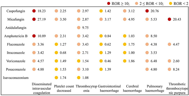
In terms of DIC, micafungin showed the strongest signal strength (ROR 27.19, 95%CI 18.49–39.98), followed by caspofungin (ROR 18.23, 95%CI 11.62–28.61) and amphotericin B (ROR 10.89, 95%CI 7.61–15.59). In terms of pulmonary haemorrhage, caspofungin had the highest signal strength (ROR 20.76, 95%CI 11.77–36.59), and for TTP, it was still micafungin that had the highest signal strength (ROR 20.43, 95%CI 8.49–49.14). It is worth noting that anidulafungin only showed a strong signal in thrombocytopenia (ROR 9.75, 95%CI 5.22–18.19). Other secondary outcome signals are mostly positive, but ROR values are generally below 10. (Fig. 2)
Fig. 2.
Subgroup analysis of different coagulation dysfunction events in different drugs
Time to onset and death outcome statistics
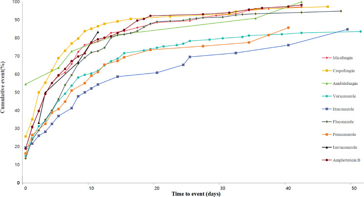
More than 70% of coagulopathy events associated with echinocandins were concentrated in the first 10 days of drug initiation, and the proportion of onset times exceeding 40 days was higher than for polyenes and triazoles. The top four shortest median onset time are: anidulafungin 0 (IQR 0-10.5), caspofungin 2.5 (IQR 0.25-7), micafungin 4 (IQR 1–9) and amphotericin B 4 (IQR 1–10). (Fig. 3)
Fig. 3.
Cumulative event (%) of coagulation events since the initiation of different drugs
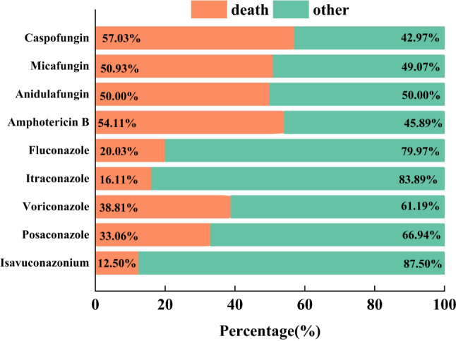
The top four drugs in terms of mortality were: caspofungin (57.03%), amphotericin B (54.11%), micafungin (50.93%) and anidulafungin (50.00%). Isavuconazole has the lowest mortality rate (12.50%). (Fig. 4)
Fig. 4.

The fatality rate related to coagulation dysfunction
Discussion
Although our results showed that anidulafungin, caspofungin and micafungin had significantly higher signal strength than triazoles and polyenes in coagulation dysfunction. At present, we can only find a small amount of research on the mechanism of micafungin leading to coagulation dysfunction, and caspofungin and anidulafungin are almost none.
The results of a case report show that the occurrence of TTP is most likely related to the use of micafungin by a mechanism that may be due to the fact that micafungin alters the function of ADAMTS13 or reduces the activity of ADAMTS13 through other pathways, which results in the circulation of von Willebrand factor (vWF), which in turn leads to the development of a platelet aggregant. ADAMTS13 works by cleaving vWF preventing it from forming large molecules, thus avoiding platelet aggregation [12]. Another view is that micafungin promotes thrombosis by causing eryptosis, which is accompanied by cell shrinkage [13] and cell membrane scrambling with phosphatidylserine translocation to the cell surface [14]. This view was confirmed in an in vitro cell trial [15]. It is worth noting that our results also showed a very strong signal between micafungen and TTP (ROR 20.43, 95%CI: 8.49–49.14) (Fig. 2). The patient in this report suffered cardiac arrest the day after the onset of TTP and subsequently died [12]. TTP is often fatal and if left untreated, usually results in death in 10–15 days [12]. Therefore, when necessary, patients using micafunzin should be monitored for laboratory indicators related to thrombosis, especially in the intensive care unit, because they are less able to describe their health than patients with mild disease. Although no relevant studies on micafungin and DIC were found, since it is clinically difficult to completely differentiate TTP from DIC [16], coupled with the fact that our results showed a very strong signal strength between micafungin and DIC (ROR 27.19, 95%CI 18.49–39.98), we believe that there is still a need to measure the coagulation parameters when using micafungin especially in patients with poor health.
In a multicentre phase IV clinical study, the AEs with the highest incidence in the micafungin group was decreased platelet count (8.2%) [17]. Results from another phase III clinical trial showed a 10% incidence of thrombocytopenia in the micafungin treatment group [10]. Our results also showed that positive signals were demonstrated between micafungin and both platelet count decreased (ROR 3.50, 95%CI: 2.36–5.19) and thrombocytopenia (ROR 2.87, 95%CI: 1.87–4.41). However, the exact mechanism is unclear.
Another case report documented that micafungin caused a patient to develop pure red cell aplasia (PRCA), which returned to normal levels after discontinuing micafungin [18]. The mechanism of its occurrence may be related to the immune response and metabolic pathways, but the exact mechanism is not known [19]. In addition, it has been reported that micafungin may cause immune complex type hemolytic anemia [20]. The main causes of drug-induced haemolytic anaemia include immunological or oxidative destruction of red blood cells (RBC). Drug-induced hemolysis of immune type can be divided into drug-dependent antibody mediated or drug-independent antibody mediated. The immune complex type is one of the drug-dependent antibody mediated types [21]. The evidence suggests that after the patient was given micafunzin, the body produced antibodies that could bind to micafunzin, and this complex caused the development of hemolytic anemia [20]. Although this type of hemolysis is very rare, it can be fatal. Therefore, as soon as micafungin is suspected of causing haemolysis in a patient, the drug should be discontinued and targeted treatment should be administered. There is a high degree of suspicion that the hemolysis caused by micafungine is closely related to bleeding at different sites. Our results also seem to support this hypothesis. The ROR (95%CI) between micafungine and gastrointestinal haemorrhage, cerebral haemorrhage and pulmonary haemorrhage were respectively: 3.17 (2.02–4.97), 4.95 (2.81–8.72), 5.53 (1.78–17.16). The results showed the strongest association between pulmonary haemorrhage and micafungin, followed by cerebral haemorrhage and gastrointestinal haemorrhage. This observation may be attributed to the location of fungal infection in the patient and the lungs are frequently susceptible to fungal infections.
In contrast, data on caspofungin and anidulafungin in coagulation dysfunction are mainly clinical trials and a few case reports, and the sample sizes are small. Results of a safety study of caspofungin showed that thrombocytopenia occurred in < 4% of patients in the caspofungin treatment group [22]. One study found the presence of platelet antibodies in a patient with caspofungin-induced thrombocytopenia by laboratory examination, and the normal bone marrow examination suggests that peripheral destruction of platelets may be the mechanism of caspofungin-induced thrombocytopenia rather than inhibition of platelet production [23]. The fact that there is an overlap between platelet count decreased and thrombocytopenia also makes it difficult for spontaneous reporters to distinguish between the two. Another study showed that caspofungin prolonged prothrombin time, activated partial thromboplastin time (aPTT), and international normalized ratio (INR) in patients with moderate-to-severe hepatic dysfunction, where the mean time of aPTT was prolonged by up to 5 s [24]. A case report shows that a small-intestine transplant patient treated with caspofungin developed DIC and eventually died [25]. In the analysis of the risk factors for linezolid-induced thrombocytopenia, the use of caspofungin was shown to be an independent risk factor [26, 27]. Our results showed a strong signal between caspofungin and pulmonary haemorrhage (ROR 20.76, 95%CI 11.78–36.59), but we didn’t find any relevant case reports, perhaps the positive signal may be related to the disease itself, just like invasive aspergillosis usually involves the lungs and can lead to bleeding in the lungs and gastrointestinal tract [28]. Nevertheless, the exact mechanism by which caspofungin causes coagulation dysfunction remains inconclusive.
Similarly, the results of a clinical trial of anidulafungin in children under 2 years showed a 10.5% incidence of thrombocytopenia, which was the only AE associated with the blood and lymphatic system. Our results similarly showed that anidulafungin showed stronger signal strength only with thrombocytopenia (ROR 9.75, 95%CI 5.22–18.19), and the two results and seem to be in agreement with each other. In another clinical trial in patients aged 2–18 years, the incidence of epistaxis in the micafungin group was 16.3%. The incidence of platelet count decreased decline was 10.20% and the incidence of DIC, cerebral haemorrhage, gastrointestinal haemorrhage and coagulopathy were all 2.04% [29]. More other post-marketing safety data were not found. Thus, we summarized the current potential mechanisms by which antimicrobials cause coagulation dysfunction: (1) Reducing synthesis of vitamin K-dependent coagulation factors II, VII, IX, and X by inhibiting vitamin K production of intestinal flora, e.g. cefoperazone [30, 31]. (2) Reducing fibrinogen by affecting liver function or gene expression, which causes coagulation dysfunction, such as tigecycline and fluconazole [32]. (3) Reducing platelet production by myelosuppression [33] or by immune-mediated increase in platelet clear [34]. (4) Drug interactions, such as fluconazole can lead to increased blood levels of superwarfarin and cause coagulation dysfunction [35]. These theories may be useful for further mechanistic studies.
An interesting topic is what is the risk of coagulation dysfunction in obese patients using echinocandins. More and more studies suggest that exposure to echinocandins is lower in obese patients compared to non-obese patients [36]. However, it has also been suggested that obese patients have a faster coagulation rate and greater antifibrinolytic capacity compared to the healthy population [37, 38]. These two conclusions may seem contradictory, and there are no studies on the risk of coagulation dysfunction in obese patients using echinocandins. This is worth exploring in depth.
In addition, considering that more than 70% AEs related with coagulation dysfunction in echinocandins occur in the first 10 days, coagulation parameters should be monitored regularly during the first week of drug use. Whether there is a causal relationship between high mortality and echinocandins needs to be explored in more rigorous studies, but it is certain that patients need to be alerted to a sudden deterioration in their health status during the use of echinocandins and amphotericin B. This is because once a coagulopathic event occurs, the patient is likely to die.
Although the study systematically predicted the risk of echinocandins and other antifungal drugs on coagulation dysfunction, there are still shortcomings. Firstly, the study does not prove a causal relationship between echinocandins and coagulation dysfunction, and the findings are only a speculation based on an algorithm. Second, the data included in the study may not fully reflect the real situation, as there are some errors or missing data in the self-reported reports. Third, the study was unable to determine the incidence of coagulation dysfunction because the total number of reports of the target patients as well as the target AEs were unknown. Lastly, the study could not rule out bias from the disease itself and drug interactions.
Conclusions
Our findings predict a higher association between echinocandins and coagulation disorders than both triazoles and polyenes. Moreover, of the three echinocandins, anidulafungin had the strongest association with coagulation dysfunction, followed by micafungin and caspofungin. The exact causality needs to be verified by further randomized controlled trials.
Electronic supplementary material
Below is the link to the electronic supplementary material.
Supplementary Material 1: Preferred terms related to coagulation dysfunction in MeDRA
Supplementary Material 2: Clinical characteristics of patients with caspofungin associated coagulation dysfunction events
Supplementary Material 3: Clinical characteristics of patients with micafungin-associated coagulation dysfunction events.
Supplementary Material 4: The calculation formula of disproportionality analysis and the criterion of positive
Supplementary Material 5: Target drugs and corresponding search terms
Supplementary Material 6: Clinical characteristics of patients with anidulafungin -associated coagulation dysfunction events.
Supplementary Material 7: Overall coagulation dysfunction signals and RORs comparing different drugs with the full database (Excluding consumer reports)
Acknowledgements
Thanks to Fan Yu and Rui Huang, two pharmacists in charge, who provided the necessary work for the early stage.
Abbreviations
- FAERS
Food and drug administration adverse event reporting system
- ROR
Reporting odds ratio
- PS
Primary suspect
- IQR
Interquartile range
- DIC
Disseminated intravascular coagulation
- TTP
Thrombotic thrombocytopenic purpura
- AE
Adverse event
- ALT
Alanine aminotransferase
- AST
Aspartate aminotransferase
- PT
Preferred term
- MedDRA
Medical dictionary for regulatory activity
- vWF
Von willebrand factor
- PRCA
Pure red cell aplasia
- RBC
Red blood cell
- aPTT
Activated partial thromboplastin time
- INR
International normalized ratio
Author contributions
Conception or design of the work: QC, and YW. Acquisition, analysis or interpretation of data: QC. Drafting the article: YW. Software: Zy Y. All authors critically reviewed the manuscript and participated in the interpretation of the results. The final manuscript was read, checked and approved by all authors.
Funding
Not applicable.
Data availability
No datasets were generated or analysed during the current study.
Declarations
Competing interests
The authors declare no competing interests.
Footnotes
Publisher’s Note
Springer Nature remains neutral with regard to jurisdictional claims in published maps and institutional affiliations.
Qian Cheng and Ye Wu contributed equally to this work.
References
- 1.Ong V, Hough G, Schlosser M, et al. Preclinical evaluation of the Stability, Safety, and efficacy of CD101, a Novel Echinocandin. Antimicrob Agents Chemother Nov. 2016;60(11):6872–9. 10.1128/aac.00701-16. 10.1128/aac.00701-16 [DOI] [PMC free article] [PubMed] [Google Scholar]
- 2.Eschenauer G, Depestel DD, Carver PL. Comparison of echinocandin antifungals. Ther Clin Risk Manag Mar. 2007;3(1):71–97. 10.2147/tcrm.2007.3.1.71. 10.2147/tcrm.2007.3.1.71 [DOI] [PMC free article] [PubMed] [Google Scholar]
- 3.de la Torre P, Reboli AC. Anidulafungin: a new echinocandin for candidal infections. Expert Rev Anti Infect Ther Feb. 2007;5(1):45–52. 10.1586/14787210.5.1.45. 10.1586/14787210.5.1.45 [DOI] [PubMed] [Google Scholar]
- 4.Bormann AM, Morrison VA. Review of the pharmacology and clinical studies of micafungin. Drug Des Devel Ther Dec. 2009;29:3:295–302. 10.2147/dddt.s3242. 10.2147/dddt.s3242 [DOI] [PMC free article] [PubMed] [Google Scholar]
- 5.Szymański M, Chmielewska S, Czyżewska U, Malinowska M, Tylicki A. Echinocandins - structure, mechanism of action and use in antifungal therapy. J Enzyme Inhib Med Chem Dec. 2022;37(1):876–94. 10.1080/14756366.2022.2050224. 10.1080/14756366.2022.2050224 [DOI] [PMC free article] [PubMed] [Google Scholar]
- 6.Eschenauer G, Depestel DD, Carver PL. Comparison of echinocandin antifungals. Ther Clin Risk Manag. 2007;3(1):71–97. 10.2147/tcrm.2007.3.1.71 [DOI] [PMC free article] [PubMed] [Google Scholar]
- 7.Mosteller RD. Simplified calculation of body-surface area. N Engl J Med Oct. 1987;22(17):1098. 10.1056/nejm198710223171717. 10.1056/nejm198710223171717 [DOI] [PubMed] [Google Scholar]
- 8.Villanueva A, Arathoon EG, Gotuzzo E, Berman RS, DiNubile MJ, Sable CA. A randomized double-blind study of caspofungin versus amphotericin for the treatment of candidal esophagitis. Clin Infect Dis Nov. 2001;1(9):1529–35. 10.1086/323401. 10.1086/323401 [DOI] [PubMed] [Google Scholar]
- 9.Roilides E, Carlesse F, Tawadrous M, et al. Safety, Efficacy and Pharmacokinetics of Anidulafungin in patients 1 Month to < 2 years of Age with Invasive Candidiasis, including Candidemia. Pediatr Infect Dis J Apr. 2020;39(4):305–9. 10.1097/inf.0000000000002568. 10.1097/inf.0000000000002568 [DOI] [PMC free article] [PubMed] [Google Scholar]
- 10.Benjamin DK Jr., Kaufman DA, Hope WW, et al. A phase 3 study of Micafungin Versus Amphotericin B Deoxycholate in infants with Invasive Candidiasis. Pediatr Infect Dis J Oct. 2018;37(10):992–8. 10.1097/inf.0000000000001996. 10.1097/inf.0000000000001996 [DOI] [PMC free article] [PubMed] [Google Scholar]
- 11.Thompson GR 3rd, Soriano A, Cornely OA, et al. Rezafungin versus caspofungin for treatment of candidaemia and invasive candidiasis (ReSTORE): a multicentre, double-blind, double-dummy, randomised phase 3 trial. Lancet Jan. 2023;7(10370):49–59. 10.1016/s0140-6736(22)02324-8. 10.1016/s0140-6736(22)02324-8 [DOI] [PubMed] [Google Scholar]
- 12.Nazzal M, Safi F, Arma F, Nazzal M, Muzaffar M, Assaly R. Micafungin-induced thrombotic thrombocytopenic purpura: a case report and review of the literature. Am J Ther Nov. 2011;18(6):e258–60. 10.1097/MJT.0b013e3181d5e401. 10.1097/MJT.0b013e3181d5e401 [DOI] [PubMed] [Google Scholar]
- 13.Lang PA, Kaiser S, Myssina S, Wieder T, Lang F, Huber SM. Role of Ca2+-activated K + channels in human erythrocyte apoptosis. Am J Physiol Cell Physiol Dec. 2003;285(6):C1553–60. 10.1152/ajpcell.00186.2003. 10.1152/ajpcell.00186.2003 [DOI] [PubMed] [Google Scholar]
- 14.Lang E, Lang F. Mechanisms and pathophysiological significance of eryptosis, the suicidal erythrocyte death. Semin Cell Dev Biol Mar. 2015;39:35–42. 10.1016/j.semcdb.2015.01.009. 10.1016/j.semcdb.2015.01.009 [DOI] [PubMed] [Google Scholar]
- 15.Peter T, Bissinger R, Signoretto E, Mack AF, Lang F. Micafungin-Induced suicidal erythrocyte death. Cell Physiol Biochem. 2016;39(2):584–95. 10.1159/000445650. 10.1159/000445650 [DOI] [PubMed] [Google Scholar]
- 16.Furlan M, Lämmle B. Aetiology and pathogenesis of thrombotic thrombocytopenic purpura and haemolytic uraemic syndrome: the role of Von Willebrand factor-cleaving protease. Best Pract Res Clin Haematol Jun. 2001;14(2):437–54. 10.1053/beha.2001.0142. 10.1053/beha.2001.0142 [DOI] [PubMed] [Google Scholar]
- 17.Ji Y, Song Y, Zhou F, et al. Efficacy and safety of micafungin for the treatment of patients with proven or probable invasive aspergillosis: a non-comparative, multicenter, phase IV, open-label study. Med (Baltimore) Dec. 2017;96(52):e9443. 10.1097/md.0000000000009443. 10.1097/md.0000000000009443 [DOI] [PMC free article] [PubMed] [Google Scholar]
- 18.Yoshida-Hiroi M, Koizumi M, Oka R, Mitsuda A, Hiroi N. First case report of acquired pure red cell aplasia associated with micafungin. Intern Med. 2011;50(9):1051–4. 10.2169/internalmedicine.50.4303. 10.2169/internalmedicine.50.4303 [DOI] [PubMed] [Google Scholar]
- 19.Yunis AA, Arimura GK, Lutcher CL, Blasquez J, Halloran M. Biochemical lesion in Dilantin-induced erythroid aplasia. Blood Nov. 1967;30(5):587–600. 10.1182/blood.V30.5.587.587 [DOI] [PubMed] [Google Scholar]
- 20.Ishii H, Sato T, Ishibashi M, et al. A case of immune complex type hemolytic anemia induced by initial micafungin administration. Int J Infect Dis Sep. 2022;122:755–7. 10.1016/j.ijid.2022.07.023. 10.1016/j.ijid.2022.07.023 [DOI] [PubMed] [Google Scholar]
- 21.Garratty G. Immune hemolytic anemia associated with drug therapy. Blood Rev Jul-Sep. 2010;24(4–5):143–50. 10.1016/j.blre.2010.06.004. 10.1016/j.blre.2010.06.004 [DOI] [PubMed] [Google Scholar]
- 22.Sable CA, Nguyen BY, Chodakewitz JA, DiNubile MJ. Safety and tolerability of caspofungin acetate in the treatment of fungal infections. Transpl Infect Dis Mar. 2002;4(1):25–30. 10.1034/j.1399-3062.2002.01004.x. 10.1034/j.1399-3062.2002.01004.x [DOI] [PubMed] [Google Scholar]
- 23.Lynch J, Wong-Beringer A. Caspofungin: a potential cause of reversible severe thrombocytopenia. Pharmacotherapy Oct. 2004;24(10):1408–11. 10.1592/phco.24.14.1408.43148. 10.1592/phco.24.14.1408.43148 [DOI] [PubMed] [Google Scholar]
- 24.Yuan SD, Wen KL, Cao YX, Huang WQ, Zhang A. Safety and efficacy of non-reduced use of caspofungin in patients with child-pugh B or C cirrhosis: a real-world study. Infect Jun. 2024;52(3):1063–72. 10.1007/s15010-023-02162-0. 10.1007/s15010-023-02162-0 [DOI] [PMC free article] [PubMed] [Google Scholar]
- 25.Köse S, Türken M, Akkoçlu G, Karaca C, Senger SS. Disseminated candidiasis developing during prophylaxis with fluconazole in a small-intestine transplant recipient. Prog Transpl Mar. 2012;22(1):110–2. 10.7182/pit2012329. 10.7182/pit2012329 [DOI] [PubMed] [Google Scholar]
- 26.Chen C, Guo DH, Cao X, et al. Risk factors for thrombocytopenia in adult Chinese patients receiving linezolid therapy. Curr Ther Res Clin Exp Dec. 2012;73(6):195–206. 10.1016/j.curtheres.2012.07.002. 10.1016/j.curtheres.2012.07.002 [DOI] [PMC free article] [PubMed] [Google Scholar]
- 27.Zhang L, Yan Y, Liao R, Dong H. Effect of platelet parameters on Linezolid-related Thrombocytopenia in Hospitalized patients. Infect Drug Resist. 2023;16:6145–54. 10.2147/idr.S408102. 10.2147/idr.S408102 [DOI] [PMC free article] [PubMed] [Google Scholar]
- 28.Ceylan B, Yilmaz M, Beköz HS, Ramadan S, Ertan Akan G, Mert A. Primary gastrointestinal aspergillosis: a case report and literature review. Infez Med Mar. 2019;1(1):85–92. [PubMed] [Google Scholar]
- 29.Roilides E, Carlesse F, Leister-Tebbe H, et al. A prospective, open-label study to assess the Safety, Tolerability and Efficacy of Anidulafungin in the Treatment of Invasive Candidiasis in Children 2 to < 18 years of age. Pediatr Infect Dis J Mar. 2019;38(3):275–9. 10.1097/inf.0000000000002237. 10.1097/inf.0000000000002237 [DOI] [PubMed] [Google Scholar]
- 30.Hua CZ, Wang HJ, Zhang Z, et al. In vitro activity and clinical efficacy of macrolides, cefoperazone-sulbactam and piperacillin/piperacillin-tazobactam against Bordetella pertussis and the clinical manifestations in pertussis patients due to these isolates: a single-centre study in Zhejiang Province, China. J Glob Antimicrob Resist Sep. 2019;18:47–51. 10.1016/j.jgar.2019.01.029. 10.1016/j.jgar.2019.01.029 [DOI] [PubMed] [Google Scholar]
- 31.Li Y, Xie J, Chen L, et al. Treatment efficacy of tigecycline in comparison to cefoperazone/ sulbactam alone or in combination therapy for carbapenenm-resistant Acinetobacter baumannii infections. Pak J Pharm Sci Jan. 2020;33(1):161–8. [PubMed] [Google Scholar]
- 32.Zhang Q, Zhou S, Zhou J. Tigecycline treatment causes a decrease in fibrinogen levels. Antimicrob Agents Chemother Mar. 2015;59(3):1650–5. 10.1128/aac.04305-14. 10.1128/aac.04305-14 [DOI] [PMC free article] [PubMed] [Google Scholar]
- 33.Stephen L. Green. Linezolid and reversible myelosuppression. JAMA. 2001;285(10):1973–4. [DOI] [PubMed] [Google Scholar]
- 34.Pascoalinho D, Vilas MJ, Coelho L, Moreira P. Linezolid-related immune-mediated severe thrombocytopenia. Int J Antimicrob Agents Jan. 2011;37(1):88–9. 10.1016/j.ijantimicag.2010.10.001. 10.1016/j.ijantimicag.2010.10.001 [DOI] [PubMed] [Google Scholar]
- 35.Kim SY, Cho SY, Lee HJ, et al. Superwarfarin intoxication of unknown etiology accompanying hemoperitoneum in a patient on fluconazole therapy. Ann Clin Lab Sci Summer. 2010;40(3):300–3. [PubMed] [Google Scholar]
- 36.Alsowaida YS, Alamer A, Thabit AK, et al. Echinocandin exposures in obese patients: a scoping review and clinical perspectives. Am J Health Syst Pharm Apr. 2023;8(8):503–17. 10.1093/ajhp/zxad021. 10.1093/ajhp/zxad021 [DOI] [PubMed] [Google Scholar]
- 37.Samuels J, Lawson PJ, Morton AP, et al. Prospective assessment of fibrinolysis in morbid obesity: tissue plasminogen activator resistance improves after bariatric surgery. Surg Obes Relat Dis Jul. 2019;15(7):1153–9. 10.1016/j.soard.2019.03.048. 10.1016/j.soard.2019.03.048 [DOI] [PMC free article] [PubMed] [Google Scholar]
- 38.De Pergola G, Pannacciulli N. Coagulation and fibrinolysis abnormalities in obesity. J Endocrinol Invest Nov. 2002;25(10):899–904. 10.1007/bf03344054. 10.1007/bf03344054 [DOI] [PubMed] [Google Scholar]
Associated Data
This section collects any data citations, data availability statements, or supplementary materials included in this article.
Supplementary Materials
Supplementary Material 1: Preferred terms related to coagulation dysfunction in MeDRA
Supplementary Material 2: Clinical characteristics of patients with caspofungin associated coagulation dysfunction events
Supplementary Material 3: Clinical characteristics of patients with micafungin-associated coagulation dysfunction events.
Supplementary Material 4: The calculation formula of disproportionality analysis and the criterion of positive
Supplementary Material 5: Target drugs and corresponding search terms
Supplementary Material 6: Clinical characteristics of patients with anidulafungin -associated coagulation dysfunction events.
Supplementary Material 7: Overall coagulation dysfunction signals and RORs comparing different drugs with the full database (Excluding consumer reports)
Data Availability Statement
No datasets were generated or analysed during the current study.


