Abstract
Background
Davoceticept (ALPN-202) is an Fc fusion of a CD80 variant immunoglobulin domain designed to mediate programmed death-ligand 1 (PD-L1)-dependent CD28 co-stimulation while inhibiting the PD-L1 and cytotoxic T-lymphocyte-associated antigen 4 (CTLA-4) checkpoints. The safety and efficacy of davoceticept monotherapy and davoceticept and pembrolizumab combination therapy in adult patients with advanced solid tumors were explored in NEON-1 and NEON-2, respectively.
Methods
In NEON-1 (n=58), davoceticept 0.001–10 mg/kg was administered intravenous either once weekly (Q1W) or once every 3 weeks (Q3W). In NEON-2 (n=29), davoceticept was administered intravenously at 2 dose levels (0.1 or 0.3 mg/kg) Q1W or Q3W with pembrolizumab (400 mg once every 6 weeks). In both studies, primary endpoints included incidence of dose-limiting toxicities (DLT); type, incidence, and severity of adverse events (AEs) and laboratory abnormalities; and seriousness of AEs. Secondary endpoints included antitumor efficacy assessed using RECIST v1.1, pharmacokinetics, anti-drug antibodies, and pharmacodynamic biomarkers.
Results
The incidence of treatment-related AEs (TRAEs) and immune-related adverse events (irAEs) was 67% (39/58) and 36% (21/58) with davoceticept monotherapy, and 62% (18/29) and 31% (9/29) with davoceticept and pembrolizumab combination, respectively. The incidence of ≥grade (Gr)3 TRAEs and ≥Gr3 irAEs was 12% (7/58) and 5% (3/58) with davoceticept monotherapy, and 24% (7/29) and 10% (3/29) with davoceticept and pembrolizumab combination, respectively. One DLT of Gr3 immune-related gastritis occurred during davoceticept monotherapy 3 mg/kg Q3W. During davoceticept combination with pembrolizumab, two Gr5 cardiac DLTs occurred; one instance each of cardiogenic shock (0.3 mg/kg Q3W, choroidal melanoma metastatic to the liver) and immune-mediated myocarditis (0.1 mg/kg Q3W, microsatellite stable metastatic colorectal adenocarcinoma), prompting early termination of both studies. Across both studies, five patients with renal cell carcinoma (RCC) exhibited evidence of clinical benefit (two partial response, three stable disease).
Conclusions
Davoceticept was generally well tolerated as monotherapy at intravenous doses up to 10 mg/kg. Evidence of clinical activity was observed with davoceticept monotherapy and davoceticept in combination with pembrolizumab, notably in RCC. However, two fatal cardiac events occurred with the combination of low-dose davoceticept and pembrolizumab. Future clinical investigation with davoceticept should not consider combination with programmed death-1-inhibitor anticancer mechanisms, until its safety profile is more fully elucidated.
Trial registration number
NEON-1 (NCT04186637) and NEON-2 (NCT04920383).
Keywords: Immune Checkpoint Inhibitor, co-inhibitory molecule, Combination therapy, Immunotherapy
WHAT IS ALREADY KNOWN ON THIS TOPIC.
WHAT THIS STUDY ADDS
NEON-1 and NEON-2 were two phase I, open-label dose-escalation trials that evaluated davoceticept as monotherapy (NEON-1), and in combination with pembrolizumab (NEON-2). Although both monotherapy and combination strategies resulted in clinical benefits including durable objective responses in some heavily pretreated patients, and davoceticept was well tolerated as monotherapy, the combination of davoceticept with pembrolizumab produced a higher incidence of Grade ≥3 adverse events, including two fatal cardiac events.
HOW THIS STUDY MIGHT AFFECT RESEARCH, PRACTICE OR POLICY.
CD28 agonism continues to have nascent therapeutic potential, as evidenced by several findings of clinical benefit in these studies, but further clinical investigation with davoceticept and perhaps other CD28 agonists should, for now, consider combinations with mechanisms besides PD-1-inhibitors given the potential for additive toxicity, until the safety profile of davoceticept and other CD28 agonists are more fully elucidated.
Introduction
Programmed death-1 (PD-1) is a receptor expressed by activated T cells that binds to programmed death-ligand-1 (PD-L1) (B7-H1)1 2 and PD-L2 (B7-DC).3 4 PD-1 suppresses T-cell functions on the engagement of PD-L1, which is expressed by a wide variety of tissues.1,4 PD-L1 is also expressed by human tumors, either constitutively or after treatment with interferon gamma (IFN-γ), as a mechanism of immune evasion.5 6 Cytotoxic T lymphocyte-associated antigen-4 (CTLA-4, CD152) is an activation-induced glycoprotein that belongs to the immunoglobulin superfamily with homology to the T-cell co-stimulatory protein CD28. It is constitutively expressed on regulatory T cells but is rapidly induced on CD8+T cells following T-cell receptor (TCR) engagement.7 8 While CD28 provides the co-stimulatory signal required for antigen-specific T-cell activation and expansion after the initial interaction between TCR and antigen-presenting cells (APCs), CTLA-4 downregulates T-cell responses by acting as a decoy receptor8 9 and competing for binding to the ligands it shares with CD28, namely CD80 (B7.1) and CD86 (B7.2).10,15 Interestingly, in addition to binding to CD28 and CTLA-4, CD80 also binds to PD-L1 in cis on the surface of APCs,16 17 which may allow for CD28 co-stimulation while interfering with PD-L1 inhibitory signaling.18,20 Inhibition of PD-1 and/or CTLA-4 by monoclonal antibodies (mAb), overcomes tumor-mediated immune suppression and leads to antitumor immune responses and improved survival in multiple cancers including advanced melanoma, renal cell carcinoma (RCC), and non-small cell lung cancer (NSCLC).21,24
PD-1 and CTLA-4 interfere with signal transduction downstream of CD28, which can be relieved by PD-1 or CTLA-4 targeting mAbs.25 26 However, these mAbs are unable to drive the CD28 co-stimulatory signal that is requisite for T-cell activation.27 In preclinical models, tumor-targeted CD28 activation by engineered expression of CD28 ligands on tumor cells produces profound antitumor immune responses.28,32 We hypothesized that conditional CD28 co-stimulation may overcome resistance to immune checkpoint blockade.
Davoceticept includes a CD80 variant immunoglobulin domain that was engineered to have increased affinity for PD-L1 and CD28 relative to wild-type CD80, while retaining its ability to bind to CTLA-4. It inhibits both the PD-L1 and CTLA-4 checkpoints and also mediates PD-L1-dependent CD28 co-stimulation.33 The clinical development program for davoceticept was initiated with two phase I, open-label dose-escalation trials: a first-in-human study of davoceticept monotherapy (NEON-1) and a study of davoceticept in combination with pembrolizumab (NEON-2). The consideration of a PD-1 inhibitor combination with davoceticept was of particular interest because PD-1 inhibition leads to PD-L1 upregulation,34,36 potentially synergizing with davoceticept’s PD-L1-dependent co-stimulatory mechanism; and in preclinical models, the combination of davoceticept and anti-PD-1 inhibited tumor growth to a greater extent than either agent alone.33
Methods
Study design (brief)
NEON-1 and NEON-2 were multicenter, open-label, phase I trials that enrolled patients at 10 and 5 sites, respectively. NEON-1 dose escalation was conducted in the USA and Australia and enrolled patients between June 2020 and January 2022; NEON-2 was initiated after the initial safety of davoceticept monotherapy had been ascertained, was conducted only in the USA, and enrolled patients between June 2021 and October 2022. Patients were moved to long-term follow-up after treatment in NEON-1 and NEON-2 was terminated. Long-term follow-up for both NEON-1 and NEON-2 was terminated in February 2023. Both trials were designed and funded by Alpine Immune Sciences, and were conducted in accordance with all local legal and regulatory requirements, as well as the general principles set forth in the International Ethical Guidelines for Biomedical Research Involving Human Subjects, Guidelines for Good Clinical Practice, and the Declaration of Helsinki. The names and reference/ID numbers of the committees that approved these studies at the trial sites are listed in the online supplemental materials. All patients provided written informed consent. NEON-2 was conducted in collaboration with Merck & Co (Rahway, New Jersey, USA).
Data were collected and analyzed by the sponsor and lead author. The manuscript was written by the lead author in collaboration with the Alpine development team. All authors approved the final version of the manuscript.
Patients
Both studies enrolled adults ≥18 years old with pathologically confirmed, metastatic solid tumors, at least one measurable lesion (as defined by RECIST v.1.1). All patients were required to have an Eastern Cooperative Oncology Group performance status of 0–1, life expectancy ≥3 months and adequate hematologic, renal, hepatic, and cardiac function. Patients enrolled in NEON-1 dose escalation had locally advanced or metastatic unresectable solid tumors that were refractory or resistant to standard therapy (including immune checkpoint inhibitors (ICIs), if approved and available) or for which standard or curative therapy was not available. NEON-2 selectively enrolled patients with ICI-sensitive advanced tumors that were eligible for treatment with PD-(L)1 ICI, were refractory or resistant to standard treatment, or for whom standard or curative therapy was not available. Following the first adverse event (AE) of Gr5 cardiogenic shock, the NEON-2 inclusion and exclusion criteria were modified to exclude patients who were at increased risk of cardiotoxicity or who harbored significant delayed pharmacodynamic (PD) effects of prior immunotherapies. Specifically, patients with prior any-grade myocarditis, significant cardiovascular events within 6 months of planned treatment administration, clinically significant atrial and/or ventricular arrhythmias and left ventricular ejection fraction <45% on screening echocardiograms were excluded. The full list of inclusion and exclusion criteria can be found in the study protocols included in the online supplemental materials.
Interventions
In NEON-1, davoceticept was administered intravenous at 0.001 mg/kg to 10 mg/kg either once weekly (Q1W) or once every 3 weeks (Q3W). In NEON-2, davoceticept was administered intravenous at 0.1 and 0.3 mg/kg Q1W or Q3W. The starting doses in NEON-2 (0.1 mg/kg Q1W or Q3W) were chosen based on the preliminary tolerability and pharmacokinetics (PK) of davoceticept monotherapy in NEON-1. Pembrolizumab was dosed as 400 mg once every 6 weeks. In both NEON-1 and NEON-2, patients were treated until one of the following occurred: a dose-limiting toxicities (DLT), an intolerable AE, confirmed progressive disease, withdrawal of consent, or study termination.
Endpoints
The primary endpoints for the dose escalation portions of NEON-1 and NEON-2 were incidence of DLTs, type, incidence severity and seriousness of AEs per Common Terminology Criteria for Adverse Events V.5.0 and type, incidence, and severity of laboratory abnormalities. Immune-related adverse events (irAEs) were defined and managed per society and consensus criteria37 38 and assessed by investigators. Secondary endpoints included antitumor efficacy as assessed using RECIST V.1.1, PK, anti-drug antibodies (ADA), and biomarkers of response to davoceticept. The DLT period for NEON-1 was 21 days and 42 days for patients on the Q1W and Q3W schedules, respectively. The DLT period for NEON-2 was initially 21 days, however, it was lengthened to 42 days after the first AE of Gr5 cardiogenic shock.
Efficacy endpoints included overall response rate (ORR), duration of response, disease control rate (DCR), progression-free survival, and overall survival. NEON-1 also included a secondary endpoint of the duration of stable disease. Efficacy was assessed every 6 weeks for the first 6 months, then every 9 weeks until 1 year, and then every 12 weeks thereafter.
Statistical analyses
The planned analyses for both NEON-1 and NEON-2 were descriptive, with descriptive statistics to describe continuous variables and frequencies and percentages to describe categorical variables. There was no statistical inference testing planned for either study.
Antitumor activity
Antitumor activity was assessed through radiologic tumor assessments conducted at baseline, during treatment, at suspected disease progression, and at the time of withdrawal from treatment. Assessment of tumor response was per RECIST V.1.1.
PK and PD biomarkers
Details regarding davoceticept concentration determination, drug saturation, ADA assessment, and analysis are provided in online supplemental materials (see online supplemental methods).
Peripheral blood mononuclear cell samples were assayed by flow cytometry for key cellular subsets and evidence of activation including staining with Ki67, CD4, and CD8. Peripheral blood cytokines were assessed by multiplexed immunoassay (Myriad RBM; Austin Texas, USA) and PD-L1 combined positive score and tumor progression score were assessed by immunohistochemistry using the 22C3 antibody.
Results
Patients and baseline characteristics
In total, 58 patients from 10 sites were enrolled in the dose escalation portion of NEON-1. Patients received davoceticept Q1W at 0.001 mg/kg (n=1), 0.01 mg/kg (n=2), 0.1 mg/kg (n=4), 0.3 mg/kg (n=6), 1 mg/kg (n=3) and 3 mg/kg (n=4); and davoceticept Q3W at 0.3 mg/kg (n=3), 1 mg/kg (n=11), 3 mg/kg (n=15) and 10 mg/kg (n=9) (online supplemental figure 1A). Three patients enrolled initially in the davoceticept 0.3 mg/kg Q1W cohort were escalated to 1 mg/kg Q1W after initial demonstration of tolerability, as allowed per-protocol. A total of 29 patients from five sites were enrolled in the NEON-2 study. Cohorts were enrolled as follows: Q1W, 0.1 mg/kg davoceticept (n=9), 0.3 mg/kg davoceticept (n=3); and Q3W regimen, 0.1 mg/kg davoceticept (n=9) and 0.3 mg/kg davoceticept (n=8) (online supplemental figure 1B). Overall, patient disposition for studies NEON-1 and NEON-2 is shown in online supplemental table 1.
The demographic and clinical characteristics of NEON-1 dose escalation and NEON-2 patients are summarized in online supplemental table 2). Briefly, the NEON-1 and NEON-2 patients were primarily men and white with a median age of 60 years. The median number of prior systemic therapies was 3, and the two most common tumor types in patients enrolled in both studies were colorectal cancer (NEON-1, 24%; NEON-2, 34%) and pancreatic cancer (NEON-1, 19%; NEON-2, 10%). NEON-1 and NEON-2 enrolled 5% and 7% of patients with RCC, respectively. The proportion of patients with prior PD-(L)1 exposure was 29% in NEON-1 (17/59), and 31% in NEON-2 (9/29).
Pharmacokinetics and drug saturation
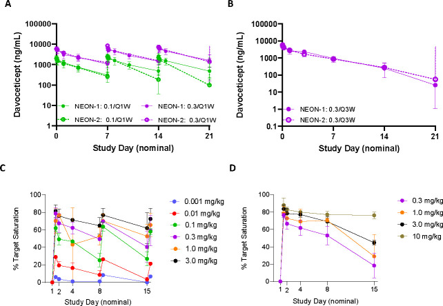
Dose-dependent increases in serum davoceticept concentrations were observed during davoceticept administration as monotherapy and in combination with pembrolizumab; PK were not significantly affected by the combination with pembrolizumab (figure 1A and B). ADAs (as defined by study-specific titer threshold of ≥1:256) were observed in 14% versus 24% of patients who received davoceticept monotherapy versus in combination with pembrolizumab, respectively, with no evidence of increasing ADA titer by dose. Further, there was no clear relationship demonstrable between ADA and safety, PK, PD, or efficacy (data not shown).
Figure 1. Pharmacokinetics and drug saturation in the NEON studies pharmacokinetics of davoceticept during monotherapy (NEON-1, solid lines) and pembrolizumab combination (NEON-2, dashed lines) by dose (●, 0.1 mg/kg davoceticept; ●, 0.3 mg/kg davoceticept) and regimen; once weekly, Q1W (A) and once every 3 weeks, Q3W (B); dose-dependent drug saturation in NEON-1 (C) and NEON-2 (D). Drug saturation of CD28 on circulating CD4+T cells by davoceticept was determined by flow cytometry pre-dose and following infusion using an antibody specific for davoceticept bound to CD28. Per cent saturation was calculated using a ratio of test article to saturated control and normalized to cycle 1 pre-dose.
Dose-dependent drug saturation, primarily reflecting occupancy of CD28 and as assessed via flow cytometry of circulating CD4+T cells, was maximal at the end of infusion during davoceticept monotherapy (days 1, 8, and 15 for Q1W and at day 1 for Q3W) and decreased over time for both Q1W (figure 1C) and Q3W (figure 1D) regimens. Drug saturation did not appear to be significantly affected by the combination with pembrolizumab (online supplemental figure 2).
Safety
Table 1 displays the AE summary of davoceticept monotherapy and in combination with pembrolizumab.
Table 1. Summary of adverse events in NEON-1 and NEON-2.
| AE, n patients (%) | NEON-1n=58 | NEON-2n=29 |
| Any grade TEAE | 58 (100) | 26 (90) |
| Any grade TRAE | 39 (67) | 18 (62) |
| Any AE of Interest | 26 (45) | 11 (38) |
| irAE | 21 (36) | 9 (31) |
| IRR | 9 (16) | 1 (3) |
| Cytokine release syndrome | 0 | 0 |
| Anaphylaxis | 0 | 0 |
| Any grade ≥3 TEAE | 32 (55) | 18 (62) |
| Any grade ≥3 TRAE | 7 (12) | 7 (24) |
| Any grade ≥3 irAE | 3 (5) | 3 (10) |
| Acute kidney injury | 1 (2) | 0 |
| Cardiogenic shock | 0 | 1 (3) |
| Gastritis | 1 (2) | 0 |
| Immune-mediated myocarditis | 0 | 1 (3) |
| Testicular pain | 1 (2) | 0 |
| Tubulointerstitial nephritis | 0 | 1 (3) |
| Urticaria | 1 (2) | 0 |
Adverse events were graded according to Common Terminology Criteria for Adverse Events V.5.0. For administration-related reactions, IRR was generally used unless more specific terms (ie, cytokine release syndrome or anaphylaxis) were definitively determined.
AEadverse eventirAEimmune-related adverse eventIRRinfusion-related reactionTEAEtreatment-emergent adverse eventTRAEtreatment-related adverse event
Davoceticept monotherapy was well-tolerated overall. TRAEs occurred in 39 patients (67%), the most common of which were fatigue (17%), infusion-related reaction (IRR) (16%), and maculopapular rash (14%) (online supplemental table 3). IRRs were all low grade (≤Gr2) and did not require treatment discontinuation. Seven patients (12%) experienced Gr3 TRAEs that included acute kidney injury, testicular pain, increased alanine aminotransferase (ALT), gastritis, increased lipase, anemia, fatigue, diarrhea, dehydration, hypokalemia, and urticaria (all n=1) (online supplemental table 4). irAEs were observed in 21 patients (36%), the majority of which were Gr1 or 2 and related to the skin (eg, macular rash, maculopapular rash, urticaria) (online supplemental table 5). In one patient with colon cancer treated with a single dose of davoceticept (3 mg/kg), a single DLT of biopsy-confirmed Gr3 immune-related gastritis occurred, which was treated with corticosteroids and resolved within 8 days. There were no Gr4 or 5 TRAEs following treatment with davoceticept monotherapy.
Combined administration of davoceticept and pembrolizumab was associated with an increased frequency of Gr3+AEs (table 1). TRAEs occurred in 18 patients (62%), the most common of which were increases in ALT, increases in aspartate aminotransferase (AST), fatigue, nausea, and fever (10% each) (online supplemental table 3). Seven patients (24%) experienced Gr3+TRAEs which included AST increase (n=2), and one each of the following: IRR, hypotension, biopsy-confirmed tubulointerstitial nephritis, cardiogenic shock (presumed to be immune-mediated myocarditis), and biopsy-confirmed immune-mediated myocarditis (online supplemental table 4). The two patients with Gr3 increases in AST were treated with corticosteroids; one was treated to resolution after 5 days and the other patient’s AST was not resolved at the time of the last follow-up. For this latter patient, ALT was also elevated and reported as a grade 2 TRAE. This patient died due to clinical progression just under 3 months from the time of the reported AST/ALT increase. The patient with Gr3 biopsy-confirmed tubulointerstitial nephritis had been treated 22 days prior with a single dose of 0.3 mg/kg davoceticept. The AE was deemed to be immune-related, was successfully treated with corticosteroids, and resolved after 3 weeks. The IRR occurred in a patient treated with davoceticept at 0.1 mg/kg, Q3W and presented as Gr3 dyspnea, Gr2 flank pain, and Gr2 hypertension that began about 1.5 hours following the start of the infusion as a Gr2 event that ultimately required hospitalization and was upgraded to a Gr3 IRR. Treatment included oxygen and corticosteroids; the IRR resolved within 3 days. The patient with Gr4 hypotension presented with Gr2 pruritic rash, a low-grade fever, malaise, and anuria for 2 days. After being hospitalized and initiating intravenous vancomycin, ceftriaxone, and azithromycin, the patient became hypotensive and required transient pressor support with norepinephrine and vasopressin and a stress dose of steroids with hydrocortisone. While there was concern for possible encephalitis and/or urinary tract infection, no definitive etiology of the hypotension was determined. On the subsequent day, the patient was weaned from pressors, the blood pressure stabilized without intervention, and the hypotension was considered resolved.
irAEs were reported in 9 (34%) patients treated with davoceticept plus pembrolizumab (online supplemental table 5). They were generally higher in grade than those reported during davoceticept monotherapy and included two Gr5 DLTs. Briefly, the first event of cardiogenic shock with suspected myocarditis developed in a female patient with choroidal melanoma who had received a single dose of davoceticept (0.3 mg/kg Q3W) and pembrolizumab. The second event of immune-mediated myocarditis occurred in a female patient with microsatellite stable colorectal adenocarcinoma who received one dose of davoceticept (0.1 mg/kg Q3W) plus pembrolizumab. Analysis of cardiac tissue obtained at autopsy from the latter patient confirmed immune-mediated myocarditis. Both patients presented with rapidly progressive symptoms approximately 2 weeks after their first and only dose of study treatment. Both patients presented with fatigue and nausea that coincided with increases in troponin-T and NT-pro-BNP in the former and troponin-I, BNP and CK in the latter, but without evidence of cytokine storm. Despite pressor support and treatment with high-dose steroids (both patients) and mycophenolate mofetil (one patient), both patients rapidly declined and passed away. Further detail of both Gr5 cardiac irAEs is provided in the accompanying article by Cavalcante et al, 2024. Following the second cardiac event, further dosing and enrollment in NEON-1 and NEON-2 were permanently discontinued.
Antitumor efficacy
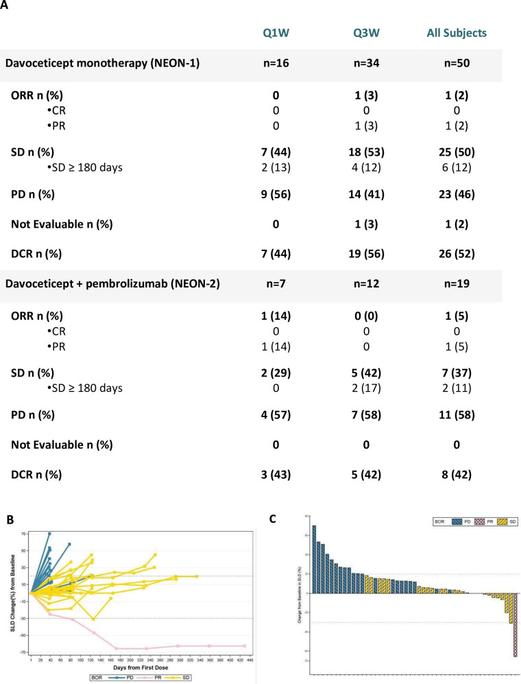
Of the 58 patients treated with davoceticept monotherapy, 50 were efficacy-evaluable (figure 2). Eight patients were not efficacy-evaluable due to a lack of on-treatment imaging, and one efficacy-evaluable patient had scans that were inadequate in quality and were therefore considered non-eval uable (figure 2A). One (2%) patient with PD-1 inhibitor-naïve papillary RCC had a deep and durable confirmed partial response (PR), and 25 patients (50%) had a best overall response (BoR) of stable disease (SD) for a DCR of 52%. Of the 25 patients with SD, 5 (10%) had SD for ≥180 days including one patient with clear cell RCC (ccRCC) with SD that lasted 12.1 months (online supplemental table 6).
Figure 2. Antitumor efficacy of davoceticept monotherapy (NEON-1) or davoceticept+pembrolizumab combination (NEON-2). Per cent change in the sum of the longest diameters during davoceticept monotherapy (NEON-1) are shown over time (panel B) and by the best change from baseline and overall response (panel C). The PRs in NEON-1 and NEON-2 were confirmed with repeat scans. BoR, best overall response; CR, complete response; DCR, disease control rate; ORR, objective response rate; PD, progressive disease; PR, partial response; Q1W, once weekly; Q3W, once every 3 weeks; SD, stable disease; SLD, sum of the longest diameters.
Of the 29 patients treated with davoceticept in combination with pembrolizumab, 19 were efficacy-evaluable with on-treatment imaging (figure 2A, online supplemental figure 3). Five patients were not efficacy-evaluable as the trial was discontinued prior to their first on-treatment scan; an additional five patients had discontinued the trial prior to their first on-treatment scan due to an AE (n=1), clinical progression (n=2) or death (n=2). One patient with PD-1-refractory ccRCC achieved a BoR of PR for an ORR of 5% and seven (37%) patients achieved a BoR of SD, for a DCR of 42%. Three (16%) patients achieved SD for ≥180 days including a PD-1 refractory patient with poorly differentiated RCC with SD that lasted 8.3 months (online supplemental figure 3). Of note, one patient with heavily pretreated metastatic castration-resistant prostate cancer demonstrated 14% tumor shrinkage that was coincident with a decrease in circulating prostate-specific antigen from 622 µg/L to 387 µg/L after only two doses of davoceticept (0.3 mg/kg, Q3W) and pembrolizumab.
A total of five patients with RCC received davoceticept (three as monotherapy, and two in combination with pembrolizumab) and all had some evidence of clinical benefit. The demographics and outcomes of these patients are summarized in online supplemental table 6 and online supplemental figure 4. The confirmed PR during davoceticept monotherapy was in a patient with treatment-naïve papillary RCC who was on treatment for about 14 months with a maximum decrease of 66% in the sum of the longest diameters (SLD) of the target lesions. The confirmed PR during davoceticept in combination with pembrolizumab was a patient with ccRCC who was on treatment for 16.6 months, with ongoing response at the time of study termination. This patient had been treated with six prior lines of therapy, including two containing PD-1 inhibitors, and had a 45% decrease in the SLD of his target lesions. While the patients with RCC enrolled varied by histology, prior treatments and davoceticept dose level, all five patients had evidence of disease control following treatment, and three (one in monotherapy, and two in combination with pembrolizumab) were still receiving treatment at the time of study termination.
Peripheral immune activation in patients treated with davoceticept (NEON-1) or davoceticept+pembrolizumab (NEON-2)
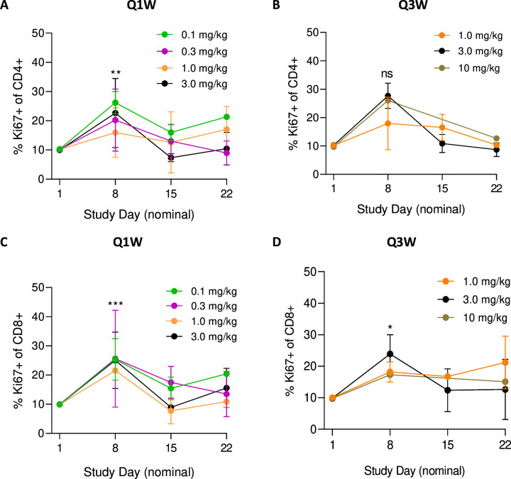
Response to anti-PD-1 therapy in melanoma and NSCLC is associated with early on-treatment changes in proliferating CD8+T cells39 40; while response to anti-CTLA-4 is associated with an expansion of both CD4+ and CD8+ T cells.41 42 Davoceticept monotherapy was associated with an early expansion of both CD4+ and CD8+ T cells that peaked at day 8, as indicated by Ki67 expression (figure 3), and by per cent change in total CD4+ and CD8+ T cells over time (online supplemental figure 5). This observation was independent of davoceticept dose and schedule (Q1W or Q3W). Peripheral T-cell activation did not appear to differ significantly among patients treated with davoceticept and pembrolizumab (data not shown).
Figure 3. Peripheral T-cell modulation in patients who received davoceticept monotherapy (NEON-1). Flow cytometric analyses of Ki67+CD4+ (A, B) and CD8+ (C, D) during treatment with davoceticept monotherapy Q1W (A, C) or Q3W (B, D). ***p=0.0005, **p=0.0017, *p=0.0313, not significant (ns). Error bars indicate stable disease where applicable. Q1W, once weekly; Q3W, once every 3 weeks.
Discussion
T cells are central to the antitumor immune response, and full T-cell activation requires binding of TCR–CD3 complex to antigen (“signal 1”), followed by engagement of co-stimulatory receptors including CD28 (“signal 2”). The binding of CD28 to multiple ligands leads to transcriptional and epigenetic changes in T cells, resulting in cellular proliferation.43 Early attempts at targeting CD28 using CD28 superagonists were associated with severe cytokine release syndrome44; and newer agents have sought to address the toxicity issue by selectively targeting CD28 within the tumor microenvironment. We hypothesized that limitations of immune checkpoint blockade could be transcended by providing an activation signal via CD28 while simultaneously blocking the inhibitory signals generated by PD-1 and CTLA-4. To mitigate safety concerns raised by CD28 superagonism, davoceticept was designed to provide CD28 co-stimulation in a PD-L1-dependent fashion with the goal of focusing CD28 activation in the tumor microenvironment (TME). As presented here, davoceticept produced dose-dependent CD28 drug saturation, underscoring successful CD28 targeting. Maximal drug saturation was observed at end of infusion in both Q1W and Q3W schedules across most dose levels.
A relatively low first-in-human dose of 0.001 mg/kg davoceticept was chosen due to the potential for unexpected toxicity of combined PD-1 and CTLA-4 blockade and CD28 agonism. Davoceticept monotherapy at doses up to 10 mg/kg was associated with a low incidence of irAEs (36% any grade, 5% Gr3+) and a single DLT (3 mg/kg Q1W dose level) that was successfully treated with resolution. It is noteworthy that at very high doses in monotherapy (10 mg/kg), no irAEs were observed, suggesting that no significant systemic CTLA-4 blockade was occurring at these doses. The incidence of Gr3+treatment-related toxicity was greater with davoceticept+pembrolizumab combination (62% Gr3+TRAEs, 10% Gr3+irAE) compared with davoceticept monotherapy, although the toxicity incidence (ie, Gr3+irAEs) appeared lower than in phase 3 trials of dual PD-1/CTLA-4 inhibition with nivolumab/ipilimumab in melanoma (58%), NSCLC (33%) and RCC (22%)21 22 24 and in a meta-analysis.45 Notably, no instances of severe cytokine release syndrome were observed during any administration of davoceticept either singly or in combination with pembrolizumab.
Data from preclinical models of cancer suggests that PD-1 and CTLA-4 functionally interact to restrain T-cell activation, and dual PD-1/CTLA-4 blockade permits development of fulminant myocarditis in genetically predisposed hosts.46 47 CD28 engagement lowers the TCR activation threshold by modulating TCR signaling downstream of the receptor.48 In this context, one may speculate that the addition of pembrolizumab to davoceticept augments T-cell signaling due to pembrolizumab-mediated upregulation of PD-L1, increasing the potential for davoceticept-mediated CD28 co-stimulation over transendocytosis of CD80 by CTLA-4; and/or pembrolizumab-mediated inhibition of PD-L2 resulting in increased T-cell proliferation and IFN-γ production.5 49 Overall, these effects may have resulted in unrestrained activation of antigen-experienced T cells in predisposed hosts, resulting in undesirable irAEs. The afore-noted cardiac toxicities prompted study discontinuation in all patients, including those with apparent ongoing antitumor responses.
Similar to observations with anti-CTLA-4,41 42 and anti-PD-1,39 40 50 response to davoceticept was associated with early increases in CD4+ and CD8+ T cells that were independent of dose and schedule. Davoceticept monotherapy resulted in a deep and durable confirmed PR in a patient with PD-1 naïve papillary RCC, and a BoR of SD in two patients with ccRCC. The two patients with ccRCC were PD-1-experienced and one had confirmed SD lasting more than 8 months. Davoceticept and pembrolizumab combination resulted in a confirmed PR in a patient with ccRCC, and durable SD in a separate patient with poorly differentiated RCC. The efficacy in RCC was seen in both ccRCC and non-ccRCC subtypes, and in both PD-1 experienced and PD-1 naïve patients (online supplemental figure 3A,B), (online supplemental table 6). Overall, the antitumor efficacy observed with davoceticept monotherapy (ORR 2%, DCR 52%) and in combination with pembrolizumab (ORR 5%, DCR 42%) was modest.
Tumor antigen-selective CD28 bispecifics, such as the anti-PSMAxCD28 bispecific co-stimulatory antibody (REGN5678), are currently in development.51 52 Like davoceticept, they are designed to decouple efficacy from toxicity by agonizing CD28 on antigen-experienced T cells in the TME. In preclinical models, this results in T-cell co-stimulation, natural killer cell-dependent elimination of PD-L1-expressing tumor cells, and activated tissue-resident memory CD8+T cells.51 52 Early reports suggested that the combination of REGN5678 and the PD-1 inhibitor, cemiplimab, was clinically active with a similar incidence of Gr3+irAEs to what was observed with the davoceticept and pembrolizumab combination.52 Strikingly, it was subsequently reported that the REGN5678 and cemiplimab combination was associated with two immune-mediated Gr5 events in a trial of advanced prostate cancer that resulted in a study halt and a redesign focused on evaluating REGN5678 along with lower doses of cemiplimab.53 Taken together, these findings suggest that stimulation of CD28 in the context of PD-1 blockade may be challenging until there is further understanding of how and where these molecules lead to untoward immune activation (ie, TME, lymph nodes, cardiac tissue), as well as what might render certain patients more susceptible to the development of severe irAEs.
In summary, the preliminary antitumor activity observed during davoceticept treatment, alone or in combination with pembrolizumab, underscores the potential value of pursuing CD28 co-stimulation strategies, particularly in RCC, to promote antitumor immunity. This finding is supported by in vitro experiments showing that CD28 co-stimulation can restore the metabolism and function of CD8+ tumor infiltrating-lymphocytes isolated from RCC tumors.54 Given the toxicity profile of davoceticept and pembrolizumab, future development efforts for davoceticept, and perhaps other CD28 agonists, should preferably for now exclude anti-PD-1 inhibitors and should be conducted with carefully designed clinical trials that include protocols for the early detection and rapid management of irAEs, particularly cardiac irAEs, early after presentation.
supplementary material
Acknowledgements
In collaboration with Merck & Co. (Rahway, New Jersey, USA)
Footnotes
Funding: These studies were funded in their entirety (except for the provision of pembrolizumab) by Alpine Immune Sciences (Seattle, Washington, USA). Merck & Co. (Rahway, New Jersey, USA) provided pembrolizumab for NEON-2. Alpine Immune Sciences led efforts for study design; in the collection, analysis, and interpretation of the data; in the writing of the report; and in the decision to submit the paper for publication. Merck & Co. was aware of and in alignment with these efforts throughout the conduct of the study and the preparation of the manuscript.
Provenance and peer review: Not commissioned; externally peer reviewed.
Patient consent for publication: Not applicable.
Ethics approval: This study involves human participants and was approved by Activated? Site ID Site Name PI IRB Name IRB TrackingY 001 START Midwest Lakhani Integreview -> Salus STMW2019.19Y 004 HonorHealth Moser WIRB Study # - 1287173, IRB tracking # - 20201807, institution tracking # - 1621084Y 006 Norton Cancer Institute Grewal WIRB Study # - 1288992, IRB tracking # - 20201807, institution tracking # - 20-N0046Y 007 Horizon Oncology Center Albany Advarra Protocol # - Pro00042805Y 008 UPMC Davar WIRB Study # - 1291478, IRB tracking # - 20201807, institution tracking # - none provided Work order number: 1-1343277-1Y 009 Providence Health Sanborn Advarra Protocol # - Pro00042805, initial approval # - SSU00149223Y 102(AUS) Linear Clinical Research Milward Bellberry HREC Initial Application No: 2020-03-207Y 103 (AUS) Alfred Health Voskoboynik The Alfred HREC/60368/Alfred-2021 (Local Reference: Project 762/19Y 227 Gabrail Cancer Center Gabrail Advarra Protocol # - Pro00042805, initial approval # - SSU00191234Y 003 Yale Sznol Advarra Protocol # - Pro00042805, initial approval # - SSU00155999Y 101(AUS) Nucleus Network Voskoboynik The AlfredProject No: 762/19. Participants gave informed consent to participate in the study before taking part.
Correction notice: This article has been corrected since it was first published online. In figure 1, Panels A and B were duplicated. The figure has now been updated to display the correct panel B.
Contributor Information
Diwakar Davar, Email: davard@upmc.edu.
Ludimila Cavalcante, Email: ludi.cavalcante@gmail.com.
Nehal Lakhani, Email: Nehal.Lakhani@startmidwest.com.
Justin Moser, Email: jmoser@honorhealth.com.
Michael Millward, Email: michael.millward@uwa.edu.au.
Meredith McKean, Email: meredith.mckean@scri.com.
Mark Voskoboynik, Email: m.voskoboynik@alfred.org.au.
Rachel E Sanborn, Email: Rachel.sanborn@providence.org.
Jaspreet S Grewal, Email: jaspreet.grewal@nortonhealthcare.org.
Ajita Narayan, Email: ajita.narayan@franciscanalliance.org.
Amita Patnaik, Email: amita.patnaik@startsa.com.
Justin F Gainor, Email: JGAINOR@MGH.HARVARD.EDU.
Mario Sznol, Email: mario.sznol@yale.edu.
Amanda Enstrom, Email: amanda.enstrom@alpineimmunesciences.com.
Lori Blanchfield, Email: Lori.Blanchfield@alpineimmunesciences.com.
Heidi LeBlanc, Email: hleblanc@gmail.com.
Heather Thomas, Email: heather.thomas@alpineimmunesciences.com.
Michael J Chisamore, Email: michael_chisamore@merck.com.
Stanford L Peng, Email: stanford.peng@alpineimmunesciences.com.
Allison Naumovski, Email: allison.naumovski@alpineimmunesciences.com.
Data availability statement
Data are available upon reasonable request.
References
- 1.Dong H, Zhu G, Tamada K, et al. B7-H1, a third member of the B7 family, co-stimulates T-cell proliferation and interleukin-10 secretion. Nat Med. 1999;5:1365–9. doi: 10.1038/70932. [DOI] [PubMed] [Google Scholar]
- 2.Freeman GJ, Long AJ, Iwai Y, et al. Engagement of the PD-1 immunoinhibitory receptor by a novel B7 family member leads to negative regulation of lymphocyte activation. J Exp Med. 2000;192:1027–34. doi: 10.1084/jem.192.7.1027. [DOI] [PMC free article] [PubMed] [Google Scholar]
- 3.Latchman Y, Wood CR, Chernova T, et al. PD-L2 is a second ligand for PD-1 and inhibits T cell activation. Nat Immunol. 2001;2:261–8. doi: 10.1038/85330. [DOI] [PubMed] [Google Scholar]
- 4.Tseng SY, Otsuji M, Gorski K, et al. B7-DC, a new dendritic cell molecule with potent costimulatory properties for T cells. J Exp Med. 2001;193:839–46. doi: 10.1084/jem.193.7.839. [DOI] [PMC free article] [PubMed] [Google Scholar]
- 5.Liang SC, Latchman YE, Buhlmann JE, et al. Regulation of PD-1, PD-L1, and PD-L2 expression during normal and autoimmune responses. Eur J Immunol. 2003;33:2706–16. doi: 10.1002/eji.200324228. [DOI] [PubMed] [Google Scholar]
- 6.Loke P, Allison JP. PD-L1 and PD-L2 are differentially regulated by th1 and th2 cells. Proc Natl Acad Sci U S A. 2003;100:5336–41. doi: 10.1073/pnas.0931259100. [DOI] [PMC free article] [PubMed] [Google Scholar]
- 7.Brunner MC, Chambers CA, Chan FK, et al. CTLA-4-mediated inhibition of early events of T cell proliferation. J Immunol. 1999;162:5813–20. [PubMed] [Google Scholar]
- 8.Walunas TL, Lenschow DJ, Bakker CY, et al. CTLA-4 can function as a negative regulator of T cell activation. Immunity. 1994;1:405–13. doi: 10.1016/1074-7613(94)90071-x. [DOI] [PubMed] [Google Scholar]
- 9.Tivol EA, Borriello F, Schweitzer AN, et al. Loss of CTLA-4 leads to massive lymphoproliferation and fatal multiorgan tissue destruction, revealing a critical negative regulatory role of CTLA-4. Immunity. 1995;3:541–7. doi: 10.1016/1074-7613(95)90125-6. [DOI] [PubMed] [Google Scholar]
- 10.Freeman GJ, Borriello F, Hodes RJ, et al. Uncovering of functional alternative CTLA-4 counter-receptor in B7-deficient mice. Science. 1993;262:907–9. doi: 10.1126/science.7694362. [DOI] [PubMed] [Google Scholar]
- 11.Freeman GJ, Gribben JG, Boussiotis VA, et al. Cloning of B7-2: a CTLA-4 counter-receptor that costimulates human T cell proliferation. Science . 1993;262:909–11. doi: 10.1126/science.7694363. [DOI] [PubMed] [Google Scholar]
- 12.Linsley PS, Brady W, Urnes M, et al. CTLA-4 is a second receptor for the B cell activation antigen B7. J Exp Med. 1991;174:561–9. doi: 10.1084/jem.174.3.561. [DOI] [PMC free article] [PubMed] [Google Scholar]
- 13.Linsley PS, Clark EA, Ledbetter JA. T-cell antigen CD28 mediates adhesion with B cells by interacting with activation antigen B7/BB-1. Proc Natl Acad Sci U S A. 1990;87:5031–5. doi: 10.1073/pnas.87.13.5031. [DOI] [PMC free article] [PubMed] [Google Scholar]
- 14.Linsley PS, Greene JL, Brady W, et al. Human B7-1 (CD80) and B7-2 (CD86) bind with similar avidities but distinct kinetics to CD28 and CTLA-4 receptors. Immunity. 1994;1:793–801. doi: 10.1016/S1074-7613(94)80021-9. [DOI] [PubMed] [Google Scholar]
- 15.van der Merwe PA, Bodian DL, Daenke S, et al. CD80 (B7-1) binds both CD28 and CTLA-4 with a low affinity and very fast kinetics. J Exp Med. 1997;185:393–404. doi: 10.1084/jem.185.3.393. [DOI] [PMC free article] [PubMed] [Google Scholar]
- 16.Butte MJ, Keir ME, Phamduy TB, et al. Programmed death-1 ligand 1 interacts specifically with the B7-1 costimulatory molecule to inhibit T cell responses. Immunity. 2007;27:111–22. doi: 10.1016/j.immuni.2007.05.016. [DOI] [PMC free article] [PubMed] [Google Scholar]
- 17.Chaudhri A, Xiao Y, Klee AN, et al. PD-L1 binds to B7-1 only in cis on the same cell surface. Cancer Immunol Res. 2018;6:921–9. doi: 10.1158/2326-6066.CIR-17-0316. [DOI] [PMC free article] [PubMed] [Google Scholar]
- 18.Sugiura D, Maruhashi T, Okazaki IM, et al. Restriction of PD-1 function by cis-PD-L1/CD80 interactions is required for optimal T cell responses. Science. 2019;364:558–66. doi: 10.1126/science.aav7062. [DOI] [PubMed] [Google Scholar]
- 19.Zhao Y, Harrison DL, Song Y, et al. Antigen-presenting cell-intrinsic PD-1 neutralizes PD-L1 in cis to attenuate PD-1 signaling in T cells. Cell Rep. 2018;24:379–90. doi: 10.1016/j.celrep.2018.06.054. [DOI] [PMC free article] [PubMed] [Google Scholar]
- 20.Zhao Y, Lee CK, Lin C-H, et al. PD-L1:CD80 cis-heterodimer triggers the co-stimulatory receptor CD28 while repressing the inhibitory PD-1 and CTLA-4 pathways. Immunity. 2019;51:1059–73. doi: 10.1016/j.immuni.2019.11.003. [DOI] [PMC free article] [PubMed] [Google Scholar]
- 21.Hellmann MD, Paz-Ares L, Bernabe Caro R, et al. Nivolumab plus ipilimumab in advanced non-small-cell lung cancer. N Engl J Med. 2019;381:2020–31. doi: 10.1056/NEJMoa1910231. [DOI] [PubMed] [Google Scholar]
- 22.Larkin J, Chiarion-Sileni V, Gonzalez R, et al. Combined nivolumab and ipilimumab or monotherapy in untreated melanoma. N Engl J Med. 2015;373:23–34. doi: 10.1056/NEJMoa1504030. [DOI] [PMC free article] [PubMed] [Google Scholar]
- 23.Larkin J, Chiarion-Sileni V, Gonzalez R, et al. Five-year survival with combined nivolumab and ipilimumab in advanced melanoma. N Engl J Med. 2019;381:1535–46. doi: 10.1056/NEJMoa1910836. [DOI] [PubMed] [Google Scholar]
- 24.Motzer RJ, Tannir NM, McDermott DF, et al. Nivolumab plus ipilimumab versus sunitinib in advanced renal-cell carcinoma. N Engl J Med. 2018;378:1277–90. doi: 10.1056/NEJMoa1712126. [DOI] [PMC free article] [PubMed] [Google Scholar]
- 25.Hui E, Cheung J, Zhu J, et al. T cell costimulatory receptor CD28 is a primary target for PD-1-mediated inhibition. Science. 2017;355:1428–33. doi: 10.1126/science.aaf1292. [DOI] [PMC free article] [PubMed] [Google Scholar]
- 26.Kamphorst AO, Pillai RN, Yang S, et al. Proliferation of PD-1+ CD8 T cells in peripheral blood after PD-1-targeted therapy in lung cancer patients. Proc Natl Acad Sci U S A. 2017;114:4993–8. doi: 10.1073/pnas.1705327114. [DOI] [PMC free article] [PubMed] [Google Scholar]
- 27.Krummel MF, Allison JP. CD28 and CTLA-4 have opposing effects on the response of T cells to stimulation. J Exp Med. 1995;182:459–65. doi: 10.1084/jem.182.2.459. [DOI] [PMC free article] [PubMed] [Google Scholar]
- 28.Chen L, Ashe S, Brady WA, et al. Costimulation of antitumor immunity by the B7 counterreceptor for the T lymphocyte molecules CD28 and CTLA-4. Cell. 1992;71:1093–102. doi: 10.1016/s0092-8674(05)80059-5. [DOI] [PubMed] [Google Scholar]
- 29.Chen L, McGowan P, Ashe S, et al. Tumor immunogenicity determines the effect of B7 costimulation on T cell-mediated tumor immunity. J Exp Med. 1994;179:523–32. doi: 10.1084/jem.179.2.523. [DOI] [PMC free article] [PubMed] [Google Scholar]
- 30.Townsend SE, Allison JP. Tumor rejection after direct costimulation of CD8+ T cells by B7-transfected melanoma cells. Science. 1993;259:368–70. doi: 10.1126/science.7678351. [DOI] [PubMed] [Google Scholar]
- 31.Townsend SE, Su FW, Atherton JM, et al. Specificity and longevity of antitumor immune responses induced by B7-transfected tumors. Cancer Res. 1994;54:6477–83. [PubMed] [Google Scholar]
- 32.Waite JC, Wang B, Haber L, et al. Tumor-targeted CD28 bispecific antibodies enhance the antitumor efficacy of PD-1 immunotherapy. Sci Transl Med. 2020;12:eaba2325. doi: 10.1126/scitranslmed.aba2325. [DOI] [PubMed] [Google Scholar]
- 33.Maurer MF, Lewis KE, Kuijper JL, et al. The engineered CD80 variant fusion therapeutic davoceticept combines checkpoint antagonism with conditional CD28 costimulation for anti-tumor immunity. Nat Commun. 2022;13:1790. doi: 10.1038/s41467-022-29286-5. [DOI] [PMC free article] [PubMed] [Google Scholar]
- 34.Haratake N, Toyokawa G, Tagawa T, et al. Positive conversion of PD-L1 expression after treatments with chemotherapy and nivolumab. Anticancer Res. 2017;37:5713–7. doi: 10.21873/anticanres.12009. [DOI] [PubMed] [Google Scholar]
- 35.Kunimasa K, Nakamura H, Nishino K, et al. Extrinsic upregulation of PD-L1 induced by pembrolizumab combination therapy in patients with NSCLC with low tumor PD-L1 expression. J Thorac Oncol. 2019;14:e231–3. doi: 10.1016/j.jtho.2019.05.026. [DOI] [PubMed] [Google Scholar]
- 36.Riaz N, Havel JJ, Makarov V, et al. Tumor and microenvironment evolution during immunotherapy with nivolumab. Cell. 2017;171:934–49. doi: 10.1016/j.cell.2017.09.028. [DOI] [PMC free article] [PubMed] [Google Scholar]
- 37.Brahmer JR, Abu-Sbeih H, Ascierto PA, et al. Society for immunotherapy of cancer (SITC) clinical practice guideline on immune checkpoint inhibitor-related adverse events. J Immunother Cancer. 2021;9:e002435. doi: 10.1136/jitc-2021-002435. [DOI] [PMC free article] [PubMed] [Google Scholar]
- 38.Brahmer JR, Lacchetti C, Thompson JA. Management of immune-related adverse events in patients treated with immune checkpoint inhibitor therapy: american society of clinical oncology clinical practice guideline summary. JOP. 2018;14:247–9. doi: 10.1200/JOP.18.00005. [DOI] [PubMed] [Google Scholar]
- 39.Huang AC, Postow MA, Orlowski RJ, et al. T-cell invigoration to tumour burden ratio associated with anti-PD-1 response. Nature New Biol. 2017;545:60–5. doi: 10.1038/nature22079. [DOI] [PMC free article] [PubMed] [Google Scholar]
- 40.Kamphorst AO, Wieland A, Nasti T, et al. Rescue of exhausted CD8 T cells by PD-1-targeted therapies is CD28-dependent. Science. 2017;355:1423–7. doi: 10.1126/science.aaf0683. [DOI] [PMC free article] [PubMed] [Google Scholar]
- 41.Das R, Verma R, Sznol M, et al. Combination therapy with anti-CTLA-4 and anti-PD-1 leads to distinct immunologic changes in vivo. J Immunol. 2015;194:950–9. doi: 10.4049/jimmunol.1401686. [DOI] [PMC free article] [PubMed] [Google Scholar]
- 42.Liakou CI, Kamat A, Tang DN, et al. CTLA-4 blockade increases ifngamma-producing CD4+icoshi cells to shift the ratio of effector to regulatory T cells in cancer patients. Proc Natl Acad Sci U S A. 2008;105:14987–92. doi: 10.1073/pnas.0806075105. [DOI] [PMC free article] [PubMed] [Google Scholar]
- 43.Esensten JH, Helou YA, Chopra G, et al. CD28 costimulation: from mechanism to therapy. Immunity. 2016;44:973–88. doi: 10.1016/j.immuni.2016.04.020. [DOI] [PMC free article] [PubMed] [Google Scholar]
- 44.Suntharalingam G, Perry MR, Ward S, et al. Cytokine storm in a phase 1 trial of the anti-CD28 monoclonal antibody TGN1412. N Engl J Med. 2006;355:1018–28. doi: 10.1056/NEJMoa063842. [DOI] [PubMed] [Google Scholar]
- 45.Xing P, Zhang F, Wang G, et al. Incidence rates of immune-related adverse events and their correlation with response in advanced solid tumours treated with NIVO or NIVO+IPI: a systematic review and meta-analysis. J Immunother Cancer. 2019;7:341. doi: 10.1186/s40425-019-0779-6. [DOI] [PMC free article] [PubMed] [Google Scholar]
- 46.Axelrod ML, Meijers WC, Screever EM, et al. T cells specific for α-myosin drive immunotherapy-related myocarditis. Nature New Biol. 2022;611:818–26. doi: 10.1038/s41586-022-05432-3. [DOI] [PMC free article] [PubMed] [Google Scholar]
- 47.Wei SC, Meijers WC, Axelrod ML, et al. A genetic mouse model recapitulates immune checkpoint inhibitor-associated myocarditis and supports a mechanism-based therapeutic intervention. Cancer Discov. 2021;11:614–25. doi: 10.1158/2159-8290.CD-20-0856. [DOI] [PMC free article] [PubMed] [Google Scholar]
- 48.Michel F, Attal-Bonnefoy G, Mangino G, et al. CD28 as a molecular amplifier extending TCR ligation and signaling capabilities. Immunity. 2001;15:935–45. doi: 10.1016/s1074-7613(01)00244-8. [DOI] [PubMed] [Google Scholar]
- 49.Shin T, Kennedy G, Gorski K, et al. Cooperative B7-1/2 (CD80/CD86) and B7-DC costimulation of CD4+ T cells independent of the PD-1 receptor. J Exp Med. 2003;198:31–8. doi: 10.1084/jem.20030242. [DOI] [PMC free article] [PubMed] [Google Scholar]
- 50.Huang AC, Orlowski RJ, Xu X, et al. A single dose of neoadjuvant PD-1 blockade predicts clinical outcomes in resectable melanoma. Nat Med. 2019;25:454–61. doi: 10.1038/s41591-019-0357-y. [DOI] [PMC free article] [PubMed] [Google Scholar]
- 51.Ramaswamy M, Kim T, Jones DC, et al. Immunomodulation of T- and NK-cell responses by a bispecific antibody targeting CD28 homolog and PD-L1. Cancer Immunol Res. 2022;10:200–14. doi: 10.1158/2326-6066.CIR-21-0218. [DOI] [PubMed] [Google Scholar]
- 52.Stein MN, Zhang J, Kelly WK, et al. Preliminary results from a phase 1/2 study of co-stimulatory bispecific psmaxcd28 antibody REGN5678 in patients (pts) with metastatic castration-resistant prostate cancer (mcrpc) J Clin Oncol. 2023;41:154. doi: 10.1200/JCO.2023.41.6_suppl.154. [DOI] [Google Scholar]
- 53.GlobeNewswire; 2023. Regeneron reports second quarter 2023 financial and operating results. [Google Scholar]
- 54.Beckermann KE, Hongo R, Ye X, et al. CD28 costimulation drives tumor-infiltrating T cell glycolysis to promote inflammation. JCI Insight. 2020;5:e138729. doi: 10.1172/jci.insight.138729. [DOI] [PMC free article] [PubMed] [Google Scholar]
Associated Data
This section collects any data citations, data availability statements, or supplementary materials included in this article.
Supplementary Materials
Data Availability Statement
Data are available upon reasonable request.


