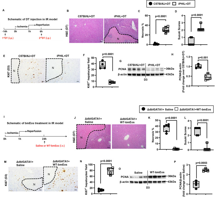
Figure 2.
Eosinophils promote liver repair after IR injury. (A-H) Male iPHIL mice and WT littermates were subjected to hepatic IR surgery. Mice were administered (i.p.) the first dose of DT at 16h prior to IR surgery and the second dose at 6h after surgery. Mice were sacrificed on day 3 (n=4/group) or day 7 (n=6/group) after IR surgery. (B, C) Liver necrosis (N, outlined areas) was evaluated and quantified on day 7 after IR surgery. (D) Liver pathology was determined by the Suzuki score on day 7 after IR surgery. (E, F) Liver tissue sections were stained for Ki67 to quantify proliferating hepatocytes on day 3 after IR surgery. (G, H) PCNA protein expression was detected by Western blotting and quantified. (I-P) Male ΔdblGATA-1 mice were subjected to hepatic IR surgery. After 24h, half of the mice were i.v. injected with bmEos (10x106) and the other half injected with saline as control. Mice were sacrificed on day 3 (n=4/group) or day 7 after IR surgery (n=6/group). (J, K) Liver necrosis (N, outlined area) was evaluated and quantified on day 7 after IR surgery. (L) Liver pathology was determined by the Suzuki score on day 7 after IR surgery. (M, N) Proliferative hepatocytes were stained for Ki67 and quantified on day 3 after IR surgery. (O, P) PCNA protein expression was detected by Western blotting and quantified. Two-tailed unpaired Student’s t-test with Welch’s correction was performed in C, D, F, H, K, L, N and P.

