Abstract
Vitamin D deficiency is a common deficiency worldwide, particularly among women of reproductive age. During pregnancy, it increases the risk of immune-related diseases in offspring later in life. However, how the body remembers exposure to an adverse environment during development is poorly understood. Herein, we explore the effects of prenatal vitamin D deficiency on immune cell proportions in offspring using vitamin D deficient mice established by dietary manipulation. We found that prenatal vitamin D deficiency alters immune cell proportions in offspring by changing the transcriptional properties of genes downstream of vitamin D receptor signaling in hematopoietic stem and progenitor cells of both the fetus and adults. Moreover, further investigations of the associations between maternal vitamin D levels and cord blood immune cell profiles from 75 healthy pregnant women and their term offspring also confirm that maternal vitamin D levels in the second trimester significantly affect immune cell proportions in the offspring. These findings imply that the differentiation properties of hematopoiesis act as long-term memories of prenatal vitamin D deficiency exposure in later life.
Subject terms: Haematopoiesis, Genetics
Introduction
Vitamin D, a micronutrient/hormone, regulates transcription by binding to its nuclear receptor, the vitamin D receptor (VDR). Humans can obtain vitamin D through photosynthesis on the skin and food intake. Approximately 50–90% of vitamin D is synthesized on the skin via sunlight exposure, while the remainder comes from the diet. Cutaneous vitamin D synthesis depends on environmental factors (geographic latitude, season, and amount of air pollution), skin type, clothing habitats, and lifestyle1. Despite country-level vitamin D fortification programs in many countries, the prevalence rate of vitamin D deficiency, especially in reproductive-age women2–9, is still high worldwide10–16. The most severe consequence of vitamin D deficiency is rickets or impaired bone formation (reviewed by Holick17). Besides its importance in bone formation, VDR signaling regulates gene transcription in nearly every tissue in our bodies, including the brain, heart, muscle, kidney, and immune system (reviewed in Pike et al.18). During development, the micronutrient status of offspring is entirely dependent on the status of the mother; therefore, developing embryos are vulnerable to adverse micronutrient conditions of deficient mothers19–23. To study the adverse consequences of prenatal vitamin D exposure, animal models with maternal dietary manipulations24–37 and knockout mouse model studies20,38–44 have been utilized. Studies using knockout mice models have shown that depletion of VDR causes a rickets-like phenotype after weaning. Both maternal dietary manipulation and knockout mouse models revealed that prenatal vitamin D deficiency can lead to immune defects in offspring. Epidemiological studies in humans have demonstrated that prenatal vitamin D deficiency has been associated with susceptibility to a number of immune-related diseases affecting their children, including asthma45–47, multiple sclerosis48, and type I diabetes49–51. These findings indicate that prenatal vitamin D deficiency disturbs immune cell development. In addition, it is possible that the hematopoietic system poses a long-term memory of exposure to vitamin D deficiency during development. However, this memory mechanism has not been extensively studied.
In this work, we show that prenatal vitamin D deficiency alters immune cell proportions of offspring during adulthood, attributed to cell fate decisions influenced by transcriptional alterations of VDR signaling pathway genes. Our data also supports that changing the cell fate of stem cells is the key component of the long-term effects of maternal vitamin D deficiency on the hematopoietic system of offspring.
Results
Lack of impact of maternal vitamin D deficient diet feeding on offspring bone development
We summarized the dietary intervention strategy in Fig. 1. We randomly assigned 6 weeks old female C57BL/6J mice to a vitamin D-sufficient diet (VDsuf, 1.0 IU/g vitamin D) and a vitamin D-deficient diet (VDdef, 0.0 IU/g vitamin D) for 5 weeks prior to mating with control diet-fed C57BL/6J male mice. The daily food intakes of each diet were comparable between the VDsuf and VDdef. To prevent hypocalcemia, the VDdef group received 1.5% calcium gluconate in their drinking water during the VDdef diet feeding. After 5 weeks of feeding VDdef, serum vitamin D (25-hydroxyvitamin D3, 25(OH)D3) concentrations of the VDdef-fed females reached the vitamin D deficient threshold (5.1 ± 3.8 ng/mL, n = 8), which is about 6.5 times lower than that of the VDsuf-fed females (33.0 ± 5.44 ng/mL, n = 10) (Supplementary Fig. 1A). After delivery, all female mice were fed VDsuf. While the number of offspring per dam was smaller in VDdef-fed females (mean = 3.82, standard deviation (sd) = 2.8, n = 16) than in VDsuf-fed females (mean = 4.7, sd = 1.7, n = 10), the difference was not significant (p = 0.32). We did not observe any obvious adverse effects of VDdef diet feeding on these female mice prior to or during their pregnancy. All offspring were fed VDsuf after weaning and maintained the diet until the sampling. The growth of VDdef-fed female offspring (VDD) and VDsuf-fed female offspring (VDS) based on their body weight were comparable for all of the time points we measured: postnatal day 1 (PD1) (n = 9 VDS and n = 11 VDD), 5, 9, and 13 weeks of age (n = 40 VDD and n = 27 VDS) (Supplementary Fig. 1B,C). While VDD males were slightly smaller than VDS males, as Seipelt et al. reported52, the difference was not statistically significant at all time points. This discrepancy could be attributed to the differences in diet used or calcium supplementation to VDdef-fed mothers. The serum vitamin D concentrations of VDD were lower than the detection limit (0.6 ng/mL, n = 3 per group) at PD1 (Supplementary Fig. 1A). While the serum vitamin D concentration of VDD was still significantly lower than that of VDS (n = 5 per group) at 5 weeks of age, their status was not deficient anymore (Supplementary Fig. 1A). We collected tissues and weighted liver, lung, kidney, thymus, spleen, and heart from the VDD and VDS at 16 weeks of age (16 wks) (n = 4–6 for each group). The tissue weights normalized by body weight were not significantly different between the groups, except the heart was heavier in male VDD compared to male VDS (p = 0.041) (Supplementary Fig. 1D). We also assessed the bone and tissue mineral density of the humerus of the left arm of the offspring using an X-ray CT system at 16 wks (n = 5–6 males for each group). Bone volume, bone mineral content, bone mineral density, tissue mineral content, tissue mineral density, and bone volume fraction were assessed, and no significant alterations were observed (Supplementary Fig. 1E).
Fig. 1.
Study design. Six-week-old female C57BL/6J mice were randomly assigned to the VDsuf or VDdef diet and fed the assigned diet for 5 weeks before mating with a control diet-fed male. 1.5% calcium gluconate water was supplemented to VDdef diet-fed group. After delivery, all F0 mice were fed VDsuf diets. The offspring were fed VDsuf diet after weaning and maintained the diet until the sampling.
Prenatal vitamin D deficiency decreases CD4+ and CD8+ T cell proportions of the offspring in peripheral blood and spleen
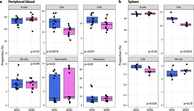
We collected peripheral blood from VDD and VDS and compared the immune cell profiles at 16 wks (n = 11 per group). We observed a significant reduction of CD4+ T cells (28.2% decrease at p = 0.0018) and CD8+ T cells (14.7% decrease at p = 0.037) in VDD males compared to VDS males (Fig. 2a). This reduction was not observed in females (Supplementary Fig. 2). We also compared the immune cell profiles of spleen (n = 6 per group) and observed the significant reduction of CD4+ T cells (18.4% decrease at p = 0.0035) and CD8+ T (11.1% decrease at p = 0.026) cells in VDD (Fig. 2b). Representative flow cytometry traces are shown in Supplementary Fig. 3.
Fig. 2.
Prenatal vitamin D deficiency reduces CD4+ and CD8+ T cells in the peripheral blood and spleen in male offspring at the adult stage. T cell proportions in the peripheral blood (a) and spleen (b) were significantly decreased in VDD offspring. Each dot on the plot represents a sample (blood, n = 11 per group; spleen, n = 6 per group). The box shows the range between the first and third quartiles. The upper and lower whiskers represent 1.5 times the interquartile range, while the black bars indicate the median. The values shown in the plot are p-values calculated using Student’s t-test.
The reduced proportion of lymphocytes in the periphery reflects the cellular composition changes in the bone marrow
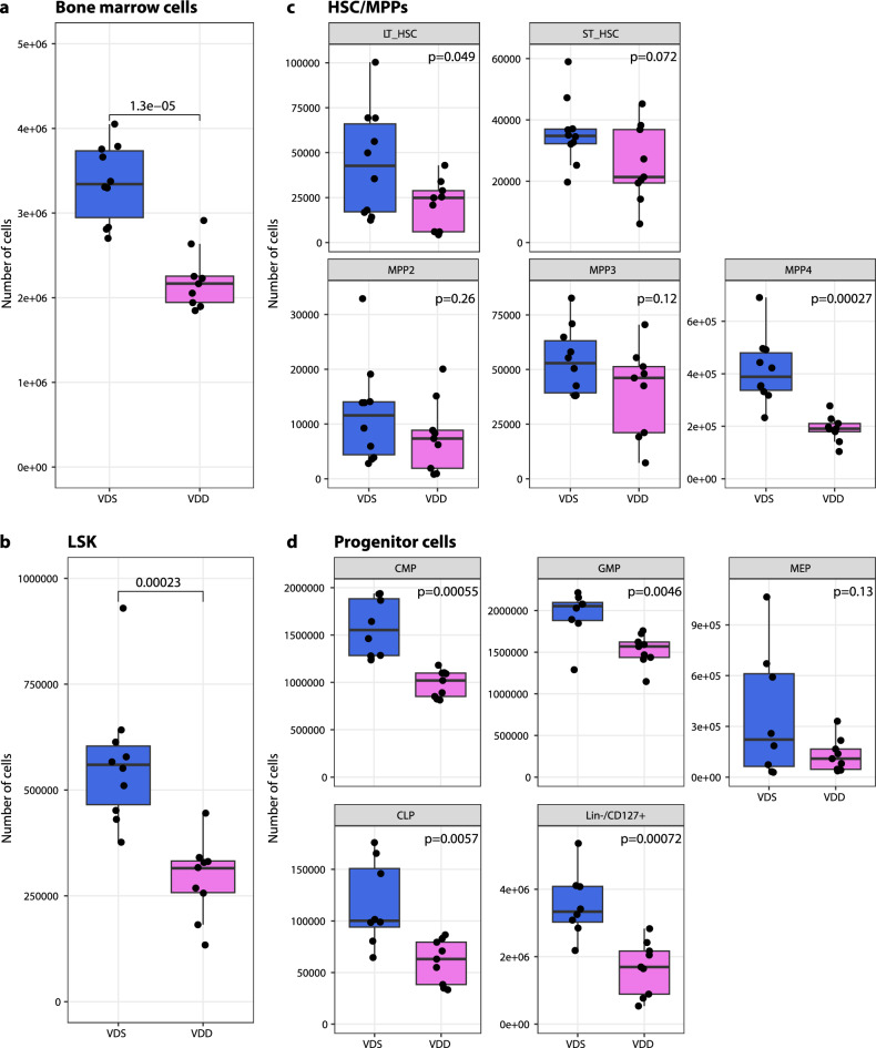
During hematopoiesis, hematopoietic stem cells (HSCs) undergo differentiation into three multipotent progenitor cells (MPP2, MPP3, and MPP4) in the bone marrow in adults53. MPP2 and MPP3 are further differentiated primarily into the myeloid lineage, while MPP4 is into the lymphoid lineage. MPP2 is biased toward producing megakaryocytes and erythrocytes, and MPP3 towards granulocytes and macrophages53. To test if the T cell reductions in the periphery are reflected in the alterations in the bone marrow, we collected mononucleated cells from the bone marrow by crushing bones (tibia, femur, iliac, sternum, and vertebrae) from the offspring at 16 wks (n = 9–10 per group). The number of bone marrow mononucleated cells collected in VDD was significantly decreased compared to VDS (3,354,105 ± 439,109 and 2,215,799 ± 353,525 in VDS (n = 10) and VDD (n = 10), respectively, p = 0.000013, Fig. 3a). We, then analyzed the proportions of HSCs, MPPs, and hematopoietic progenitor cells using the previously reported definitions53,54; lineage negative (Lin−, CD3−/CD4−/CD8−/B220−/Ter119−/CD11b−/Gr-1−/CD127−), Lin−/Sca-1+/c-Kit+ (LSK), long-term hematopoietic stem cell (LT-HSC, LSK/Flk2−/CD150+/CD48−), short-term hematopoietic stem cell (ST-HSC, LSK/Flk2−/CD150−/CD48−), multipotent progenitor 2 (MPP2, LSK/Flk2−/CD150+/CD48+), multipotent progenitor 3 (MPP3, LSK/Flk2−/CD150−/CD48+), multipotent progenitor 4 (MPP4, LSK/Flk2 +), common myeloid progenitor cells (CMP, Lin−/c-Kit+/CD34+/CD16/32−), common lymphoid progenitor cells (CLP, Lin−/c-Kit+/Flk2+/CD127+), granulocyte-monocyte progenitor cells (GMP, Lin−/c-Kit+/CD34+/CD16/32+), megakaryocyte-erythrocyte progenitor cells (MEP, Lin−/c-Kit+/CD34−/CD16/32−), and Lin−/CD127+ (early T cell progenitor). We observed a significant reduction (48.9% decrease) in the absolute number of LSK (565,312 ± 153,178 and 289,052 ± 91,984 in VDS and VDD, respectively, p = 0.00023, Fig. 3b). While the p-value (p = 0.078) didn’t pass our significant threshold, the proportion (%) of LSK in the live bone marrow mononucleated cells was also decreased in VDD mice (0.19 ± 0.04% and 0.16 ± 0.05% in VDS (n = 10) and VDD (n = 9), respectively, Supplementary Fig. 4a). As the absolute number of LSK decreased in VDD, the absolute numbers of HSCs, MPPs, and progenitors were also decreased (Fig. 3c,d). Among the MPPs, MPP4 was the only MPP that significantly reduced in both absolute numbers (413,267 ± 127,529 and 190,922 ± 49,401 in VDS (n = 10) and VDD (n = 9), respectively, p = 0.0027, Fig. 3c) and the proportion of the live bone marrow mononucleated cells (0.14 ± 0.03% and 0.10 ± 0.02% in VDS and VDD, respectively, p = 0.0096, Supplementary Fig. 4b). Among the progenitor cells, the absolute numbers of CMP, GMP, CLP, and Lin−/CD127+ were significantly reduced in VDD (n = 9) compared to VDS (n = 9) (Fig. 3d). Interestingly, while the proportional averages of Lin−/CD127+ (p = 0.07) and CLP (p = 0.19) were decreased, the proportion of GMPs was increased in VDD compared to VDS (50.1% increase, p = 0.001, Supplementary Fig. 4c). Representative flow cytometry traces are shown in Supplementary Fig. 5.
Fig. 3.
Prenatal vitamin D deficiency decreased the number of bone marrow cells and LSKs and concomitantly reduced lymphoid lineage cells at the adult stage. Prenatal vitamin D deficiency reduced the total number of bone marrow cells (n = 9 for VDD and n = 10 for VDS) (a) and the number of LSKs in bone marrow (n = 9 per group) (b). The differences in total HSC, long-term and short-term HSC, and three MPPs in the bone marrow (n = 9 for VDD and n = 10 for VDS) (c) and hematopoietic progenitor cells in the bone marrow (n = 9 per group) of VDD and VDS mice are displayed in the box plots (d). A significant decrease in MPP4, CLP, and Lin−/CD127+ cells was observed in VDD mice, indicating that prenatal vitamin D deficiency disproportionately affects the production of hematopoietic lineages. Each dot on the plot represents a sample. The box shows the range between the first and third quartiles. The upper and lower whiskers represent 1.5 times the interquartile range, while the black bars indicate the median. The values shown in the plot are p-values calculated using Student’s t-test.
Prenatal vitamin D deficiency alters gene expression profiles of MPP4 cells
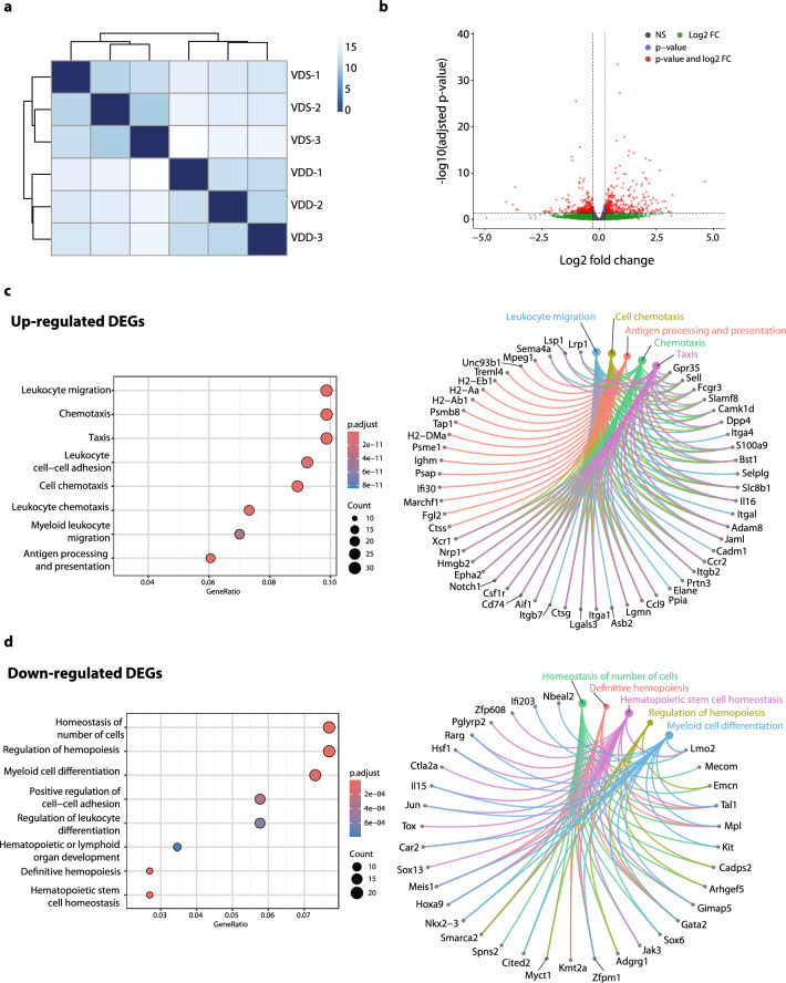
We performed transcriptome analysis on bone marrow MPP4 from VDD and VDS to see the transcriptional alterations at 16 weeks of age (n = 3 per group). The sequencing status and quality of each sample are summarized in Supplementary Data 1. All samples have passed primary quality checks. A hierarchical clustering analysis showed a clear dissociation, suggesting genome-wide transcriptional alterations in VDD (Fig. 4a). We identified 612 differentially expressed genes (DEGs) with at least 1.2-fold change and a false discovery rate adjusted p-value (FDR-adj p-value) < 0.05 (Fig. 4b, Supplementary Data 2). Of those, 345 were upregulated, and 267 were downregulated. The Gene Ontology (GO) enrichment analysis of upregulated genes revealed enrichment of genes related to leukocyte migration and chemotaxis (Fig. 4c left), while downregulated genes showed enrichment in the homeostasis of cell numbers and regulation of hemopoiesis (Fig. 4d left, Supplementary Data 3). The Gene-Concept Network analysis revealed that hematopoietic transcription factors, including Lmo2, Tal1, Gata2, and Mpl, were downregulated and were found to overlap in the top five enriched GO terms (Fig. 4d right). Transcription factor (TF) Perturbations Followed by Expression analysis (https://maayanlab.cloud/Enrichr/) revealed that the detected DEGs were enriched in the genes perturbed in LSK of the hematopoietic transcription factor knockout models in the same directions. The upregulated genes were enriched in NFIX (GSE45492)55 and SRF (GSE23556)56 knockouts upregulated genes (adjusted p-value = 3.92E−47 and 9.15E−29, respectively) and downregulated genes were enriched in NFIX and SRF knockout downregulated genes (adjusted p-value = 1.98E−36 and 4.55E−12, respectively) (Supplementary Fig. 6). Gene Set Enrichment Analysis (GSEA) showed leukocyte differentiation (GO:0002521, enrichment score = 0.385, q-value = 0.010), lymphocyte differentiation (GO:0030098, enrichment score = 0.412, q-value = 0.026), T cell differentiation (GO:0030217, enrichment score = 0.435, q-value = 0.023) were enriched (Supplementary Data 4). This result suggests that prenatal vitamin D deficiency induces long-term transcriptional alterations in hematopoietic stem cells that persist into adulthood.
Fig. 4.
Bulk RNA-seq analyses reveal the transcriptional alterations in MPP4. Bulk RNA-seq analysis identified significant differences in the transcriptional profiles of MPP4 between VDD and VDS (n = 3 per group). (a) The heatmap and dendrogram show a distinct clustering between VDD and VDS. (b) The volcano plot displays the − log10 adjusted p-values and log2 fold-change differences of the identified DEGs. Each dot represents a gene, and significant DEGs are indicated by red dots. (c) The GO enrichment analysis reveals that the up-regulated DEGs between VDD and VDS MPP4 are enriched in Leukocyte migration and chemotaxis-related genes. The dot plot shows the significance and the gene ratios of the top eight GO terms (left), and the Cnet plot indicates the identified DEGs of the top five GO terms (right). (d) The GO enrichment analysis reveals that the down-regulated DEGs between VDD and VDS MPP4 are enriched in the regulation of hematopoiesis-related genes. The dot plot shows the significance and the gene ratios of the top eight GO terms (left), and the Cnet plot indicates the identified DEGs of the top five GO terms (right).
Prenatal vitamin D deficiency alters cellular compositions of the embryonic day 14.5 (e14.5) embryonic liver, suggesting immune cell proportion changes start during development
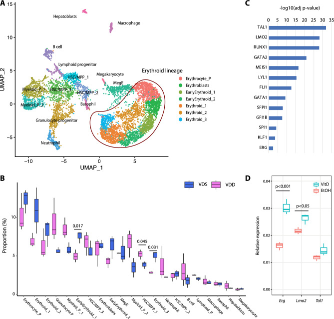
To assess if the cell composition alteration started at the embryonic stage, we performed single-cell RNA-seq (scRNA-seq) on the e14.5 fetal liver of both VDS and VDD embryos using the 10× Genomics Chromium platform (n = 3 per group). Multiplexing with cell hash antibodies was used to reduce the technical batch effect. The obtained sequences were aligned by the 10× Genomics software, CellRanger, and the matrix was demultiplexed and analyzed by Seurat, an R-package57–59. After eliminating low-quality cells (< 1000 genes/cell, < 5000 reads/cell, and > 10% mitochondrial reads/cell) and cells without cell hashing information, we have identified 21 different cell clusters in a total of 6947 cells. We identified the cell types of each cell cluster based on the marker gene expression status (Fig. 5a, Supplementary Data 5). We detected three HSC/MPP populations, lineage-specific progenitor cells, erythroid lineage cells, and hepatoblast cells in e14.5 liver. Concordant with the previous report60, more than half of the cells were identified as erythroid lineage cells. The cellular composition analysis showed that one of the HSC/MPP populations (HSC/MPP 1) was significantly increased in VDD embryos, and two erythroid lineage cells (Early Erythroid 1 and Erythroid 3) were decreased (Fig. 5b), suggesting the cell composition changes started at least at the embryonic stage. A pseudo bulk RNA-seq analysis on the scRNAseq datasets showed that down-regulated genes were enriched in the genes regulated by hematopoietic system transcription factors (Fig. 5c, Supplementary Data 6–8). Among these transcription factors, Tal1, Lmo2, Meis1, and Erg were also identified as VDD downregulated genes in MPP4 at the adult stage (Supplementary Data 2). To test whether vitamin D alters the expression of these genes, we measured the expression of Tal1, Lmo2, and Erg, the known hematopoietic transcription factors, by quantitative RT-PCR on an embryonic stem cell-derived hematopoietic progenitor cell line, HPC-761, after treating 1-alpha-25-dihydroxyvitamin D3, a ligand of VDR, or ethanol (solvent) for 24 h by quantitative RT-PCR (Fig. 4d). We observed a significant increase of Lmo2 (p = 0.034) and Erg (p = 0.005) expression. This result suggests that the gene expression alterations observed at e14.5 were attributed to the dysregulation of hematopoietic transcription factors, such as Lmo2 and Erg, regulated by VDR signaling pathways.
Fig. 5.
Prenatal vitamin D deficiency alters cellular compositions of the embryonic liver, suggesting immune cell proportion changes start during development. (a) UMAP representation of single-cell RNA-seq gene expression data and cellular lineage identification of E14.5 fetal liver (n = 3 per group). (b) The boxplots indicate prenatal vitamin D deficiency alters cellular compositions of E14.5 fetal liver. (c) Genes downregulated in VDD E14.5 fetal liver are enriched in the genes regulated by hematopoietic transcription factors. (d) Treating HPC7 cells with 1-alpha-25-dihydroxyvitamin D3 significantly increases gene expression levels of Erg and Lmo2, suggesting these genes are regulated by VDR (n = 3 per treatment group).
Maternal serum vitamin D status in the second trimester is positively associated with the CD8+ T cell proportion in the cord blood
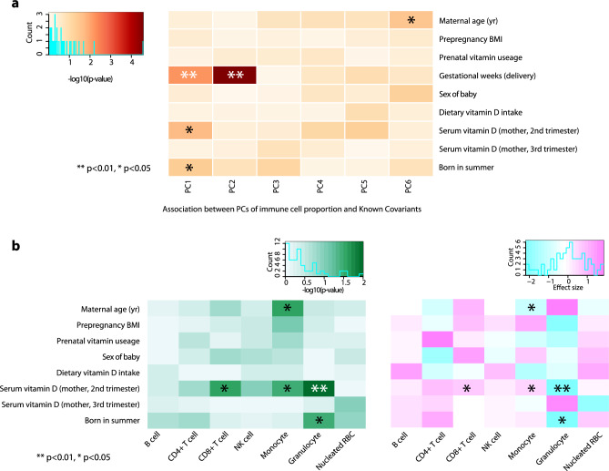
To test the associations between maternal vitamin D levels and immune cell proportions of offspring, we assessed 75 mother–offspring pairs who were recruited as participants of the Birth Cohort Gene and Environment Interaction Study of TMDU (BC-GENIST) project at the Tokyo Medical and Dental University, Bunkyo, Tokyo, Japan62,63. We measured the concentration of serum 25(OH)D3 at two time points: time point 1 (T1), intended to represent the second trimester from week 10 to week 29, and time point 2 (T2), to represent the third trimester from week 33 to week 40 of gestational age. The immune cell proportions were estimated from the bulk DNA methylation profiles of the cord blood of the fetus using a Bioconductor package FlowSorted.CordBloodCombined.450k64–67. We excluded one participant who did not have T1 serum 25(OH)D3 status from the analysis. The demographic, clinical, and phenotypic information for the study participants is provided in Table 1. All participants are healthy pregnant Japanese women without smoking or drinking during their pregnancy. No participants had hypertension. The average maternal age at delivery was 34.2 (sd 4.0) years old, with pre-pregnancy BMI 17.1–29.2 (average 20.73, sd 2.6). 70.3% of women took prenatal vitamin supplements, and the average estimated daily dietary vitamin D intake was 5.1 (sd 4.4) µg/day68,69. 34 offspring were males (45.9%), and the average gestational week of the delivery was 39.1 (sd 1.2). The average concentration of serum 25(OH)D3 was 25.2 (sd 11.8) ng/mL in the second trimester (average gestation week 19.0 (sd 4.5), T1) and 28.0 ng/mL (sd 14.4) in the third trimester (average gestation week 35.9 (sd 0.9), T2). 30 participants were vitamin D deficient (serum 25(OH)D3 < 20 ng/mL) at T1 (40.5%), and 23 were vitamin D deficient at T2 (31.8%). Of those, eight participants were vitamin D deficient at both T1 and T2. While we observed significant associations between the serum 25(OH)D3 levels and sampling season (Summer and Winter) (p = 0.047 and p < 0.001, T1 and T2, respectively), no significant associations were observed in maternal age, pre-pregnancy BMI, sex of fetuses, prenatal vitamin supplements usage and dietary vitamin D intake. The results of the univariate model fitting are shown in Supplementary Data 9. To analyze the associations between maternal serum 25(OH)D3 levels and immune cell profiles of the cord blood, we calculated principal components (PCs) of immune cell profiles using an R stats function, prcomp, and assessed the contributions of each covariate. Gestation weeks at delivery showed the most significant contributions to PC1 (p = 0.0063 and adjusted r2 = 0.087) and PC2 (p = 0.000027 and adjusted r2 = 0.208), followed by maternal serum 25(OH)D3 at T1 to PC1 (p = 0.021 and adjusted r2 = 0.059) and being born in summer (p = 0.049 and adjusted r2 = 0.04) (Fig. 6a). We then assessed the associations of covariates to each cell type. We observed that the gestational weeks at delivery were negatively associated with the proportions of CD4 T cells (estimate − 0.027 and p = 0.00006) and B cells (estimate − 1.12 and p = 0.00002) and positively associated with proportions of granulocytes (estimate 0.028 and p = 0.0014). Maternal serum 25(OH)D3 at T1 was positively associated with proportions of CD8 T cells (estimate 0.0007 and p = 0.0216) and monocytes (estimate 0.0005 and p = 0.0349) and negatively associated with proportions of granulocytes (estimate − 0.0024 and p = 0.0365) (Supplementary Fig. 7). These associations were still significant after adjusting for the sex of the fetus and gestational weeks at delivery, season of T1, and the gestational week at T1 (Fig. 6b, Supplementary Data 10). These findings indicate that maternal vitamin D levels, especially in the second trimester, affect the immune cell proportions in the offspring at birth.
Table 1.
Demographic information of BC-GENIST cohort.
| Total N (%) | 74 (100.0) | |
|---|---|---|
| Maternal age (yr) | Mean (SD) | 34.2 (4.0) |
| Pre-pregnancy BMI | Mean (SD) | 20.7 (2.6) |
| Prenatal vitamin usage, N (%) | Yes | 52 (70.3) |
| Fetal sex, N (%) | Female | 40 (54.1) |
| Gestational age at delivery (weeks) | Mean (SD) | 39.1 (1.2) |
| Gestational age at T1 | Mean (SD) | 19.0 (4.5) |
| Serum vitamin D concentration (ng/mL) at T1 | Mean (SD) | 25.2 (11.8) |
| Gestational age at T2 | Mean (SD) | 35.9 (0.9) |
| Serum vitamin D concentration (ng/mL) at T2 | Mean (SD) | 28.0 (14.4) |
| Estimated dietary vitamin D intake per day | Mean (SD) | 5.1 (4.4) |
| Maternal serum vitamin D deficiency levels, N (%)☨ | Deficient | 8 (10.8) |
| Insufficient | 20 (27.0) | |
| Sufficient | 46 (62.2) | |
| Estimated immune proportions from cord blood DNA methylation profiles | ||
| CD8 T cell | Mean (SD) | 5.23(3.35) |
| CD4 T cell | Mean (SD) | 19.25(7.15) |
| NK cell | Mean (SD) | 0.71(1.24) |
| B cell | Mean (SD) | 6.67(2.84) |
| Monocyte | Mean (SD) | 3.15(2.27) |
| Granulocyte | Mean (SD) | 61.46(9.2) |
| Nucleated red blood cell (nRBC) | Mean (SD) | 4.09(3.26) |
☨Deficient, both T1 and T2 serum vitamin D concentrations were less than 20 ng/mL; insufficient, one of the measurements was less than 20 ng/mL and the other was less than 30 ng/mL.
Fig. 6.
Maternal serum vitamin D status in the second trimester is positively associated with the CD8+ T cell proportion in the cord blood. (a) The heatmap shows that the gestational week at the delivery has the strongest associations with immune cell composition variations assessed by the principal component (PC), followed by maternal serum vitamin D (2nd trimester) and being born in the summer season. (b) After adjusting for the sex of the fetus, gestational age, the season of T1, and the gestational week at T1, maternal serum vitamin D (2nd trimester) maintains significant associations with immune cell composition, specifically positive association with proportions of CD8+ T cell and monocytes, and negative association with granulocytes. Asterisks indicate the significance (**p < 0.01 and *p < 0.05, Student’s t-test). The left panel shows − log10(p-value), and the right panel shows the direction of the associations.
Discussion
In this study, we found that prenatal vitamin D deficiency by maternal dietary intervention reduced peripheral T-cells in both the blood and spleen in adults and this reduction was linked to the decreased number of bone marrow cells and the proportional alterations of hematopoietic progenitor cells in the bone marrow. Our results demonstrate that cellular composition alterations could be the long-term memories of prenatal exposure at the adult stage.
Besides insufficient nutritional intake or photosynthesis on the skin, genetic mutations significantly contribute to vitamin D deficiency. In humans, mutations in genes of the vitamin D biosynthetic pathway are the most common cause of heritable vitamin D-dependent rickets (VDDR), which can be treated with active form vitamin D supplementation, whereas VDR mutation results in hereditary vitamin D-resistant rickets (HVDR), which could partially be ameliorated with Ca2+ supplementation70,71. Animal models with gene knockout have been studied to understand the functions of genes and diseases associated with them. A VDR Knockout mouse model study revealed that VDR null mice are developed without any significant defects before birth; however, they developed a rickets-like phenotype after weaning, and most of the animals died within 15 weeks after birth42, suggesting VDR is critical in the growth and bone formation in the post-weaning stage. Subsequent studies using the same VDR knockout mouse line feeding normal chow diet identified that while VDR knockout mice showed immune defects, no significant differences in the numbers or percentages of red and white cells compared to their wildtype littermates were observed38,39. Interestingly, correcting hypocalcemia by feeding a lactose-rich or polyunsaturated fat-rich diet was able to restore the immune abnormalities observed in VDR knockout mice39. Another research group independently developed another VDR knockout mouse line in C57BL/6 background and reported similar phenotypes, vitamin D-dependent rickets type II with alopecia after weaning44. Studies using this mouse line revealed that this line also developed immune abnormalities41,72. Additionally, the nonobese diabetic (NOD) mouse line with a VDR knockout showed that although diabetes onset is not accelerated by VDR deletion, NOD VDR knockout mice develop rickets and have lower numbers of natural killer T-cells and CD4+CD25+ T-cells43. These findings from independent knockout mouse studies indicate that VDR-depleted animals are phenotypically normal at birth but develop hypocalcemia within the first month of life and immune abnormalities. The active form 1-α-25-dihydroxyvitamin D [1,25(OH)2D] is synthesized from its precursor 25 hydroxyvitamin D [25(OH)D] via catalytic action of the 25(OH)D-1alpha-hydroxylase [1α(OH)ase] enzyme, encoded by Cyp27b1. Mice deficient in 1α(OH)ase developed hypocalcemia, skeletal abnormalities characteristics of rickets, female infertile, and a reduction in CD4 and CD8 peripheral T lymphocytes after weaning20. A zebrafish study showed that loss of Cyp27b1-mediated biosynthesis by gene knockdown significantly reduced runx1 expression and hematopoietic stem and progenitor cell productions70. In this study, we found that prenatal vitamin D deficiency-exposed offspring at the adult stage did not show significant alterations in bone morphology, suggesting that vitamin D depletion during development does not affect embryonic bone development and bone morphology later in life. On the other hand, the immune phenotype of prenatal vitamin D deficiency-exposed offspring we observed was much more severe than those of VDR knockout models and similar to the Cyp27b1 deletion models. Since we observed this phenotype in offspring at 16 weeks of age, while VDR knockout mice typically do not survive beyond 15 weeks42, age might be another factor in developing this phenotype. However, our findings suggest that the immune phenotypes in offspring exposed to prenatal vitamin D deficiency may be caused by a lack of ligands rather than receptor malfunctions during the developmental phase.
A large biological difference between our study and the Cyp27b1 deletion models is the availability of 1,25(OH)2D after weaning. All offspring were fed VDS diets after weaning, and the serum vitamin D status of the VDD offspring became normal at 5 weeks of age. Therefore, the alterations we observed at 16 weeks of age were not associated with the vitamin D deficiency status at the time of measurement. However, it does indicate that prenatal vitamin D deficiency irreversibly alters bone marrow development in the offspring. This aligns with a previous study conducted on rats73. The study found that the rats with prenatal vitamin D deficiency by maternal dietary manipulation showed significant reductions in the total number of nucleated bone marrow cells and in the colony-forming unit (CFU) content with a corresponding increase in cell cycle rate73. The authors measured the CFU content by transplanting nucleated bone marrow cells to AJ mice purchased from Jackson Laboratory (vitamin D-sufficient diet-fed mice) and found that while 1,25(OH)2D3- or 24,25(OH)D3-treatment on vitamin D deficient rats in postnatal life corrected the serum calcium and phosphate, the treatments did not reverse the CFU content alterations. Their findings clearly demonstrate that prenatal and early-life vitamin D deficiency could cause irreversible effects on HSC development. However, we cannot fully rule out the possibility that alterations in hematopoietic stem cell niches might also be irreversibly altered. HSC niches in different developmental stages play critical roles in the maintenance and differentiation of HSCs (reviewed by Gao et al.74). It would be beneficial to explore this possibility further in upcoming research.
As a transcriptional regulation mechanism, 1,25(OH)2D binds to the VDR, which forms a heterodimer with the retinoid X receptor and regulates gene expression by binding to vitamin D responsive elements in genomic DNA. In this study, the transcription factor network analysis on e14.5 fetal liver scRNA-seq revealed that Tal1, Lmo2, Runx1, Gata2, and Erg downstream genes were significantly enriched in VDD downregulated genes. We confirmed that the expressions of Erg and Lmo2 are regulated by VDR, as the treatment with 1-alpha-25-dihydroxyvitamin D3 (1,25(OH)2D3) led to an increase in gene expression in fetal hematopoietic progenitor cells, HPC7. A mutant and chimeric embryo study reported that ERG is a direct upstream regulator of Runx1 and Gata2 in fetal livers75, suggesting prenatal vitamin D deficiency exposure perturbs the transcription profiles of the hematopoietic cells during development via modulating hematopoiesis transcription factors regulated by VDR. Interestingly, this transcription factor expression perturbation persists in the adult stage even though these mice were fed a VDsuf diet and their vitamin D was fully recovered. Using bulk RNA-seq analysis, we identified 612 DEGs between VDD and VDS MPP4. Downregulated DEGs were enriched in hematopoietic-related pathways, and many hematopoietic transcription factors, including Tal1, Lmo2, Runx1, Gata1, and Erg, were downregulated. Gene Enrichment Analysis showed that the identified DEGs were enriched in the DEGs of Nfix and Srf knockout mouse LSKs in the same direction. Nfix and Srf are hematopoietic transcription factors. Nfix is associated with hematopoietic stem and progenitor cells (HSPCs) survival, and Srf regulates hematopoietic stem cell adhesions. Although both Nfix and Srf were not identified as DEGs, their expression levels were lower in VDD compared to VDS. It has been reported that loss of Nfix expression in HSPCs of murine adult bone marrow is concomitant with the reduced expression of genes associated with HSPCs survival, such as Erg, Mecom, and Mpl55. These same genes were also significantly downregulated in VDD MPP4. However, the authors reported no selective loss of B, T, or myeloid cells was observed in Nfix-depleted HSPCs55. On the other hand, Ragu et al. reported that while depleting Srf, increased % of the LSK in the bone marrow, Srf knockout mice showed a significant decline in white blood cell counts, which was primarily due to decreased numbers of circulating B and T cells56. Our results suggest prenatal vitamin D deficiency modulates the Nfix and Srf pathways in the long term.
Our study also revealed that higher maternal vitamin D levels during the second trimester were associated with increased proportions of CD8+ T cells and decreased granulocytes in cord blood in humans. The associations were maintained after adjusting for the season of the second trimester, the gestational week at the maternal blood draw, the sex of the fetus, and the gestational week at birth. Hematopoietic development consists of primitive and definitive hematopoiesis76. In mice, the definitive hematopoiesis starts in the aorta-gonad mesonephros (AGM) region at approximately E10.5, then moves to the liver around E12.5 and the bone marrow around E17.577,78. After birth, the bone marrow is the only site where HSCs are maintained and expanded. In human development, the transition of the hematopoietic stem cell production site from the fetal liver to bone marrow occurs in the second trimester78,79 when we observed significant associations. Recently, Elgormus et al. reported that newborn serum vitamin D levels were negatively correlated with neutrophil-to-lymphocyte ratios (NLR) in newborn offspring80. The most common white blood cell is the granulocyte, composed of three distinct types: neutrophils, eosinophils, and basophils. Among them, neutrophil is the most abundant type. Our results and their finding concordantly indicate that the immune cell compositions, especially granulocyte (neutrophil) and lymphocyte, of the offspring are influenced by vitamin D levels in early life. Of note, cord blood NLR has been proposed as an indicator for the diagnosis of early neonatal sepsis combined with other laboratory tests and clinical manifestations81–83. The associations between neonatal sepsis and cord blood vitamin D levels have been well reported. A meta-analysis of 18 studies revealed that low maternal and cord blood vitamin D levels were significantly associated with the incidence of neonatal sepsis84. Additionally, a study on 4340 neonates appropriate for gestational age found a negative correlation between cord blood NLR and fetal malnutrition. This indicates that cord blood NLR could be utilized as a marker for fetal malnutrition85. For successful supplementation during pregnancy to reduce adverse outcomes, considering the amount and timing of vitamin D supplementation is crucial47,86,87. Our results indicate that the maternal serum vitamin D level in the second trimester, not the third trimester, is significantly associated with immune cell alterations. Therefore, the timing of supplementation, such as early or preconception, could be a crucial factor for successful interventions to mitigate risks of offspring later in life.
In conclusion, this study demonstrates that prenatal vitamin D deficiency reduces the number and the proportion of MPP4 cells in the bone marrow, changing the expression status of the genes that may be directly and indirectly regulated by VDR, and this alteration results in reduced T cell proportions in peripheral blood and spleens of the offspring. The association between T cell proportion and maternal vitamin D status was also observed in our BC-GENIST mother–offspring cohorts. We and others have reported that micronutrient deficiency changed the cellular compositions of adult mature organs and may contribute to the phenotypes using animal dietary manipulation models23,37,88. These findings indicate that the cell fate decision alteration of stem cells could be the key component of the long-term memory of prenatal micronutrient deficiency, which links to disease risks later in life. Nevertheless, this study has several limitations. A study conducted in Greece on mother-infant pairs revealed that insufficient levels of vitamin D (< 50 nmol/L) in pregnant mothers were linked to an increased occurrence of micronucleated cells in binucleated T-cells89. Micronuclei are early indicators of genetic effects used to test the relationship between exposure to genotoxic substances and cancer. However, our study did not explore whether maternal VDD increases DNA damage risks in offspring. Sexual dimorphism is another covariate we must explore. Since female mice did not show significant alterations in immune cell proportions in adults, we only performed transcriptional analysis on male mice. Both males and females may have prenatal and neonatal immune cell proportional alterations. Furthermore, there could be transcriptional changes in certain genes in adult females, but these changes may not affect cell fate decisions. These possibilities should be further investigated in future studies.
Methods
Ethics approval and inclusion to participate
All experiments involving the use of animals were approved and conducted in accordance with the ethical guidelines of the Albert Einstein College of Medicine Institutional Animal Care and Use Committee (IACUC, protocol # 20160710) and ARRIVE guidelines. Furthermore, all experiments involving human participants in the Birth Cohort Gene and Environment Interaction Study of TMDU (BC-GENIST) were approved by the Institutional Review Board of Tokyo Medical and Dental University (No. G2000-181, 29 July 2014) and are carried out in compliance with the Declaration of Helsinki and the International Conference of Harmonization Guidelines for Good Clinical Practice.
Maternal vitamin D deficiency mouse model
We purchased 5-week-old C57BL/6J female mice from the Jackson Laboratory. After 1 week of acclimation, we started dietary manipulations at 6 weeks old. Female mice (F0) were fed vitamin D deficient (VDdef) or sufficient (VDsuf) diets for 5 weeks before mating with the control diet-fed male mice and throughout the subsequent pregnancy. To avoid the paternal VDdef treatment effects, the males did not stay in the female cage for more than 3 days. VDdef (0.0 IU/g vitamin D) and nutrient-matched VDsuf (1.0 IU/g vitamin D) diets were obtained from Research Diets Inc. (10 kcal%fat, 20 kcal%protein and 70 kcal%carbohydrate). The detailed ingredients of each diet are listed in Supplementary Data 11. VDdef-fed females were supplemented with 1.5% calcium gluconate water for drinking water. After delivery, all F0 mice were fed VDsuf diets. The offspring were fed VDsuf diet after weaning and maintained the diet until the sampling.
Serum vitamin D level of mouse serum samples
Serum vitamin D (25(OH)D) levels of mouse serum samples were assessed by commercially available ELISA kits (Eagle Biosciences, Inc. Amherst NH, or Abcam, Cambridge, MA) according to the manufacturer’s instructions. The signal was detected with BioTek Synergy 4 Microplate reader (Agilent Technologies), and the results were analyzed using a 4-parameter logistic regression algorithm (http://www.elisaanalysis.com/app). The measurements were performed as duplicates.
Immune cell profiling on peripheral blood and spleen
Immune cell profiling was analyzed in both males and females using flow cytometry (FACS Aria2, BD Biosciences) after fluorescent dye conjugate antibodies staining. The obtained data were analyzed with FlowJo_10.6.1_CL (https://www.flowjo.com/). Peripheral blood samples were collected using submandibular vein bleeding methods. The spleen was obtained after euthanizing the animals with carbon dioxide inhalation. The spleen samples were dissociated on 70 µm filters. The obtained single-cell suspensions were stained with antibodies of immune cell surface marker proteins to identify cell types. Representative flow cytometry traces are shown in Supplementary Fig. 3. The antibodies used in this study are listed in Supplementary Data 12.
Isolating multipotent progenitor cells from bone marrow
Bone marrow cells from male animals were collected by crushing bones (tibia, Femur, iliac, sternum, and vertebrae). The red cells were lysed with lysis buffer (150 mM NH4Cl, 1 mM KHCO3, and 0.1 mM EDTA). HSC/MPP fractions were defined by the previously reported definition53. MPP4, lymphoid-primed hematopoietic multipotent progenitor cells were isolated for gene expression analysis. Representative flow cytometry traces are shown in Supplementary Fig. 5.
Cell culture
Hematopoietic progenitor cell line HPC-7 was kindly gifted by Dr. Britta Will at Albert Einstein College of Medicine. HPC-7 cells were maintained at density of 1–10 × 105/mL in Iscove’s modified Dulbecco’s medium (Invitrogen) supplemented with 50–100 ng/mL of mouse stem cell factor (Gemini Bio-Products), 1 mM Sodium Pyruvate, 6.9 ng/mL α-Monothioglycerol (SIGMA-Aldrich), 5% of bovine calf serum and Penicillin–Streptomycin. To examine the effect of vitamin D treatment on gene expression levels, the HPC-7 cells were treated with 0.1 µM of 1alpha,25-Dihydroxyvitamin D3 (SIGMA-Aldrich) or 0.1% (vol/vol) ethanol (solvent) for 24 h.
RNA-seq library construction and sequencing
We performed RNA-seq on FACS isolate MPP4 cells (lymphoid primed-multipotent progenitor cells). Total RNA was isolated with AllPrep DNA/RNA micro kit (QIAGEN). After we depleted ribosomal RNAs from total RNA, we generated the RNA-seq libraries using KAPA RNA HyperPrep with RiboErase kit (Roche). The generated libraries were sequenced on an Illumina NOVA-seq sequencer (Novogene Co., Ltd., USA).
RNA-seq alignment
After checking the quality of the sequencing files using FastQC90 and trimming low-quality reads and adapter sequences using Cutadapt91, the obtained sequences were aligned to the mouse mm10 reference genome with the gencode M15 gene annotation using STAR aligner92. The quality of the library was assessed with RSeQC93. The obtained transcript counts were analyzed with DESeq2. We identified significant differentially expressed genes (DEGs) that showed two times higher or lower expression with a false discovery rate-adjusted p-value less than 0.05. The detailed sequencing and alignment status are listed in Supplementary Data 1.
Enrichment analysis for Gene Ontology (GO)
We used the Gene Ontology (GO) enrichment analyses and Gene Set Enrichment Analyses (GSEA) of a Bioconductor package ClusterProfiler94 to see the functional enrichment of DEGs. Q-values < 0.05 were considered significant.
Quantitative RT-PCR
Total RNA samples were isolated with AllPrep DNA/RNA micro kit, and cDNA libraries were synthesized using SuperScript III transcriptase (Invitrogen) with random hexamer. The real-time PCR was performed with Roche LightCycler 480 SYBR GREEN I Master mix on a LightCycler 480 system (Roche). The thermal cycle was programmed for 5 min at 95 °C for initial denaturation, followed by 45 cycles of 10 s at 95 °C for denaturation, 20 s at 60 °C for annealing, and 30 s at 72 °C for extension and quantification. The PCR products were assessed by a melting curve analysis (65–95 °C). The CT method and Gapdh as an internal control calculated relative gene expression abundance between samples. The sequences of primers used in this study are listed in Supplementary Data 13.
Single-cell RNA sequencing library preparation
We used e14.5 male mouse liver (n = 3 per group) to study transcriptional alteration at single-cell resolution. The e14.5 mouse liver samples were dissociated on 70 µm filters, then each sample was stained with unique cell hashing antibodies (BioLegend). 3000–4000 cells per sample were targeted on the 10× Genomics Chromium platform. Single-cell mRNA libraries were built using the Chromium Next GEM Single Cell 3′ Library Construction V3 Kit, libraries sequenced on an Illumina NOVA-seq. Sequencing data were aligned to mm10 mouse reference using the Cell Ranger 3.0.2 pipeline (10× Genomics). Counting cell hashing tag were performed using CITE-seq Count version 1.3.495.
scRNA-seq data processing, batch correction, clustering, cell-type labeling, and data visualization
All scRNA-seq analysis and data visualization were performed using an R package, Seurat57–59. After demultiplexing based on the cell hashing tag information, low-quality cells (< 1000 genes/cell, < 5000 reads/cell, and > 10% mitochondrial reads/cell) were eliminated from the further analyses. Data integration and identifying cell clusters were carried out after performing SCTransform59. Cell types of each cell cluster were identified based on the expression of the marker genes96. Proportions of assigned cell types were analyzed using Student’s t-test. p-values < 0.05 were considered significant. Differentially expressed genes of each cell type between VDD and VDS were identified as at least 25% of cells expressed the gene, the log fold change greater than 0.5, and the FDR adjusted p-value less than 0.05. GO enrichment analysis was performed using a Bioconductor package ClusterProfiler94 with q-values < 0.05 considered significant. The enrichment status of the transcription factors was assessed using the CHEA transcription factor targets dataset97 and TF Perturbations Followed by Expression functions provided by Enrichr (http://amp.pharm.mssm.edu/Enrichr)98.
Measurement of bone mineral density
Bone mineral density was measured humerus of the offspring at dissection. After the dissection, the left arms (n = 5–6 animals for each group) were scanned using an X-ray CT system, Inveon Multimodality scanner (Siemens). The CT x-rays were generated by an 80 kV peak voltage difference between the cathode and tungsten target at 0.5 mA current and 200 ms exposure time. The arm samples were placed on the 38 mm width bed tandemly. The CT field of view was 5.5 cm by 8.5 cm with an overall resolution without magnification of 60 μm. A Scout View was performed before the start of the CT Acquisition to ensure the correct positioning of the subject in the field of view. Image analysis was performed using MicroView (https://microview.parallax-innovations.com/).
Serum vitamin D level of serum samples from pregnant women
Healthy pregnant women were recruited as participants in The Birth Cohort Gene and Environment Interaction Study of TMDU (BC-GENIST) project at the Tokyo Medical and Dental University, Bunkyo, Tokyo, Japan62,63. Written informed consent was obtained from the participants, and the study was approved by the Institutional Review Board of Tokyo Medical and Dental University (No. G2000-181, 29 July 2014). In this study, all participants were healthy Japanese females aged 27–42 years old without smoking or drinking alcohol during their pregnancy. We used the following demographic information of the participants in this study; maternal age at delivery, prepregnant body mass index, prenatal vitamin supplements usage, sex of the fetus, gestation weeks, and estimated daily vitamin D intake. The daily vitamin D intake was estimated 3-day food record questionnaire. Maternal serum samples were collected twice, around 20 and 36 gestational weeks, and aliquoted samples were stored at − 150 °C until use. Serum 25(OH)D3 levels were measured using a modified LC-APCI-MS/MS method99. As previously described, this method involves the use of deuterated 25(OH)D3 (d6-25(OH)D3) as an internal standard compound and the selection of a precursor and product ion with an MS/MS multiple reaction monitoring (MRM) method. The internal standard d6-25(OH)D3 (0.5 ng/10 μL) was added to serum (40 μL) and precipitated with acetonitrile (200 μL). After evaporation of the supernatant, the precipitant was dissolved with ethyl acetate (400 μL) and distilled water (200 μL) with vigorous shaking. The ethyl acetate phase was removed and evaporated. Extracted vitamin D metabolites from serum were derivatized by 4-[2-(6,7-dimethoxy-4-methyl-3-oxo-3,4-dihydroquinoxalyl)ethyl]-1,2,4-triazoline-3,5-dione (DMEQ-TAD) to obtain high sensitivity by increasing ionization efficiency100. Separation was carried out using a reverse-phase C18 analytical column (CAPCELL PAK C18 UG120, 5 μm; (4.6 I.D. × 250 mm) (SHISEIDO, Tokyo, Japan) with a solvent system consisting of (A) acetonitrile, (B) distilled water (0–5 min A = 30%, 5–34 min (A) = 30 → 70%, and 34–37 min (A) = 70 → 100%) as the mobile phase and a flow rate of 1.0 mL/min. All MS data were collected in the positive ion mode, and quantitative analysis was carried out using MS/MS-MRM of the precursor/product ion for DMED-TAQ-25(OH)D3 (m/z; 746.5/468.1) and DMED-TAQ-d6-25(OH)D3 (m/z; 752.5/468.1) with a dwell time of 200 ms (AB Sciex LLC., Framingham, MA, USA).
Estimation of immune cell profiles of human cord blood
We used a modified deconvolution approach to estimate the immune cell profiles of human cord blood from the bulk DNA methylation profiles101. Bulk DNA methylation profiles of cord blood were accessed using Infinium HumanMethylation450 BeadChip, and the estimation was performed using a Bioconductor package FlowSorted.CordBloodCombined.450k64.
Statistical analysis
Mouse phenotype results were analyzed using Student’s t-test. The associations between maternal serum vitamin D levels and immune cell profiles in human cord blood were tested using one-way ANOVA. p-values < 0.05 were considered significant. Differences in gene expression between VDD-F1 and VDS-F1 in RNA-seq were analyzed using DESeq2 with FDR-adjusted p-value < 0.05. R v4.0 (https://www.r-project.org/) was used for most of the analyses.
Supplementary Information
Acknowledgements
The authors thank Dr. Britta Will at Albert Einstein College of Medicine for generously providing the HPC7 cells. Additionally, we would like to acknowledge the MicroPET Facility, supported by The M. Donald Blaufox Laboratory for Molecular Imaging and NIH (1S10RR029545 “MicroPET/SPECT/CT Animal Imaging Device”), the Flow Cytometry Core Facility, and the Genomic Core Facility at Albert Einstein College of Medicine.
Author contributions
Conceptualization and methodology, G.L., U.G.S, J.M.G., and M.S.; investigation, K.U., N.S., S.S.C., M.N., K.Y., B.S.B., L.C., R.D.T., W.R.K., D.R., and M.S.; formal analysis and software, K.U., N.S., S.S.C., M.N., K.Y., and M.S.; resources, N.M. and N.S.; writing—original draft, M.S.; writing—review and editing, all authors. Supervision, G.L., U.G.S, J.M.G., and M.S.
Funding
This work was supported by the Human Genomic Pilot Grant; Department of Genetics, Albert Einstein College of Medicine (M.S.), internal Texas A&M AgriLife Research (M.S.), the National Institutes of Health under award number R01HL145302 (M.S.) and R01DK136989 (M.S.), Nanken-Kyoten, Tokyo Medical and Dental University, under award number 2021-kokusai 02 (M.S.), and Mishima Kaiun Memorial Fund (M.S.). This work was also supported by Jane A. and Myles P. Dempsey and by NIH grant R35CA253127 (to U.G.S.). U.G.S. holds the Edward P. Evans Endowed Professorship in Myelodysplastic Syndromes at Albert Einstein College of Medicine. The Endowed Professorship was supported by a grant from the Edward P. Evans Foundation.
Data availability
The authors declare that all data supporting the findings of this study are available within the article and its supplementary information files, except for human cord blood analyses, which may contain sensitive information. All sequencing data, RNA-seq and scRNA-seq of this study, is deposited in NCBI’s Gene Expression Omnibus GEO database under the accession number GSE242043, and processed data and code used in this study are available upon request.
Competing interests
The authors declare no competing interests.
Footnotes
Publisher's note
Springer Nature remains neutral with regard to jurisdictional claims in published maps and institutional affiliations.
These authors contributed equally: Koki Ueda, Shu Shien Chin and Noriko Sato.
Supplementary Information
The online version contains supplementary material available at 10.1038/s41598-024-70911-8.
References
- 1.Mostafa, W. Z. & Hegazy, R. A. Vitamin D and the skin: Focus on a complex relationship: A review. J. Adv. Res.6, 793–804 (2015). 10.1016/j.jare.2014.01.011 [DOI] [PMC free article] [PubMed] [Google Scholar]
- 2.Bodnar, L. M. et al. Maternal vitamin D deficiency increases the risk of preeclampsia. J. Clin. Endocrinol. Metab.92, 3517–3522 (2007). 10.1210/jc.2007-0718 [DOI] [PMC free article] [PubMed] [Google Scholar]
- 3.van der Pligt, P. et al. Associations of maternal vitamin D deficiency with pregnancy and neonatal complications in developing countries: A systematic review. Nutrients10, 640 (2018). 10.3390/nu10050640 [DOI] [PMC free article] [PubMed] [Google Scholar]
- 4.Zosky, G. R. et al. Vitamin D deficiency at 16 to 20 weeks’ gestation is associated with impaired lung function and asthma at 6 years of age. Ann. Am. Thorac. Soc.11, 571–577 (2014). 10.1513/AnnalsATS.201312-423OC [DOI] [PubMed] [Google Scholar]
- 5.Weinert, L. S. & Silveiro, S. P. Maternal-fetal impact of vitamin D deficiency: A critical review. Matern. Child Health J.19, 94–101 (2015). 10.1007/s10995-014-1499-7 [DOI] [PubMed] [Google Scholar]
- 6.Hart, P. H. et al. Vitamin D in fetal development: Findings from a birth cohort study. Pediatrics135, e167–e173 (2015). 10.1542/peds.2014-1860 [DOI] [PubMed] [Google Scholar]
- 7.Gilani, S. & Janssen, P. Maternal vitamin D levels during pregnancy and their effects on maternal-fetal outcomes: A systematic review. J. Obstet. Gynaecol. Can.10.1016/j.jogc.2019.09.013 (2019). 10.1016/j.jogc.2019.09.013 [DOI] [PubMed] [Google Scholar]
- 8.Mirzakhani, H. et al. Early pregnancy vitamin D status and risk of preeclampsia. J. Clin. Investig.126, 4702–4715 (2016). 10.1172/JCI89031 [DOI] [PMC free article] [PubMed] [Google Scholar]
- 9.Rostami, M. et al. Effectiveness of prenatal vitamin D deficiency screening and treatment program: A stratified randomized field trial. J. Clin. Endocrinol. Metab.103, 2936–2948 (2018). 10.1210/jc.2018-00109 [DOI] [PubMed] [Google Scholar]
- 10.Cashman, K. D. Vitamin D requirements for the future-lessons learned and charting a path forward. Nutrients10, 533 (2018). 10.3390/nu10050533 [DOI] [PMC free article] [PubMed] [Google Scholar]
- 11.Roth, D. E. et al. Vitamin D supplementation in pregnancy and lactation and infant growth. N. Engl. J. Med.379, 535–546 (2018). 10.1056/NEJMoa1800927 [DOI] [PMC free article] [PubMed] [Google Scholar]
- 12.Cashman, K. D. Vitamin D deficiency: Defining, prevalence, causes, and strategies of addressing. Calcif. Tissue Int.106, 14–29 (2020). 10.1007/s00223-019-00559-4 [DOI] [PubMed] [Google Scholar]
- 13.Aspelund, T. et al. Effect of genetically low 25-hydroxyvitamin d on mortality risk: Mendelian randomization analysis in 3 large European cohorts. Nutrients11, 74 (2019). 10.3390/nu11010074 [DOI] [PMC free article] [PubMed] [Google Scholar]
- 14.Forrest, K. Y. Z. & Stuhldreher, W. L. Prevalence and correlates of vitamin D deficiency in US adults. Nutr. Res.31, 48–54 (2011). 10.1016/j.nutres.2010.12.001 [DOI] [PubMed] [Google Scholar]
- 15.Liu, X., Baylin, A. & Levy, P. D. Vitamin D deficiency and insufficiency among US adults: Prevalence, predictors and clinical implications. Br. J. Nutr.119, 928–936 (2018). 10.1017/S0007114518000491 [DOI] [PubMed] [Google Scholar]
- 16.Lips, P. Worldwide status of vitamin D nutrition. J. Steroid Biochem. Mol. Biol.121, 297–300 (2010). 10.1016/j.jsbmb.2010.02.021 [DOI] [PubMed] [Google Scholar]
- 17.Holick, M. F. Resurrection of vitamin D deficiency and rickets. J. Clin. Investig.116, 2062–2072 (2006). 10.1172/JCI29449 [DOI] [PMC free article] [PubMed] [Google Scholar]
- 18.Pike, J. W., Meyer, M. B., Lee, S.-M., Onal, M. & Benkusky, N. A. The vitamin D receptor: Contemporary genomic approaches reveal new basic and translational insights. J. Clin. Investig.127, 1146–1154 (2017). 10.1172/JCI88887 [DOI] [PMC free article] [PubMed] [Google Scholar]
- 19.Yetgin, S. & Ozsoylu, S. Myeloid metaplasia in vitamin D deficiency rickets. Scand. J. Haematol.28, 180–185 (1982). 10.1111/j.1600-0609.1982.tb00512.x [DOI] [PubMed] [Google Scholar]
- 20.Panda, D. K. et al. Targeted ablation of the 25-hydroxyvitamin D 1alpha -hydroxylase enzyme: Evidence for skeletal, reproductive, and immune dysfunction. Proc. Natl. Acad. Sci. USA98, 7498–7503 (2001). 10.1073/pnas.131029498 [DOI] [PMC free article] [PubMed] [Google Scholar]
- 21.Maka, N. et al. Vitamin D deficiency during pregnancy and lactation stimulates nephrogenesis in rat offspring. Pediatr. Nephrol.23, 55–61 (2008). 10.1007/s00467-007-0641-9 [DOI] [PubMed] [Google Scholar]
- 22.Gezmish, O. & Black, M. J. Vitamin D deficiency in early life and the potential programming of cardiovascular disease in adulthood. J. Cardiovasc. Transl. Res.6, 588–603 (2013). 10.1007/s12265-013-9475-y [DOI] [PubMed] [Google Scholar]
- 23.Foong, R. E. et al. The effects of in utero vitamin D deficiency on airway smooth muscle mass and lung function. Am. J. Respir. Cell Mol. Biol.53, 664–675 (2015). 10.1165/rcmb.2014-0356OC [DOI] [PubMed] [Google Scholar]
- 24.Hawes, J. E. et al. Maternal vitamin D deficiency alters fetal brain development in the BALB/c mouse. Behav. Brain Res.286, 192–200 (2015). 10.1016/j.bbr.2015.03.008 [DOI] [PubMed] [Google Scholar]
- 25.Wu, J., Zhong, Y., Shen, X., Yang, K. & Cai, W. Maternal and early-life vitamin D deficiency enhances allergic reaction in an ovalbumin-sensitized BALB/c mouse model. Food Nutr. Res.10.29219/fnr.v62.1401 (2018). 10.29219/fnr.v62.1401 [DOI] [PMC free article] [PubMed] [Google Scholar]
- 26.Belenchia, A. M., Johnson, S. A., Ellersieck, M. R., Rosenfeld, C. S. & Peterson, C. A. In utero vitamin D deficiency predisposes offspring to long-term adverse adipose tissue effects. J. Endocrinol.234, 301–313 (2017). 10.1530/JOE-17-0015 [DOI] [PMC free article] [PubMed] [Google Scholar]
- 27.Belenchia, A. M. et al. Maternal vitamin D deficiency during pregnancy affects expression of adipogenic-regulating genes peroxisome proliferator-activated receptor gamma (PPARγ) and vitamin D receptor (VDR) in lean male mice offspring. Eur. J. Nutr.57, 723–730 (2018). 10.1007/s00394-016-1359-x [DOI] [PMC free article] [PubMed] [Google Scholar]
- 28.Reichetzeder, C. et al. Maternal vitamin D deficiency and fetal programming–lessons learned from humans and mice. Kidney Blood Press. Res.39, 315–329 (2014). 10.1159/000355809 [DOI] [PubMed] [Google Scholar]
- 29.Nicholas, C. et al. Maternal vitamin D deficiency programs reproductive dysfunction in female mice offspring through adverse effects on the neuroendocrine axis. Endocrinology157, 1535–1545 (2016). 10.1210/en.2015-1638 [DOI] [PMC free article] [PubMed] [Google Scholar]
- 30.Liu, N. Q. et al. Dietary vitamin D restriction in pregnant female mice is associated with maternal hypertension and altered placental and fetal development. Endocrinology154, 2270–2280 (2013). 10.1210/en.2012-2270 [DOI] [PubMed] [Google Scholar]
- 31.Xue, J., Schoenrock, S. A., Valdar, W., Tarantino, L. M. & Ideraabdullah, F. Y. Maternal vitamin D depletion alters DNA methylation at imprinted loci in multiple generations. Clin. Epigenet.8, 107 (2016). 10.1186/s13148-016-0276-4 [DOI] [PMC free article] [PubMed] [Google Scholar]
- 32.Fernandes de Abreu, D. A. et al. Prenatal vitamin D deficiency induces an early and more severe experimental autoimmune encephalomyelitis in the second generation. Int. J. Mol. Sci.13, 10911–10919 (2012). 10.3390/ijms130910911 [DOI] [PMC free article] [PubMed] [Google Scholar]
- 33.Maia-Ceciliano, T. C. et al. Maternal vitamin D-restricted diet has consequences in the formation of pancreatic islet/insulin-signaling in the adult offspring of mice. Endocrine54, 60–69 (2016). 10.1007/s12020-016-0973-y [DOI] [PubMed] [Google Scholar]
- 34.Nascimento, F. A. M., Ceciliano, T. C., Aguila, M. B. & Mandarim-de-Lacerda, C. A. Maternal vitamin D deficiency delays glomerular maturity in F1 and F2 offspring. PLoS One7, e41740 (2012). 10.1371/journal.pone.0041740 [DOI] [PMC free article] [PubMed] [Google Scholar]
- 35.Nascimento, F. A. M., Ceciliano, T. C., Aguila, M. B. & Mandarim-de-Lacerda, C. A. Transgenerational effects on the liver and pancreas resulting from maternal vitamin D restriction in mice. J. Nutr. Sci. Vitaminol.59, 367–374 (2013). 10.3177/jnsv.59.367 [DOI] [PubMed] [Google Scholar]
- 36.Liang, Y. et al. Vitamin D deficiency worsens maternal diabetes induced neurodevelopmental disorder by potentiating hyperglycemia-mediated epigenetic changes. Ann. N. Y. Acad. Sci.10.1111/nyas.14535 (2020). 10.1111/nyas.14535 [DOI] [PubMed] [Google Scholar]
- 37.Lundy, K. et al. Vitamin D deficiency during development permanently alters liver cell composition and function. Front. Endocrinol. (Lausanne)13, 860286 (2022). 10.3389/fendo.2022.860286 [DOI] [PMC free article] [PubMed] [Google Scholar]
- 38.O’Kelly, J. et al. Normal myelopoiesis but abnormal T lymphocyte responses in vitamin D receptor knockout mice. J. Clin. Investig.109, 1091–1099 (2002). 10.1172/JCI0212392 [DOI] [PMC free article] [PubMed] [Google Scholar]
- 39.Mathieu, C. et al. In vitro and in vivo analysis of the immune system of vitamin D receptor knockout mice. J. Bone Miner. Res.16, 2057–2065 (2001). 10.1359/jbmr.2001.16.11.2057 [DOI] [PubMed] [Google Scholar]
- 40.Yu, S. & Cantorna, M. T. Epigenetic reduction in invariant NKT cells following in utero vitamin D deficiency in mice. J. Immunol.186, 1384–1390 (2011). 10.4049/jimmunol.1002545 [DOI] [PMC free article] [PubMed] [Google Scholar]
- 41.Yu, S. & Cantorna, M. T. The vitamin D receptor is required for iNKT cell development. Proc. Natl. Acad. Sci. USA105, 5207–5212 (2008). 10.1073/pnas.0711558105 [DOI] [PMC free article] [PubMed] [Google Scholar]
- 42.Yoshizawa, T. et al. Mice lacking the vitamin D receptor exhibit impaired bone formation, uterine hypoplasia and growth retardation after weaning. Nat. Genet.16, 391–396 (1997). 10.1038/ng0897-391 [DOI] [PubMed] [Google Scholar]
- 43.Gysemans, C. et al. Unaltered diabetes presentation in NOD mice lacking the vitamin D receptor. Diabetes57, 269–275 (2008). 10.2337/db07-1095 [DOI] [PubMed] [Google Scholar]
- 44.Li, Y. C. et al. Targeted ablation of the vitamin D receptor: An animal model of vitamin D-dependent rickets type II with alopecia. Proc. Natl. Acad. Sci. USA94, 9831–9835 (1997). 10.1073/pnas.94.18.9831 [DOI] [PMC free article] [PubMed] [Google Scholar]
- 45.Camargo, C. A. et al. Maternal intake of vitamin D during pregnancy and risk of recurrent wheeze in children at 3 y of age. Am. J. Clin. Nutr.85, 788–795 (2007). 10.1093/ajcn/85.3.788 [DOI] [PMC free article] [PubMed] [Google Scholar]
- 46.Brehm, J. M. et al. Serum vitamin D levels and severe asthma exacerbations in the Childhood Asthma Management Program study. J. Allergy Clin. Immunol.126, 52–8.e5 (2010). 10.1016/j.jaci.2010.03.043 [DOI] [PMC free article] [PubMed] [Google Scholar]
- 47.Weiss, S. T. et al. Prenatal vitamin D supplementation to prevent childhood asthma: 15-year results from the Vitamin D Antenatal Asthma Reduction Trial (VDAART). J. Allergy Clin. Immunol.153, 378–388 (2024). 10.1016/j.jaci.2023.10.003 [DOI] [PMC free article] [PubMed] [Google Scholar]
- 48.Mirzaei, F. et al. Gestational vitamin D and the risk of multiple sclerosis in offspring. Ann. Neurol.70, 30–40 (2011). 10.1002/ana.22456 [DOI] [PMC free article] [PubMed] [Google Scholar]
- 49.Mulligan, M. L., Felton, S. K., Riek, A. E. & Bernal-Mizrachi, C. Implications of vitamin D deficiency in pregnancy and lactation. Am. J. Obstet. Gynecol.202(429), e1-9 (2010). [DOI] [PMC free article] [PubMed] [Google Scholar]
- 50.Stene, L. C., Ulriksen, J., Magnus, P. & Joner, G. Use of cod liver oil during pregnancy associated with lower risk of Type I diabetes in the offspring. Diabetologia43, 1093–1098 (2000). 10.1007/s001250051499 [DOI] [PubMed] [Google Scholar]
- 51.Vitamin D supplement in early childhood and risk for Type I (insulin-dependent) diabetes mellitus. The EURODIAB Substudy 2 Study Group. Diabetologia42, 51–54 (1999). [DOI] [PubMed]
- 52.Seipelt, E. M. et al. Prenatal maternal vitamin D deficiency sex-dependently programs adipose tissue metabolism and energy homeostasis in offspring. FASEB J.34, 14905–14919 (2020). 10.1096/fj.201902924RR [DOI] [PubMed] [Google Scholar]
- 53.Pietras, E. M. et al. Functionally distinct subsets of lineage-biased multipotent progenitors control blood production in normal and regenerative conditions. Cell Stem Cell17, 35–46 (2015). 10.1016/j.stem.2015.05.003 [DOI] [PMC free article] [PubMed] [Google Scholar]
- 54.Challen, G. A., Boles, N., Lin, K.K.-Y. & Goodell, M. A. Mouse hematopoietic stem cell identification and analysis. Cytom. A75, 14–24 (2009). 10.1002/cyto.a.20674 [DOI] [PMC free article] [PubMed] [Google Scholar]
- 55.Holmfeldt, P. et al. Nfix is a novel regulator of murine hematopoietic stem and progenitor cell survival. Blood122, 2987–2996 (2013). 10.1182/blood-2013-04-493973 [DOI] [PMC free article] [PubMed] [Google Scholar]
- 56.Ragu, C. et al. The transcription factor Srf regulates hematopoietic stem cell adhesion. Blood116, 4464–4473 (2010). 10.1182/blood-2009-11-251587 [DOI] [PubMed] [Google Scholar]
- 57.Satija, R., Farrell, J. A., Gennert, D., Schier, A. F. & Regev, A. Spatial reconstruction of single-cell gene expression data. Nat. Biotechnol.33, 495–502 (2015). 10.1038/nbt.3192 [DOI] [PMC free article] [PubMed] [Google Scholar]
- 58.Butler, A., Hoffman, P., Smibert, P., Papalexi, E. & Satija, R. Integrating single-cell transcriptomic data across different conditions, technologies, and species. Nat. Biotechnol.36, 411–420 (2018). 10.1038/nbt.4096 [DOI] [PMC free article] [PubMed] [Google Scholar]
- 59.Hafemeister, C. & Satija, R. Normalization and variance stabilization of single-cell RNA-seq data using regularized negative binomial regression. Genome Biol.20, 296 (2019). 10.1186/s13059-019-1874-1 [DOI] [PMC free article] [PubMed] [Google Scholar]
- 60.Wang, X. et al. Comparative analysis of cell lineage differentiation during hepatogenesis in humans and mice at the single-cell transcriptome level. Cell Res.30, 1109–1126 (2020). 10.1038/s41422-020-0378-6 [DOI] [PMC free article] [PubMed] [Google Scholar]
- 61.Pinto do, O. P., Kolterud, A. & Carlsson, L. Expression of the LIM-homeobox gene LH2 generates immortalized steel factor-dependent multipotent hematopoietic precursors. EMBO J.17, 5744–5756 (1998). 10.1093/emboj/17.19.5744 [DOI] [PMC free article] [PubMed] [Google Scholar]
- 62.Pavethynath, S. et al. Metabolic and immunological shifts during mid-to-late gestation influence maternal blood methylation of CPT1A and SREBF1. Int. J. Mol. Sci.20, 1066 (2019). 10.3390/ijms20051066 [DOI] [PMC free article] [PubMed] [Google Scholar]
- 63.Sato, N. et al. Placenta mediates the effect of maternal hypertension polygenic score on offspring birth weight: A study of birth cohort with fetal growth velocity data. BMC Med.19, 260 (2021). 10.1186/s12916-021-02131-0 [DOI] [PMC free article] [PubMed] [Google Scholar]
- 64.Lucas, A. et al. FlowSorted.CordBloodCombined.450k. Bioconductor10.18129/b9.bioc.flowsorted.cordbloodcombined.450k (2019). 10.18129/b9.bioc.flowsorted.cordbloodcombined.450k [DOI] [Google Scholar]
- 65.Gervin, K. et al. Systematic evaluation and validation of reference and library selection methods for deconvolution of cord blood DNA methylation data. Clin. Epigenet.11, 125 (2019). 10.1186/s13148-019-0717-y [DOI] [PMC free article] [PubMed] [Google Scholar]
- 66.Salas, L. A. et al. An optimized library for reference-based deconvolution of whole-blood biospecimens assayed using the Illumina HumanMethylationEPIC BeadArray. Genome Biol.19, 64 (2018). 10.1186/s13059-018-1448-7 [DOI] [PMC free article] [PubMed] [Google Scholar]
- 67.Koestler, D. C. et al. Improving cell mixture deconvolution by identifying optimal DNA methylation libraries (IDOL). BMC Bioinform.17, 120 (2016). 10.1186/s12859-016-0943-7 [DOI] [PMC free article] [PubMed] [Google Scholar]
- 68.Imai, C. et al. Diet quality and its relationship with weight characteristics in pregnant Japanese women: A single-center birth cohort study. Nutrients15, 1827 (2023). 10.3390/nu15081827 [DOI] [PMC free article] [PubMed] [Google Scholar]
- 69.Imai, C. et al. Application of the nutrient-rich food index 9.3 and the dietary inflammatory index for assessing maternal dietary quality in japan: A single-center birth cohort study. Nutrients13, 2854 (2021). 10.3390/nu13082854 [DOI] [PMC free article] [PubMed] [Google Scholar]
- 70.Cortes, M. et al. Developmental vitamin D availability impacts hematopoietic stem cell production. Cell Rep.17, 458–468 (2016). 10.1016/j.celrep.2016.09.012 [DOI] [PMC free article] [PubMed] [Google Scholar]
- 71.Fraser, D. et al. Pathogenesis of hereditary vitamin-D-dependent rickets. An inborn error of vitamin D metabolism involving defective conversion of 25-hydroxyvitamin D to 1 alpha,25-dihydroxyvitamin D. N. Engl. J. Med.289, 817–822 (1973). 10.1056/NEJM197310182891601 [DOI] [PubMed] [Google Scholar]
- 72.Yu, S., Bruce, D., Froicu, M., Weaver, V. & Cantorna, M. T. Failure of T cell homing, reduced CD4/CD8alphaalpha intraepithelial lymphocytes, and inflammation in the gut of vitamin D receptor KO mice. Proc. Natl. Acad. Sci. USA105, 20834–20839 (2008). 10.1073/pnas.0808700106 [DOI] [PMC free article] [PubMed] [Google Scholar]
- 73.Wientroub, S., Hagan, M. P. & Reddi, A. H. Reduction of hematopoietic stem cells and adaptive increase in cell cycle rate in rickets. Am. J. Physiol.243, C303–C306 (1982). 10.1152/ajpcell.1982.243.5.C303 [DOI] [PubMed] [Google Scholar]
- 74.Gao, X., Xu, C., Asada, N. & Frenette, P. S. The hematopoietic stem cell niche: From embryo to adult. Development145, dev139691 (2018). 10.1242/dev.139691 [DOI] [PMC free article] [PubMed] [Google Scholar]
- 75.Taoudi, S. et al. ERG dependence distinguishes developmental control of hematopoietic stem cell maintenance from hematopoietic specification. Genes Dev.25, 251–262 (2011). 10.1101/gad.2009211 [DOI] [PMC free article] [PubMed] [Google Scholar]
- 76.Orkin, S. H. & Zon, L. I. Hematopoiesis: An evolving paradigm for stem cell biology. Cell132, 631–644 (2008). 10.1016/j.cell.2008.01.025 [DOI] [PMC free article] [PubMed] [Google Scholar]
- 77.Baron, M. H., Isern, J. & Fraser, S. T. The embryonic origins of erythropoiesis in mammals. Blood119, 4828–4837 (2012). 10.1182/blood-2012-01-153486 [DOI] [PMC free article] [PubMed] [Google Scholar]
- 78.Soares-da-Silva, F., Peixoto, M., Cumano, A. & Pinto-do-Ó, P. Crosstalk between the hepatic and hematopoietic systems during embryonic development. Front. Cell Dev. Biol.8, 612 (2020). 10.3389/fcell.2020.00612 [DOI] [PMC free article] [PubMed] [Google Scholar]
- 79.Tavian, M. & Péault, B. Embryonic development of the human hematopoietic system. Int. J. Dev. Biol.49, 243–250 (2005). 10.1387/ijdb.041957mt [DOI] [PubMed] [Google Scholar]
- 80.Elgormus, Y., Okuyan, O. & Uzun, H. The relationship between hematological indices as indicators of inflammation and 25-hydroxyvitamin D3 status in newborns. BMC Pediatr.23, 83 (2023). 10.1186/s12887-023-03903-8 [DOI] [PMC free article] [PubMed] [Google Scholar]
- 81.Xin, Y. et al. Accuracy of the neutrophil-to-lymphocyte ratio for the diagnosis of neonatal sepsis: A systematic review and meta-analysis. BMJ Open12, e060391 (2022). 10.1136/bmjopen-2021-060391 [DOI] [PMC free article] [PubMed] [Google Scholar]
- 82.Sumitro, K. R., Utomo, M. T. & Widodo, A. D. W. Neutrophil-to-lymphocyte ratio as an alternative marker of neonatal sepsis in developing countries. Oman Med. J.36, e214 (2021). 10.5001/omj.2021.05 [DOI] [PMC free article] [PubMed] [Google Scholar]
- 83.Panda, S. K., Nayak, M. K., Rath, S. & Das, P. The utility of the neutrophil-lymphocyte ratio as an early diagnostic marker in neonatal sepsis. Cureus13, e12891 (2021). [DOI] [PMC free article] [PubMed] [Google Scholar]
- 84.Workneh Bitew, Z., Worku, T. & Alemu, A. Effects of vitamin D on neonatal sepsis: A systematic review and meta-analysis. Food Sci. Nutr.9, 375–388 (2021). 10.1002/fsn3.2003 [DOI] [PMC free article] [PubMed] [Google Scholar]
- 85.Can, E. & Can, C. The value of neutrophil-to-lymphocyte ratio (NLR) and platelet-to-lymphocyte ratio (PLR) parameters in analysis with fetal malnutrition neonates. J. Perinat. Med.47, 775–779 (2019). 10.1515/jpm-2019-0016 [DOI] [PubMed] [Google Scholar]
- 86.Hollis, B. W. & Wagner, C. L. Substantial vitamin D supplementation is required during the prenatal period to improve birth outcomes. Nutrients14, 899 (2022). 10.3390/nu14040899 [DOI] [PMC free article] [PubMed] [Google Scholar]
- 87.Kabuyanga, R. K. et al. Effect of early vitamin D supplementation on the incidence of preeclampsia in primigravid women: A randomised clinical trial in Eastern Democratic Republic of the Congo. BMC Pregnancy Childbirth24, 107 (2024). 10.1186/s12884-024-06277-6 [DOI] [PMC free article] [PubMed] [Google Scholar]
- 88.Chen, F. et al. Prenatal retinoid deficiency leads to airway hyperresponsiveness in adult mice. J. Clin. Investig.124, 801–811 (2014). 10.1172/JCI70291 [DOI] [PMC free article] [PubMed] [Google Scholar]
- 89.O’Callaghan-Gordo, C. et al. Vitamin D insufficient levels during pregnancy and micronuclei frequency in peripheral blood T lymphocytes mothers and newborns (Rhea cohort, Crete). Clin. Nutr.36, 1029–1035 (2017). 10.1016/j.clnu.2016.06.016 [DOI] [PubMed] [Google Scholar]
- 90.Andrews/Babraham Institute, S. FastQC: A quality control tool for high throughput sequence data. https://www.bioinformatics.babraham.ac.uk/projects/fastqc/ (2010).
- 91.Martin, M. Cutadapt removes adapter sequences from high-throughput sequencing reads. EMBnet J.17, 10 (2011). 10.14806/ej.17.1.200 [DOI] [Google Scholar]
- 92.Dobin, A. et al. STAR: Ultrafast universal RNA-seq aligner. Bioinformatics29, 15–21 (2013). 10.1093/bioinformatics/bts635 [DOI] [PMC free article] [PubMed] [Google Scholar]
- 93.Wang, L., Wang, S. & Li, W. RSeQC: Quality control of RNA-seq experiments. Bioinformatics28, 2184–2185 (2012). 10.1093/bioinformatics/bts356 [DOI] [PubMed] [Google Scholar]
- 94.Yu, G., Wang, L.-G., Han, Y. & He, Q.-Y. clusterProfiler: An R package for comparing biological themes among gene clusters. OMICS16, 284–287 (2012). 10.1089/omi.2011.0118 [DOI] [PMC free article] [PubMed] [Google Scholar]
- 95.Stoeckius, M. et al. Cell hashing with barcoded antibodies enables multiplexing and doublet detection for single cell genomics. Genome Biol.19, 224 (2018). 10.1186/s13059-018-1603-1 [DOI] [PMC free article] [PubMed] [Google Scholar]
- 96.Gao, S. et al. Identification of HSC/MPP expansion units in fetal liver by single-cell spatiotemporal transcriptomics. Cell Res.32, 38–53 (2022). 10.1038/s41422-021-00540-7 [DOI] [PMC free article] [PubMed] [Google Scholar]
- 97.Lachmann, A. et al. ChEA: Transcription factor regulation inferred from integrating genome-wide ChIP-X experiments. Bioinformatics26, 2438–2444 (2010). 10.1093/bioinformatics/btq466 [DOI] [PMC free article] [PubMed] [Google Scholar]
- 98.Chen, E. Y. et al. Enrichr: Interactive and collaborative HTML5 gene list enrichment analysis tool. BMC Bioinform.14, 128 (2013). 10.1186/1471-2105-14-128 [DOI] [PMC free article] [PubMed] [Google Scholar]
- 99.Nishikawa, M. et al. Generation of 1,25-dihydroxyvitamin D3 in Cyp27b1 knockout mice by treatment with 25-hydroxyvitamin D3 rescued their rachitic phenotypes. J. Steroid Biochem. Mol. Biol.185, 71–79 (2019). 10.1016/j.jsbmb.2018.07.012 [DOI] [PubMed] [Google Scholar]
- 100.Higashi, T., Awada, D. & Shimada, K. Simultaneous determination of 25-hydroxyvitamin D2 and 25-hydroxyvitamin D3 in human plasma by liquid chromatography-tandem mass spectrometry employing derivatization with a Cookson-type reagent. Biol. Pharm. Bull.24, 738–743 (2001). 10.1248/bpb.24.738 [DOI] [PubMed] [Google Scholar]
- 101.Houseman, E. A. et al. DNA methylation arrays as surrogate measures of cell mixture distribution. BMC Bioinform.13, 86 (2012). 10.1186/1471-2105-13-86 [DOI] [PMC free article] [PubMed] [Google Scholar]
Associated Data
This section collects any data citations, data availability statements, or supplementary materials included in this article.
Supplementary Materials
Data Availability Statement
The authors declare that all data supporting the findings of this study are available within the article and its supplementary information files, except for human cord blood analyses, which may contain sensitive information. All sequencing data, RNA-seq and scRNA-seq of this study, is deposited in NCBI’s Gene Expression Omnibus GEO database under the accession number GSE242043, and processed data and code used in this study are available upon request.


