Abstract
Serial CA125 and second line transvaginal ultrasound (TVS) screening in the UKCTOCS indicated a shift towards detection of earlier stage ovarian cancer (OvCa), but did not yield a significant mortality reduction. There remains a need to establish additional biomarkers that can complement CA125 for even earlier and at a larger proportion of new cases. Using a cohort of plasma samples from 219 OvCa cases (59 stage I/II and 160 stage III/IV) and 409 female controls and a novel Sensitivity Maximization At A Given Specificity (SMAGS) method, we developed a blood-based metabolite-based test consisting of 7 metabolites together with CA125 for detection of OvCa. At a 98.5% specificity cutpoint, the metabolite test achieved sensitivity of 86.2% for detection of early-stage OvCa and was able to capture 64% of the cases with low CA125 levels (< 35 units/mL). In an independent test consisting of 65 early-stage OvCa cases and 141 female controls, the metabolite panel achieved sensitivity of 73.8% at a 91.4% specificity and captured 13 (44.8%) out of 29 early-stage cases with CA125 levels < 35 units/mL. The metabolite test has utility for ovarian cancer screening, capable of improving upon CA125 for detection of early-stage disease.
Supplementary Information
The online version contains supplementary material available at 10.1186/s40364-024-00629-2.
Keywords: Metabolites, Biomarkers, Early detection, Ovarian cancer
To the editor
Currently, over 70% of patients with ovarian cancer present with advanced stage (III-IV) disease, which contributes to dismal long-term survival rates of less than 30%. Five-year survival rates up to 70–90% can be achieved with conventional surgery and chemotherapy, when disease is localized to the ovary (stage I) or pelvis (stage II) [1, 2]. A two-stage strategy using the Risk of Ovarian Cancer Algorithm (ROCA) whereby rising CA125 prompts transvaginal ultrasound (TVS) has been applied for screening and shown to achieve adequate specificity [3]. However, a recent United Kingdom-based randomized controlled trial reported that no significant reduction in ovarian or tubal cancer deaths was observed in the multimodal screening (longitudinal CA125 and second line TVS) or ultrasound screening (TVS first and second-line test) groups compared with the no screening group, which may be attributed to a modest stage-shift of 10–14% [4]. There remains a need for additional circulating marker(s) to improve lead-time detection of disease that would complement the performance shortcomings of CA125.
Using mass spectrometry technology (see Supplemental Methods), we assessed the predictive performance of polyamines diacetylspermine (DAS), acetylspermidine (AcSpmd), diacetylspermidine (DiAcSpmd), and N-(3-acetamidopropyl)pyrrolidin-2-one (N3AP) as well as a previously validated 3-marker panel (3MetP: DAS + N3AP + CA125) [5] for detection of OvCa using plasma samples from an NCI-sponsored EDRN reference set consisting of 219 newly diagnosed OvCa cases (59 stage I + II and 160 stage III + IV) as well as 409 healthy controls (Table S1). The 3MetP had an AUC of 0.97 (95% CI: 0.95–0.99) for detection of OvCa, and an AUC of 0.95 (95% CI: 0.91–0.98) when considering early-stage disease (Table S2-4). Among individuals below the clinical cut-off for CA125 (< 35 units/mL), the 3MetP had an AUC of 0.81 (95% CI: 0.70–0.93) (Figure S1).
In our prior study, we demonstrated that, in addition to acetylated polyamines, carbohydrate antigens NANA, NAcMan, and NAcLac as well as the oncometabolite HBA were elevated in plasma of OvCa cases compared to patients with benign pelvic masses [6]. These four metabolites were also found to be significantly (Wilcoxon rank sum test 2-sided p < 0.050) elevated in OvCa cases compared to healthy controls with AUC estimates ranging from 0.57–0.91 (Table S2-3).
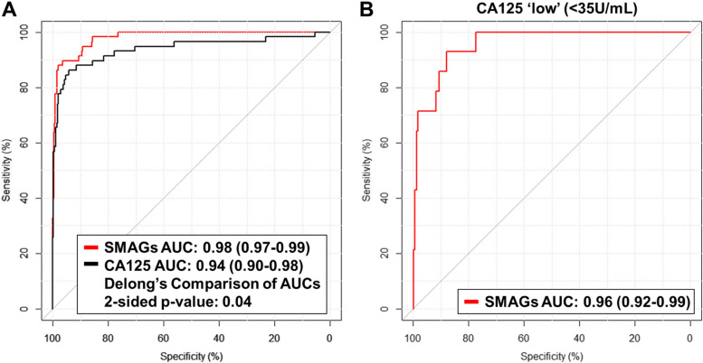
Using a novel Sensitivity Maximization At A Given Specificity (SMAGS) method (see Supplemental Methods), we developed a model consisting of 7 metabolites plus CA125 that yielded an AUC of 0.98 (95% CI: 0.97–0.99) for early-stage disease (Fig. 1A; Table S5). At a 98.5% specificity cutoff, the SMAGS-derived model had sensitivity of 86.2%, correctly identifying 50 of 58 early-stage OvCa cases, which was improved compared to a sensitivity of 75.9% for CA125 alone identifying 44 of 58 early-stage OvCa cases (Table S6). Moreover, the SMAGS-derived model captured 64% of the 14 early stage OvCa cases with CA125 < 35 units/mL, with an AUC estimate of 0.96 (95% CI: 0.92–0.99) (Fig. 1B; Table S7).
Fig. 1.
Performance estimates of the SMAGS model for detection of early-stage ovarian cancer in the EDRN Reference Set. A AUC curves for the SMAGS model and CA125 for detection of early-stage ovarian cancer. B AUC curves for the SMAGS model for detection of early-stage ovarian cancer among individuals with CA125 levels < 35 units/mL
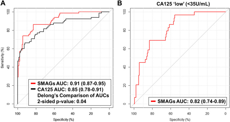
The SMAGS-derived model was validated in an independent set of plasma samples from 65 early stage (I + II) OvCa cases and 141 healthy female controls (Testing Set). The SMAGS-derived model had an AUC of 0.91 (95% CI: 0.87–0.95) for early-stage OvCa, which was improved compared to CA125 alone (AUC: 0.85 (95% CI: 0.78–0.91); 2-sided p-value: 0.04) (Fig. 2A; Figure S2). Using the same cut point developed in the EDRN reference set, the SMAGS-derived model achieved a sensitivity of 73.8% and specificity of 91.4%. In comparison, CA125 at the clinical cutoff of 35 units/mL had 55.4% sensitivity and specificity of 97.2% (Tables S6).
Fig. 2.
Performance estimates of the SMAGs model for detection of early-stage ovarian cancer in the independent Test Set. A AUC curves for the SMAGs model and CA125 for detection of early-stage ovarian cancer. B AUC curves for the SMAGs model for detection of early-stage ovarian cancer among individuals with CA125 levels < 35 units/mL
Among the 29 early-stage OvCa cases with CA125 levels < 35 units/mL, the SMAGS-derived model had an AUC of 0.82 (95% CI: 0.74–0.89) (Fig. 2). At the cut point, the SMAGS-derived captured 13 of the 28 early-stage OvCa cases (44.8% sensitivity) that would otherwise have been missed by CA125 (Tables S7).
The blood-based metabolite test provides a potential clinical tool for identifying women at high-risk of harboring OvCa and that would benefit from surveillance and screening with TSV or MRI for earlier detection of disease, which is anticipated to result in mortality reduction due to OvCa [7, 8]. Given the low incidence of ovarian cancer (11.4 in every 100,000 women) in the general population, the blood-based metabolite test may best be suited for detection of OvCa among higher-risk women presenting with non-specific symptoms such as pelvic/abdominal pain [9, 10] or those with BRCA pathological variants [11].
Supplementary Information
Acknowledgements
N/A
Authors’ contributions
Conceptulization: Johannes F. Fahrmann, Ehsan Irajizad. Methodology: Johannes F. Fahrmann, Seyyed Mahmood Ghasemi, Ranran Wu, Ehsan Irajizad. Validation: Johannes F. Fahrmann, Ehsan Irajizad. Formal Analysis: Johannes F. Fahrmann, Seyyed Mahmood Ghasemi, Ehsan Irajizad. Investigation: Johannes F. Fahrmann, Ehsan Irajizad. Resources: Joseph Celestino, Karen Lu, Zhen Lu, Charles Drescher, Samir Hanash, Robert C. Bast. Data Curation: Johannes F. Fahrmann, Ranran Wu. Writing—original draft preparation: Johannes F. Fahrmann, Seyyed Mahmood Ghasemi, Ehsan Irajizad. Writing—review and editing: Chae Y Han, Ranran Wu, Jennifer B. Dennison, Jody Vykoukal, Joseph Celestino, Karen Lu, Zhen Lu, Charles Drescher, Kim-Anh Do, Samir Hanash, Robert C. Bast.
Funding
This project was supported by MD Anderson SPORE in Ovarian Cancer Caeer Enhancement Program Award (JFF), the National Cancer Institute Early Detection Research Network U01 CA200462 (RCB), the MD Anderson Ovarian SPOREs P50 CA83639 and P50CA217685 (RCB), National Cancer Institute, Department of Health and Human Services; the Cancer Prevention Research Institute of Texas (RP160145) (RCB); Golfer’s Against Cancer, the Mossy Foundation, the Anne and Henry Zarrow Foundation, the Roberson Endowment, National Foundation for Cancer Research, UT MD Anderson Women’s Moon Shot, and generous donations from Stuart and Gaye Lynn Zarrow, Karen and Barry Elson, and Arthur and Sandra Williams.
Availability of data and materials
No datasets were generated or analysed during the current study.
Declarations
Ethics approval and consent to participate
The study is a retrospective analysis of blood specimens that were obtained preoperatively with informed consent under IRB/ethical committees approved protocols at the University of Texas M.D. Anderson Cancer Center (MDACC, LAB04-0687) and at the Fred Hutchinson Cancer Research Center (FHCRC, IRB 4563) [12]. Control plasma were obtained from women who did not develop cancer while participating in the Normal Risk Ovarian Screening Study (NROSS) trial coordinated by MDACC [13] or were healthy donors at the FHCC under improved IRB protocols.
Consent for publication
Not applicable.
Competing interests
Dr. Bast receives royalties from Fujirebio Diagnostics, Inc, for the discovery of CA125.
Footnotes
Publisher’s Note
Springer Nature remains neutral with regard to jurisdictional claims in published maps and institutional affiliations.
References
- 1.Badgwell D, Bast RC Jr. Early detection of ovarian cancer. Dis Markers. 2007;23(5–6):397–410. 10.1155/2007/309382 [DOI] [PMC free article] [PubMed] [Google Scholar]
- 2.Siegel RL, Miller KD, Jemal A. Cancer statistics, 2017. CA Cancer J Clin. 2017;67(1):7–30. 10.3322/caac.21387 [DOI] [PubMed] [Google Scholar]
- 3.Buys SS, Partridge E, Black A, Johnson CC, Lamerato L, Isaacs C, et al. Effect of screening on ovarian cancer mortality: the Prostate, Lung, Colorectal and Ovarian (PLCO) cancer screening randomized controlled trial. JAMA. 2011;305(22):2295–303. 10.1001/jama.2011.766 [DOI] [PubMed] [Google Scholar]
- 4.Menon U, Gentry-Maharaj A, Burnell M, Singh N, Ryan A, Karpinskyj C, et al. Ovarian cancer population screening and mortality after long-term follow-up in the UK Collaborative Trial of Ovarian Cancer Screening (UKCTOCS): a randomised controlled trial. Lancet. 2021;397(10290):2182–93. 10.1016/S0140-6736(21)00731-5 [DOI] [PMC free article] [PubMed] [Google Scholar]
- 5.Fahrmann JF, Irajizad E, Kobayashi M, Vykoukal J, Dennison JB, Murage E, et al. A MYC-driven plasma polyamine signature for early detection of ovarian cancer. Cancers (Basel). 2021;13(4):913. 10.3390/cancers13040913 [DOI] [PMC free article] [PubMed] [Google Scholar]
- 6.Irajizad E, Han CY, Celestino J, Wu R, Murage E, Spencer R, et al. A blood-based metabolite panel for distinguishing ovarian cancer from benign pelvic masses. Clin Cancer Res. 2022;28(21):4669–76. 10.1158/1078-0432.CCR-22-1113 [DOI] [PMC free article] [PubMed] [Google Scholar]
- 7.van Nagell JR Jr, DePriest PD, Ueland FR, DeSimone CP, Cooper AL, McDonald JM, et al. Ovarian cancer screening with annual transvaginal sonography: findings of 25,000 women screened. Cancer. 2007;109(9):1887–96. 10.1002/cncr.22594 [DOI] [PubMed] [Google Scholar]
- 8.Engbersen MP, Van Driel W, Lambregts D, Lahaye M. The role of CT, PET-CT, and MRI in ovarian cancer. Br J Radiol. 2021;94(1125): 20210117. 10.1259/bjr.20210117 [DOI] [PMC free article] [PubMed] [Google Scholar]
- 9.Dilley J, Burnell M, Gentry-Maharaj A, Ryan A, Neophytou C, Apostolidou S, et al. Ovarian cancer symptoms, routes to diagnosis and survival - population cohort study in the “no screen” arm of the UK Collaborative Trial of Ovarian Cancer Screening (UKCTOCS). Gynecol Oncol. 2020;158(2):316–22. 10.1016/j.ygyno.2020.05.002 [DOI] [PMC free article] [PubMed] [Google Scholar]
- 10.Goff BA, Mandel LS, Melancon CH, Muntz HG. Frequency of symptoms of ovarian cancer in women presenting to primary care clinics. JAMA. 2004;291(22):2705–12. 10.1001/jama.291.22.2705 [DOI] [PubMed] [Google Scholar]
- 11.Walker M, Jacobson M, Sobel M. Management of ovarian cancer risk in women with BRCA1/2 pathogenic variants. CMAJ. 2019;191(32):E886–93. 10.1503/cmaj.190281 [DOI] [PMC free article] [PubMed] [Google Scholar]
- 12.Young Han C, Bedia JS, Yang WL, Hawley SJ, Bergan L, Hopper M, et al. Autoantibodies, antigen-autoantibody complexes and antigens complement CA125 for early detection of ovarian cancer. Br J Cancer. 2024;130:861–8. 10.1038/s41416-023-02560-z [DOI] [PMC free article] [PubMed] [Google Scholar]
- 13.Han CY, Lu KH, Corrigan G, Perez A, Kohring SD, Celestino J, et al. Normal risk ovarian screening study: 21-year update. J Clin Oncol. 2024;42(10)Jco2300141. [DOI] [PMC free article] [PubMed]
Associated Data
This section collects any data citations, data availability statements, or supplementary materials included in this article.
Supplementary Materials
Data Availability Statement
No datasets were generated or analysed during the current study.


