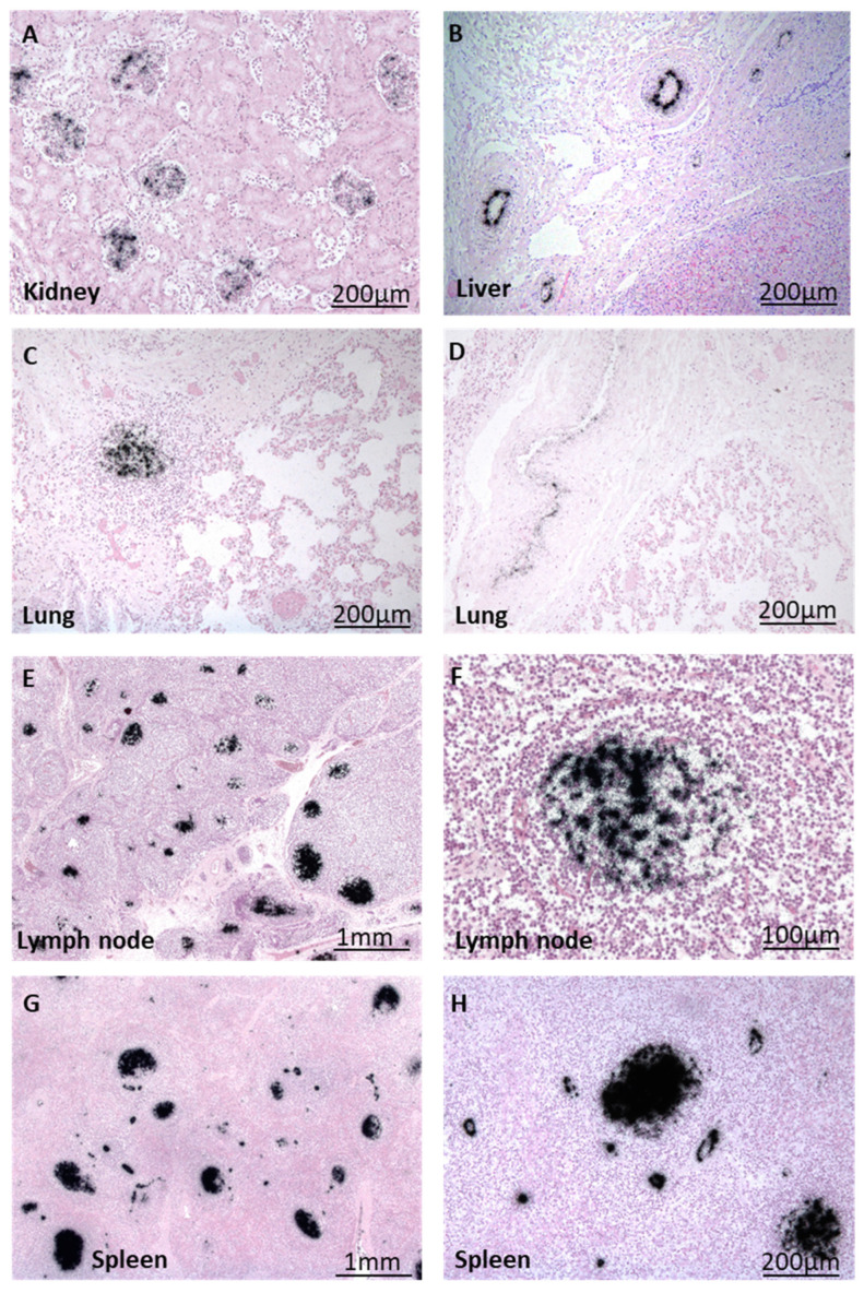
Figure 5.
Visualization of B19V DNA (black) via radioactive ISH in different organs of patient B. (A) Localization of B19V DNA within kidney glomeruli and (B) arterioles of liver tissue. (C,D) B19V-positive immune cells and arterioles in the lung. (E) B19V DNA is present in numerous immune cells of lymph nodes and endothelial cells, with a close-up shown in (F). (G) B19V DNA-positive follicles and vessels in splenic tissue, with a close-up shown in (H).

