Abstract
Simple Summary
Alterations in the SMARCB1/INI1 expression pattern have been detected in many tumors, including chordomas. We studied a large group of patients with conventional spinal chordomas, and the aims were to assess the differences in the immunohistochemical expression of SMARCB1/INI1 and the underlying alterations in the SMARCB1 gene and to investigate the correlation between clinicopathological features and patient survival. Partial SMARCB1/INI1 loss was identified in several patients, and this pattern correlated with mobile spine location and inadequate surgical margins. Moreover, mobile spine tumor location and inadequate surgical margins negatively impacted disease-free survival. The complete loss of SMARCB1/INI1 is currently ongoing as a target for molecular therapy; therefore, the partial loss of SMARCB1/INI1 in tumors could also have therapeutic implications.
Abstract
The partial loss of SMARCB1/INI1 expression has recently been reported in skull base conventional chordomas, with possible therapeutic implications. We retrospectively analyzed 89 patients with conventional spinal chordomas to investigate the differences in the immunohistochemical expression of SMARCB1/INI1 and the underlying genetic alterations in the SMARCB1 gene. Moreover, we assessed the correlation of clinicopathological features (age, gender, tumor size, tumor location, surgical margins, Ki67 labelling index, SMARCB1/INI1 pattern, previous surgery, previous treatment, type of surgery, and the Charlson Comorbidity Index) with patient survival. Our cohort included 51 males and 38 females, with a median age at diagnosis of 61 years. The median tumor size at presentation was 5.9 cm. The 5-year overall survival (OS) and 5-year disease-free survival (DFS) rates were 90.8% and 54.9%, respectively. Partial SMARCB1/INI1 loss was identified in 37 (41.6%) patients with conventional spinal chordomas (27 mosaic and 10 clonal). The most frequent genetic alteration detected was the monoallelic deletion of a portion of the long arm of chromosome 22, which includes the SMARCB1 gene. Partial loss of SMARCB1/INI1 was correlated with cervical–thoracic–lumbar tumor location (p = 0.033) and inadequate surgical margins (p = 0.007), possibly due to the high degree of tumor invasiveness in this site. Among all the considered clinicopathological features related to patient survival, only tumor location in the sacrococcygeal region and adequate surgical margins positively impacted DFS. In conclusion, partial SMARCB1/INI1 loss, mostly due to 22q deletion, was detected in a significant number of patients with conventional spinal chordomas and was correlated with mobile spine location and inadequate surgical margins.
Keywords: conventional chordoma, SMARCB1/INI1, SMARCB1 gene, FISH analysis
1. Introduction
Chordomas are rare malignant neoplasms that develop from embryonic remnants of the notochord. They exhibit distinct histotypes (conventional, poorly differentiated, and dedifferentiated) with different clinical behavior [1]. Conventional chordoma accounts for approximately 95% of cases [1,2]. Chordomas are locally destructive tumors characterized by very slow growth, with possible local recurrence and metastases. The 5- and 10-year OS rates are estimated to be 68.4% and 39.2%, respectively, and the 5- and 10-year DFS rates are 80.9% and 60.1%, respectively [3]. The diagnostic hallmark of chordomas is the nuclear expression of the brachyury protein [1,4]. Complete loss of the SMARCB1/INI1 nuclear protein has also been reported as a peculiar feature of poorly differentiated chordoma [3,5,6]. Recently, the partial loss of SMARCB1/INI1 protein expression has been detected in conventional chordomas localized in the skull base [7]. SMARCB1/INI1 is a tumor suppressor encoded by the SMARCB1 gene (SWI/SNF-related, matrix-associated, actin-dependent regulator of chromatin, subfamily B, member 1), which is located on the long arm of chromosome 22 (22q11.23). This protein is part of the multisubunit ‘SWItch/Sucrose NonFermentable ATP-dependent chromatin remodelling complex’ (SWI/SNF), which regulates different cellular mechanisms, including gene expression and cell proliferation and differentiation [8,9]. Abnormal expression of SMARCB1/INI1 has been detected extensively in different tumor types, and three distinct expression patterns have been identified: complete loss, partial loss, and reduced expression [10,11]. However, the type of abnormal expression pattern and the type of mutation in the SMARCB1 gene do not always match; in some cases, no DNA or RNA changes are detected [10]. Among tumors with focal expression of SMARCB1/INI1, different types of genetic alterations have been described, the most frequent being the monoallelic deletion of a portion of the long arm of chromosome 22, which includes the SMARCB1 gene [7,10]. However, several studies have revealed that SMARCB1/INI1-deficient tumors, despite being very different from each other in location and type, generally share an aggressive clinical course with high local recurrence rates and a prognosis that is often poor [11,12,13,14].
From a treatment perspective, chordoma appears to be resistant to common chemotherapy, and clinical studies are currently ongoing to treat some of these forms with new targeted molecules, including tyrosine kinase inhibitors, CDK4 inhibitors, and immunotherapy based on monoclonal antibodies [2,3,15]. Specifically, the complete loss of SMARCB1/INI1 expression is considered a marker for the evaluation of the effectiveness of Enhancer of Zeste Homolog 2 (EZH2) inhibitors (Tazemetostat) [15,16]. The most frequent cytogenetic abnormalities observed in conventional chordomas are monosomy of chromosome 1 and copy number gains of chromosomes 2, 6, and 7 [1,3]. Loss of chromosome 22 and/or genetic alterations in the SMARCB1 gene seem to be rare [17,18,19].
This study aimed to compare SMARCB1/INI1 protein expression patterns in spinal conventional chordomas with genetic alterations detectable in the SMARCB1 gene by FISH, clinicopathological features, OS, and DFS.
2. Materials and Methods
A retrospective study of 89 patients with conventional spinal chordoma diagnosed at the Anatomy and Pathological Histology Unit of the Rizzoli Orthopedic Institute from 2010 to 2019 was carried out. In order to perform morphological, immunohistochemical, and molecular analyses, a formalin-fixed paraffin-embedded (FFPE) tumor tissue sample of adequate size and quality was used, after selection by pathologists (MG and AR). The diagnosis of all the original tumor slides was confirmed independently by two pathologists (MG and AR) via the immunohistochemical expression of brachyury and pan-cytokeratin AE1/AE3. The clinicopathological parameters investigated were: age, gender, tumor size, tumor location, surgical margins, Ki67 labelling index, SMARCB1/INI1 pattern, previous surgery, previous treatment, type of surgery, and comorbidities. The surgical margins were classified according to the Enneking classification [20] and to the Weinstein–Boriani–Biagini (WBB) system [21]. The comorbidities were evaluated by the Charlson Comorbidity Index (CCI) [22]. Ethical committee approval was obtained from the Comitato Etico di Area Vasta Emilia Centro on 27/04/2023 (protocol # CE AVEC: 312/2023/Oss/IOR). As a comparison group, 4 patients with poorly differentiated chordoma were included in the analysis.
Immunohistochemical staining was performed using an automated immunostainer following the manufacturer’s guidelines (Ventana BenchMark-Ventana Medical Systems, Tucson, AZ, USA), with a mouse monoclonal anti-INI-1 antibody at a concentration of 0.4 μg/mL (MRQ-27; Cell Marque, Rocklin, CA, USA) and a rabbit monoclonal primary anti-Ki-67 antibody at a concentration of 0.2 μg/mL (clone 30-9, Ventana). The immunohistochemical evaluation was executed independently by two pathologists to determine the percentage of proliferating cells (Ki67 labelling index) and to select only samples with partial SMARCB1/INI1 expression and a minimum 10% cut-off of neoplastic nuclei. Regarding SMARCB1/INI1, both patients with mosaic expression (defined by the presence of negative nuclei mixed with positive nuclei) and patients with clonal expression (characterized by the presence of a completely negative high-magnification field alongside a fully positive high-magnification field) were considered eligible; homogeneous nuclear staining in the background of inflammatory cells, stromal fibroblasts, normal epithelial cells, and/or vascular endothelial cells were used as an internal control.
FISH for the SMARCB1 gene was performed using a commercial SPEC SMARCB1/22q12 Dual color CE/IVD Probe (ZytoVision, Bremerhaven, Germany). The analysis was performed on conventional chordomas with focal SMARCB1/INI1 expression and four poorly differentiated chordomas. The probe included a 545 kb sequence mapped to the 22q11.23 region (ZyGreen fluorochrome label) harboring the SMARCB1 gene and a 335 kb sequence mapped to the 22q12.1–q12.2 region (ZyOrange fluorochrome label) harboring the KREMEN1 gene, which was used as an internal control region to detect large chromosome 22q deletions. FISH was performed on interphase nuclei using the Histology FISH accessory kit (Dako, Glostrup, Denmark) according to the manufacturer’s protocol [23], as previously described [7]. For each slide, a minimum of 100 intact nuclei within the tumor area previously marked by the pathologist were scored using a BX41 fluorescence microscope (Olympus, Tokyo, Japan) at 100× magnification, and visible alteration in at least 10% of the cells was considered a positive result. Nuclei with no signal or signals in overlapping nuclei were considered non-informative and were not analyzed. A Color View III CCD camera soft imaging system (Olympus) was used to capture images, which were subsequently analyzed with CytoVision imaging software version 7.5 (Leica Biosystem Richmond Inc., Richmond, IL, USA). The presence of two green signals and two orange signals in a 1:1 ratio was considered the normal copy number pattern; any FISH signals differing from this pattern were classified as altered. The detection of one green signal and one orange signal indicated a monoallelic co-deletion of SMARCB1 and the control region, which was classified as a monoallelic 22q large deletion, and the presence of additional copies of both green and orange signals indicated a copy number gain (CNG) of chromosome 22.
OS was defined as the time between the date of diagnosis and the date of death or the last follow-up, and DFS was defined as the time between the first disease relapse or metastasis and the last follow-up. Descriptive statistics were used to report patient and clinical characteristics. All the continuous data were expressed as the means and the standard deviations of the means; the categorical data were expressed as frequencies and percentages. Fisher’s chi-square exact test was used to analyze dichotomous variables. Pearson’s chi-square exact test was performed to investigate categorical variables. Kaplan–Meier survival analyses with the log-rank test were performed to assess the influence of the different parameters on OS and DFS. For all the tests, p < 0.05 was considered as statistically significant. All the statistical analyses were performed using SPSS v.19.0 (IBM Corp., Armonk, NY, USA).
3. Results
Table 1 summarizes the main clinicopathological features of 89 patients with conventional spinal chordomas.
Table 1.
Clinicopathological features of 89 patients with conventional spinal chordomas.
| Parameters | All Samples (n = 89) |
|---|---|
| Gender (N, %) | |
| Male | 51 (57.3%) |
| Female | 38 (42.7%) |
| Age (median, range in years) | 61 (17–86) |
| Age (N, %) | |
| ≤60 years | 42 (47.2%) |
| >60 years | 47 (52.8%) |
| Tumor size (N, %) | |
| <5 cm | 36 (40.4%) |
| ≥5 cm | 39 (43.9%) |
| Not available | 14 (15.7%) |
| Tumor localization | |
| Cervical–thoracic–lumbar region | 43 (48.3%) |
| Sacrococcygeal region | 46 (51.7%) |
| Surgical margin | |
| Adequate | 45 (50.6%) |
| Inadequate | 25 (28.1%) |
| Not available | 19 (21.3%) |
| Ki-67 index (median, range) | 3 (1–12) |
| Ki-67 index (N, %) | |
| ≤3% | 43 (48.3%) |
| >3% | 37 (41.6%) |
| Not evaluable | 9 (10.1%) |
| SMARCB1/INI1 immunohistochemical expression (N, %) | |
| Positive | 52 (58.4%) |
| Positive/negative | 37 (41.6%) |
| Previous surgery | |
| No | 53 (59.6%) |
| Yes | 21 (23.6%) |
| Not available | 15 (16.9%) |
| Previous treatment | |
| No | 59 (66.3%) |
| Yes | 14 (15.7%) |
| Not available | 16 (18%) |
| Type of surgery | |
| En bloc resection | 54 (60.7%) |
| Other surgery | 16 (18%) |
| No surgery | 19 (21.3%) |
| Charlson Comorbidity Index (CCI) | |
| Mean (SD) | 4.1 (0.260) |
The dataset included 51 (57.3%) males and 38 (42.7%) females, with a median age at diagnosis of 61 years (range 17–86). Clinically, 43 (48.3%) tumors were located in the cervical–thoracic–lumbar region (mobile spine), while 46 (51.7%) were located in the sacrococcygeal region. The median tumor size at presentation was 5.9 cm (range 1.4–16 cm). The mean CCI of the population was 4.1. Twenty-one patients (23.6%) underwent previous surgical treatment, and 14 patients (15.7%) underwent previous systemic therapy and/or radiotherapy for the same tumor.
Among the 70 patients who underwent surgical resection, 45 patients (50.6%) had adequate surgical margins (wide and radical), while 25 (28.1%) had inadequate surgical margins (intralesional and marginal), according to the Enneking classification [20] (Table S1). Among the remaining 19 inoperable patients, 12 were treated with carbon ion therapy, 3 with proton therapy, and 1 with radiation and chemotherapy; for 3 patients only biopsy information was available without follow-up data. Of the cases with inadequate margins, nine cases were localized at the cervical region, seven cases were localized at the thoracic–lumbar region (six patients were previously treated with surgery at other centers), and nine cases were localized at the sacrococcygeal region (three patients were previously treated with surgery at other centers). When feasible, a classification according to the WBB system [21] was performed and all 10 tumors analyzed had very large extensions with both extra-osseous and intracanal components (Table 2), which did not allow resection with wide margins.
Table 2.
The WBB classification of patients with surgical inadequate margins.
| Case Number | Tumor Localization | WBB Classification | Revision Surgery |
|---|---|---|---|
| 1 | L3 | layers A–E; zones 12–1 | NO |
| 2 | sacrum | n.a. | NO |
| 7 | C4–C5 | layers C–E, zones 8–5 | NO |
| 15 | sacrum | n.a. | NO |
| 19 | sacrum | n.a. | NO |
| 25 | L5 | n.a. | YES |
| 29 | C2 | layers A–E; zones 11–7 | NO |
| 34 | C3 | layers A–E; zones 2–8 | NO |
| 35 | C2 | layers A–E; zones 9–4 | NO |
| 40 | L3 | n.a. | YES |
| 42 | sacrum | n.a. | YES |
| 44 | C2–C3 | layers A–E; zones 6–2 | NO |
| 45 | L4–L5 | n.a. | YES |
| 48 | L2 | n.a. | YES |
| 52 | sacrum | n.a. | YES |
| 58 | T2–T3 | n.a. | YES |
| 64 | T9 | layers A–E; zones 9–1 | YES |
| 66 | C2 | layers A–E; zones 7–4 | NO |
| 68 | C2 | layers A–E; zones 11–5 | NO |
| 71 | C5–C6 | n.a. | YES |
| 72 | coccyx | n.a. | NO |
| 73 | sacrum | n.a. | YES |
| 78 | sacrum | n.a. | NO |
| 79 | C1–C2 | layers A–E; zones 6–3 | NO |
| 89 | sacrum | n.a. | NO |
n.a. = not applicable, because of localization on the sacrococcygeal region or because of the absence of pre-operative imaging.
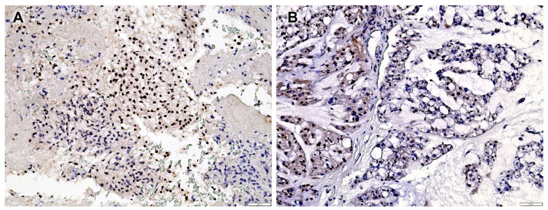
The median Ki-67 labelling index was 3% (range 1–12%), excluding nine non-evaluable cases (absence of positive internal controls in normal bone marrow cells). The SMARCB1/INI1 immunohistochemical analyses revealed a partial loss of SMARCB1/INI1 (range 10–80%) in 37 (41.6%) patients, while 52 (58.4%) patients exhibited complete protein expression in all neoplastic cells (Table S1). In the 37 patients with focal SMARCB1/INI1 loss, 2 different staining patterns were identified: 27 cases had a mosaic expression pattern (with mixed negative and positive nuclei), while 10 cases had a clonal expression pattern (with separate fully negative and fully positive high-magnification fields) (Figure 1A,B). The four poorly differentiated chordomas exhibited complete loss of SMARCB1/INI1 in all the evaluated neoplastic cells.
Figure 1.
(A) Case n.25 showing clonal expression of SMARCB1/INI1; (B) case n.44 showing mosaic expression of SMARCB1/INI1.
Partial loss of the immunohistochemical expression of SMARCB1/INI1 was significantly associated with localization in the cervical–thoracic–lumbar region (p = 0.033) and inadequate surgical margins (p = 0.007). No significant associations were found with gender, age at diagnosis, tumor size, or Ki67 index (Table 3).
Table 3.
Clinicopathological features according to SMARCB1/INI1 immunohistochemical expression.
| SMARCB1/INI1 + (n = 52) |
SMARCB1/INI +/− (n = 37) |
p-Value | |
|---|---|---|---|
|
Gender (N, %) Male Female |
|||
| 28 (53.8%) | 23 (62.2%) | 0.516 | |
| 24 (46.2%) | 14 (37.8%) | ||
| Age (median, range in years) | 61.5 (28–86) | 59 (17–79) | 0.511 |
| Age (N, %) | |||
| ≤60 years | 22 (42.3%) | 20 (54.1%) | 0.291 |
| >60 years | 30 (57.7%) | 17 (45.9%) | |
| Tumor size (N, %) | |||
| <5 cm | 20 (38.5%) | 16 (43.2%) | |
| ≥5 cm | 20 (38.5%) | 19 (51.4%) | 0.818 |
| Not available | 12 (23%) | 2 (5.4%) | |
| Tumor localization | |||
| Cervical–thoracic–lumbar region | 20 (38.5%) | 23 (62.2%) | 0.033 |
| Sacrococcygeal region | 32 (61.5%) | 14 (37.8%) | |
| Surgical margin | |||
| Adequate | 30 (57.7%) | 15 (40.5%) | |
| Inadequate | 8 (15.3%) | 17 (46%) | 0.007 |
| Not available | 14 (27%) | 5 (13.5%) | |
|
Ki-67 index (median, range in percentage) |
3 (1–12%) | 3 (1–9%) | 0.459 |
| Ki-67 index (N, %) | |||
| ≤3% | 26 (50%) | 17 (46%) | |
| >3% | 24 (46.2%) | 13 (35%) | 0.817 |
| Not evaluable | 2 (3.8%) | 7 (19%) |
Statistically significant p values are shown in red color.
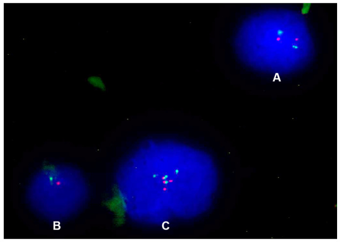
The FISH analysis performed on 37 conventional spinal chordoma patients with focal SMARCB1/INI1 loss revealed three possible molecular patterns (Figure 2).
Figure 2.
(A) Normal nucleus, with two signals for the control region (orange) and two signals for the SMARCB1 gene (green); (B) nucleus with monoallelic deletion, with only one signal for the control region (orange) and only one signal for the SMARCB1 gene (green); (C) nucleus with CNG, with three or more signals for both the control region (orange) and SMARCB1 gene (green).
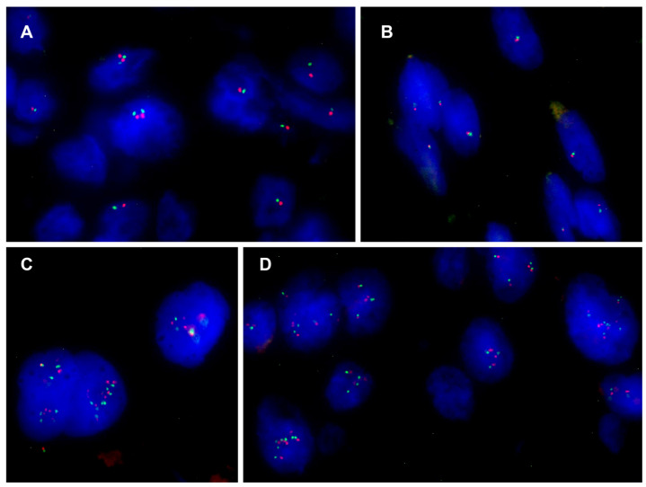
Monoallelic deletion of the SMARCB1 gene associated with co-deletion of the control region was observed in 16 cases of conventional chordoma (range 26–94%) (Figure 3A,B); 5 of these also had nuclei with additional copies of both signals (Figure 3C,D). One case exhibited only nuclei with CNG and none with deletions. Due to poor tissue quality, 20 samples did not show hybridized signals and were considered inadequate for FISH scoring (Table S1). Considering the two different staining patterns of focal SMARCB1/INI1 expression, all 10 cases with mosaic patterns had a monoallelic 22q deletion (range 30–94%), 3 of these cases also had nuclei with CNG of both signals; 5 of 6 cases with clonal patterns had a monoallelic 22q deletion (range 26–81%); 2 of these cases also had nuclei with extra copies of SMARCB1 and the control region, whereas 1 case had only nuclei with CNG of both signals.
Figure 3.
(A,B) Nuclei with monoallelic co-deletion of the SMARCB1 gene and the control region from cases n.58 and n.21, respectively; (C,D) nuclei with CNG from cases n.37 and n.66, respectively.
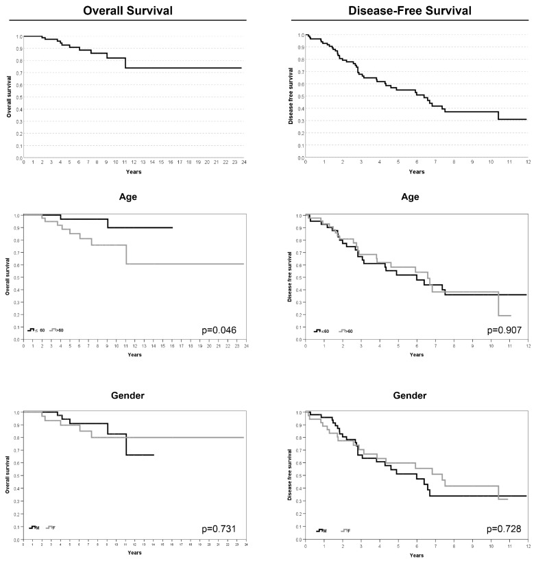
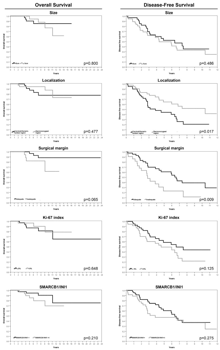
In the four cases of poorly differentiated chordoma, FISH analyses revealed biallelic SMARCB1 deletions in two cases, a monoallelic deletion in one case, and a pattern with a monoallelic SMARCB1 deletion associated with an additional control region signal in one case. The average follow-up duration after treatment completion was 66 months (range 2–148). The 5-year OS and 5-year DFS rates were 90.8% (SE 3.6%) and 54.9% (SE 6%), respectively. Univariate analysis revealed worse overall survival for patients older than 60 years (p = 0.046). The risk of local recurrence or metastasis was greater for patients with a tumor in the cervical–thoracic–lumbar region (p = 0.017), for those with inadequate surgical margins (p = 0.009), and for patients who underwent a previous surgery for the same tumor (p < 0.0005) (Table 4; Figure 4 and Figure 5). Moreover, the presence of comorbidities significantly affected both OS and DFS, as shown in Table 4 and Table 5.
Table 4.
Results from univariate Kaplan–Meier models for OS and DFS.
| 5 Years—OS % (SE) | p-Value | 5 Years—DFS % (SE) | p-Value | |
|---|---|---|---|---|
| Entire sample | 90.8% (3.6%) | 54.9% (6%) | ||
| Gender (N, %) | ||||
| Male | 91% (5%) | 0.731 | 51.1% (8.1%) | 0.728 |
| Female | 89.7% (5.6%) | 59.8% (8.8%) | ||
| Age (N, %) | ||||
| ≤60 years | 96.8% (3.2%) | 0.046 | 51.9% (8.3%) | 0.907 |
| >60 years | 85.1% (6.2%) | 58.3% (8.5%) | ||
| Tumor size (N, %) | ||||
| <5 cm | 90.5% (5.2%) | 0.800 | 52.7% (9%) | 0.486 |
| ≥5 cm | 94.4% (5.4%) | 49.7% (9.3%) | ||
| Tumor localization | ||||
| Cervical–thoracic–lumbar region | 87.7% (5.8%) | 0.477 | 44.2% (8.5%) | 0.017 |
| Sacrococcygeal region | 94.6% (3.7%) | 64.8% (8.1%) | ||
| Surgical margin | ||||
| Adequate | 96.8% (3.2%) | 0.065 | 61% (8%) | 0.009 |
| Inadequate | 82.2% (9.3%) | 23.2% (10.4%) | ||
| Ki-67 index (N, %) | ||||
| ≤3% | 89.6% (5.7%) | 0.648 | 60.5% (7.9%) | 0.125 |
| >3% | 96.7% (3.3%) | 47.3% (9.1%) | ||
| SMARCB1/INI1 immunohistochemical expression (N, %) | ||||
| Positive | 94.8% (3.6%) | 0.210 | 58.6% (8.8%) | 0.275 |
| Positive/negative | 85.5% (6.8%) | 49.4% (9.1%) | ||
| Previous surgery | ||||
| No | 88.9% (4.8%) | 0.98 | 66.3% (7.5%) | <0.0005 |
| Yes | 93.7% (7.4%) | 25.3% (10.4%) | ||
| Previous treatment | ||||
| No | 90.6% (4.5%) | 0.858 | 54.4% (7.4%) | 0.56 |
| Yes | 90.0% (9.5%) | 58.4% (14.5%) | ||
| Type of surgery | ||||
| En bloc resection | 88.7% (4.8%) | 0.693 | 61.6% (7.2%) | 0.899 |
| Other | 90.0% (9.5%) | 44.7% (17.1%) | ||
| Charlson Comorbidity Index (CCI) | ||||
| ≤4 | 92.3% | 0.076 | 63.0% (7.3%) | 0.011 |
| >4 | 83.8% | 39.3% (10.2%) |
Statistically significant p values are shown in red color.
Figure 4.
Kaplan–Meier survival analyses for age and gender features.
Figure 5.
Kaplan–Meier survival analyses for size and tumor localization, surgical margins, Ki-67 index, and SMARCB1/INI1 immunohistochemical expression.
Table 5.
Univariate analysis for CCI as continuous variable.
| 5 years—OS | p-Value | HR | 95.0% CI | ||
| Inferior | Superior | ||||
| CCI | 0.043 | 1.694 | 1.018 | 2.820 | |
| 5 years—DFS | p-Value | HR | 95.0% CI | ||
| Inferior | Superior | ||||
| CCI | 0.078 | 1.222 | 0.978 | 1.528 | |
Statistically significant p values are shown in red color.
The results of the multivariate analysis demonstrated that the inadequate surgical margin and an age older than 60 years significantly impaired the OS (Table 6). The risk of local recurrence or metastases was increased by a higher Ki67 index, by an inadequate surgical margin, and by a high CCI: with the same surgical margin and Ki67 scores, the increase of 1 unit of the CCI increases the risk by 40.5% (Table 7). It should be noted that the CCI includes the age, and all patients older than 60 years have a CCI higher than 4.
Table 6.
Multivariate analysis for overall survival.
| p-Value | HR | 95.0% CI | |||
|---|---|---|---|---|---|
| Inferior | Superior | ||||
| Phase 1 | CCI | 0.788 | 1.110 | 0.519 | 2.373 |
| margin (1 vs. 0) * | 0.006 | 29.965 | 2.619 | 342.854 | |
| age (˃ 60 vs. ≤60) | 0.050 | 19.600 | 1.001 | 383.640 | |
| Phase 2 | margin (1 vs. 0) * | 0.006 | 30.049 | 2.634 | 342.745 |
| age (˃ 60 vs. ≤60) | 0.012 | 24.592 | 2.019 | 299.586 | |
* 0 = adequate margin; 1 = inadequate margin. Statistically significant p values are shown in red color.
Table 7.
Multivariate analysis for disease-free survival.
| p-Value | HR | 95.0% CI | |||
|---|---|---|---|---|---|
| Inferior | Superior | ||||
| Phase 1 | Ki67 | 0.037 | 1.216 | 1.012 | 1.461 |
| margin (1 vs. 0) * | 0.036 | 2.501 | 1.060 | 5.904 | |
| localization | 0.233 | 0.530 | 0.187 | 1.504 | |
| previous surgery | 0.321 | 1.489 | 0.678 | 3.270 | |
| CCI | 0.008 | 1.526 | 1.119 | 2.079 | |
| type of surgery (other) | 0.556 | 0.681 | 0.189 | 2.447 | |
| type of Surgery (en bloc resection) | 0.868 | 0.913 | 0.313 | 2.664 | |
| Phase 2 | Ki67 | 0.033 | 1.216 | 1.016 | 1.455 |
| margin (1 vs. 0) * | 0.026 | 2.598 | 1.119 | 6.032 | |
| localization | 0.210 | 0.548 | 0.214 | 1.403 | |
| previous surgery | 0.279 | 1.529 | 0.709 | 3.298 | |
| CCI | 0.004 | 1.513 | 1.141 | 2.007 | |
| Phase 3 | Ki67 | 0.032 | 1.216 | 1.017 | 1.453 |
| margin (1 vs. 0) * | 0.018 | 2.771 | 1.195 | 6.429 | |
| localization | 0.203 | 0.547 | 0.216 | 1.383 | |
| CCI | 0.004 | 1.517 | 1.143 | 2.013 | |
| Phase 4 | Ki67 | 0.061 | 1.188 | 0.992 | 1.421 |
| margin (1 vs. 0) * | 0.019 | 2.667 | 1.173 | 6.059 | |
| CCI | 0.004 | 1.502 | 1.142 | 1.976 | |
* 0 = adequate margin; 1 = inadequate margin. Statistically significant p values are shown in red color.
4. Discussion
Conventional spinal chordoma is a rare, slow-growing, locally aggressive malignant neoplasm [1,2]. In recent years, an increasing number of tumors, including poorly differentiated chordomas, have been found to exhibit complete loss of SMARCB1/INI1 protein expression. In many patients, molecular analyses of the SMARCB1 gene revealed a biallelic deletion [3,11]. Recently, conventional skull base chordomas have also been investigated by immunohistochemistry, and partial loss of SMARCB1/INI1 was identified [7]. In our study, the immunohistochemical pattern of SMARCB1/INI1 in conventional spinal chordomas was analyzed for the first time, and partial loss of SMARCB1/INI1 was observed in 41.6% of cases. In particular, two distinct expression patterns were detected, mosaic and, less frequently, clonal, confirming what has been previously reported on conventional skull base chordomas [7]. From a molecular perspective, several types of genetic alterations have been described among tumors with focal expression, but the most frequent is the monoallelic deletion of a portion of the long arm of chromosome 22 (involving SMARCB1) [7,10,16]. However, the genomic studies in the literature revealed that the loss of chromosome 22 or the monoallelic deletion of SMARCB1 is rare in conventional spinal chordomas [17,18]. In our series, we genetically investigated only conventional chordomas with impaired SMARCB1/INI1 pattern expression, and in 43.2% of the feasible cases, a monoallelic co-deletion of the SMARCB1 gene and the control region was observed. To evaluate the SMARCB1 locus at chromosome 22q, we used FISH analysis with a CE-IVD probe. Due to cross-hybridization of chromosome 22 alpha satellites to other centromeric regions, a probe mapped to the 22q12.1-q12.2 region was used as an internal control, which has already been proven to be a reliable control for investigating large deletions [24]. Heterozygous partial deletion of the long arm of chromosome 22 was confirmed as the main molecular mechanism underlying the focal expression of the SMARCB1/INI1 protein. Specifically, the chordomas with mosaic SMARCB1/INI1 expression showed mainly monoallelic 22q deletion, whereas the cases with clonal SMARCB1/INI1 expression were associated with different types of genetic patterns. Nuclei with additional copies of the SMARCB1 gene and 22q12 control region were also frequently detected in several subclones of cases with deletion, confirming a previously described event [7,16,19]. However, point mutations in SMARCB1 were not investigated in our study, and epigenetic alterations or post-translational modifications might play an additional role in interpreting the large genetic variability associated with the phenotypic expression of SMARCB1/INI1. We observed that partial loss of SMARCB1/INI1 was significantly associated with the cervical–thoracic–lumbar region (p = 0.033) and inadequate surgical margins (p = 0.007), suggesting that partial loss of the protein might be associated with increased clinical aggressiveness. A possible reason for the correlation between partial SMARCB1/INI1 loss and inadequate margins could be the major extra-osseous and intracanal involvement of the tumors in the mobile spine, thus increasing the difficulty in obtaining adequate surgical margins. Indeed, 37.5% of patients with inadequate surgical margins were treated for local recurrence of the tumor. The statistical analysis, moreover, indicated the localization in the mobile spine and the presence of surgical inadequate margins as negative prognostic factors in terms of the disease-free survival (p = 0.017 and p = 0.009, respectively), unlike the cases located in the skull base, where no correlations were found between the partial loss of SMARCB1/INI1 and the clinicopathological parameters evaluated [7]. The multivariate analyses revealed the most crucial factors to be monitored for patient prognosis. The presence of inadequate surgical margins was confirmed as the prevalent risk factor both for OS and DFS; moreover, an age older than 60 years also significantly impaired the OS, whereas DFS was also associated with a high Ki67 index and by a high CCI.
Due to the difficulty in surgically eradicating tumors and the known resistance of chordoma to common chemotherapies [25,26], new molecular targets are being investigated to properly treat these tumors [15]. Increasing knowledge of SMARCB1/INI1 function has enabled the identification of specific targets, including the EZH2 gene. This target is a catalytic subunit of the polycomb repressive complex 2 (PRC2), which plays a role in the chromatin regulation, in cell fate determination, and in cellular differentiation and is often up-regulated in tumors with a loss of SMARCB1/INI1 [8,27,28]. An increase in EZH2 expression correlates with tumor aggressiveness [28], and specifically, this mechanism has been associated with the progression of chordomas [29]. Thus, clinical trials on inhibitors of the EZH2 enzyme are currently underway in tumors with complete loss of SMARCB1/INI1 expression, including poorly differentiated chordomas (ClinicalTrials.gov Identifiers: NCT02601950 and NCT05407441) [30,31,32]. These trials show the safety tolerability and effectiveness of the drug, with the possibility of use in other types of malignancies [2,3,28]; specifically, the potential use of EZH2 inhibitors could also be promising for patients with partial SMARCB1/INI1 loss, but it needs further exploration.
5. Conclusions
In conclusion, we retrospectively analyzed 89 cases of conventional spinal chordoma, and two distinct expression patterns (mosaic and clonal) of partial SMARCB1/INI1 loss were observed. The most frequent molecular alteration detected in conventional chordoma was the monoallelic deletion of the 22q locus (including SMARCB1 gene). Partial loss of SMARCB1/INI1 was significantly associated with location in the mobile spine and inadequate surgical margins. Inadequate surgical margins, a high Ki67 index, a high CCI, and an age older than 60 years were also associated with a worse prognosis. Treatments with inhibitors of the EZH2 enzyme are currently ongoing in tumors with complete loss of SMARCB1/INI1 expression; therefore, tumors with partial loss of SMARCB1/INI1 could also have therapeutic implications.
Acknowledgments
We are grateful to Muscolo skeletal tumor biobank – biobanca dei tumori muscoloscheletrici (BIOTUM)—member of the CRB-IOR—which provided us with the biological samples. The authors thank Cristina Ghinelli for her help in the graphic design and Monica Contoli for recovery of archival material.
Supplementary Materials
The following supporting information can be downloaded at: https://www.mdpi.com/article/10.3390/cancers16162808/s1.
Author Contributions
Conceptualization, A.R.; formal analysis, E.P.; investigation, M.M., S.C., G.G. and S.B.; resources, G.B., A.G. and R.G.; data curation, C.G., L.E.N., C.A., A.R, M.M., M.G. and C.F.; writing—original draft, M.M., S.C., C.G. and E.P.; writing—review and editing, A.R., M.G., S.B., G.M., G.G., S.C., M.M., C.G., C.F. and R.G.; funding acquisition, S.A. All authors have read and agreed to the published version of the manuscript.
Institutional Review Board Statement
The study was conducted according to the guidelines of the Declaration of Helsinki and approved by the Ethics Committee of Area Vasta Emilia Centro on 27/04/2023 (protocol # CE AVEC: 312/2023/Oss/IOR).
Informed Consent Statement
Informed consent was obtained from all subjects involved in the study.
Data Availability Statement
The original contributions presented in the study are included in the article. Further inquiries can be directed to the corresponding authors.
Conflicts of Interest
The authors declare no conflicts of interest.
Funding Statement
This research was supported by funds for selected research topics from the Fondazione CARISBO Project (#19344).
Footnotes
Disclaimer/Publisher’s Note: The statements, opinions and data contained in all publications are solely those of the individual author(s) and contributor(s) and not of MDPI and/or the editor(s). MDPI and/or the editor(s) disclaim responsibility for any injury to people or property resulting from any ideas, methods, instructions or products referred to in the content.
References
- 1.WHO Classification of Tumours Editorial Board, editor. WHO Classification of Tumours 5th Edition: Soft Tissue and Bone Tumours. 5th ed. WHO Press; Geneva, Switzerland: 2020. [Google Scholar]
- 2.Wedekind M.F., Widemann B.C., Cote G. Chordoma: Current status, problems, and future directions. Curr. Probl. Cancer. 2021;45:100771. doi: 10.1016/j.currproblcancer.2021.100771. [DOI] [PubMed] [Google Scholar]
- 3.Ulici V., Hart J. Chordoma. Arch. Pathol. Lab. Med. 2022;146:386–395. doi: 10.5858/arpa.2020-0258-RA. [DOI] [PubMed] [Google Scholar]
- 4.Vujovic S., Henderson S., Presneau N., Odell E., Jacques T.S., Tirabosco R., Boshoff C., Flanagan A.M. Brachyury, a crucial regulator of notochordal development, is a novel biomarker for chordomas. J. Pathol. 2006;209:157–165. doi: 10.1002/path.1969. [DOI] [PubMed] [Google Scholar]
- 5.Shih A.R., Chebib I., Deshpande V., Dickson B.C., Iafrate A.J., Nielsen G.P. Molecular characteristics of poorly differentiated chordoma. Genes Chromosomes Cancer. 2019;58:804–808. doi: 10.1002/gcc.22782. [DOI] [PubMed] [Google Scholar]
- 6.Mobley B.C., McKenney J.K., Bangs C.D., Callahan K., Yeom K.W., Schneppenheim R., Hayden M.G., Cherry A.M., Gokden M., Edwards M.S.B., et al. Loss of SMARCB1/INI1 expression in poorly differentiated chordomas. Acta Neuropathol. 2010;120:745–753. doi: 10.1007/s00401-010-0767-x. [DOI] [PubMed] [Google Scholar]
- 7.Righi A., Cocchi S., Maioli M., Zoli M., Guaraldi F., Carretta E., Magagnoli G., Pasquini E., Melotti S., Vornetti G., et al. SMARCB1/INI1 loss in skull base conventional chordomas: A clinicopathological and molecular analysis. Front. Oncol. 2023;13:1160764. doi: 10.3389/fonc.2023.1160764. [DOI] [PMC free article] [PubMed] [Google Scholar]
- 8.Kalimuthu S.N., Chetty R. Gene of the month: SMARCB1. J. Clin. Pathol. 2016;69:484–489. doi: 10.1136/jclinpath-2016-203650. [DOI] [PMC free article] [PubMed] [Google Scholar]
- 9.Centore R.C., Sandoval G.J., Soares L.M.M., Kadoch C., Chan H.M. Mammalian SWI/SNF Chromatin Remodeling Complexes: Emerging Mechanisms and Therapeutic Strategies. Trends Genet. 2020;36:936–950. doi: 10.1016/j.tig.2020.07.011. [DOI] [PubMed] [Google Scholar]
- 10.Kohashi K., Oda Y. Oncogenic roles of SMARCB1/INI1 and its deficient tumors. Cancer Sci. 2017;108:547–552. doi: 10.1111/cas.13173. [DOI] [PMC free article] [PubMed] [Google Scholar]
- 11.Pawel B.R. SMARCB1-deficient Tumors of Childhood: A Practical Guide. Pediatr. Dev. Pathol. 2018;21:6–28. doi: 10.1177/1093526617749671. [DOI] [PubMed] [Google Scholar]
- 12.Parker N.A., Al-Obaidi A., Deutsch J.M. SMARCB1/INI1-deficient tumors of adulthood. F1000Research. 2020;9:662. doi: 10.12688/f1000research.24808.2. [DOI] [PMC free article] [PubMed] [Google Scholar]
- 13.Chitguppi C., Rabinowitz M.R., Johnson J., Bar-Ad V., Fastenberg J.H., Molligan J., Berman E., Nyquist G.G., Rosen M.R., Evans J.E., et al. Loss of SMARCB1 Expression Confers Poor Prognosis to Sinonasal Undifferentiated Carcinoma. J. Neurol. Surg. Part B Skull Base. 2020;81:610–619. doi: 10.1055/s-0039-1693659. [DOI] [PMC free article] [PubMed] [Google Scholar]
- 14.Wang J., Andrici J., Sioson L., Clarkson A., Sheen A., Farzin M., Toon C.W., Turchini J., Gill A.J. Loss of INI1 expression in colorectal carcinoma is associated with high tumor grade, poor survival, BRAFV600E mutation, and mismatch repair deficiency. Hum. Pathol. 2016;55:83–90. doi: 10.1016/j.humpath.2016.04.018. [DOI] [PubMed] [Google Scholar]
- 15.Chen S., Ulloa R., Soffer J., Alcazar-Felix R.J., Snyderman C.H., Gardner P.A., Patel V.A., Polster S.P. Chordoma: A Comprehensive Systematic Review of Clinical Trials. Cancers. 2023;15:5800. doi: 10.3390/cancers15245800. [DOI] [PMC free article] [PubMed] [Google Scholar]
- 16.Wen X., Cimera R., Aryeequaye R., Abhinta M., Athanasian E., Healey J., Fabbri N., Boland P., Zhang Y., Hameed M. Recurrent loss of chromosome 22 and SMARCB1 deletion in extra-axial chordoma: A clinicopathological and molecular analysis. Genes Chromosomes Cancer. 2021;60:796–807. doi: 10.1002/gcc.22992. [DOI] [PMC free article] [PubMed] [Google Scholar]
- 17.Choy E., MacConaill L.E., Cote G.M., Le L.P., Shen J.K., Nielsen G.P., Iafrate A.J., Garraway L.A., Hornicek F.J., Duan Z. Genotyping cancer-associated genes in chordoma identifies mutations in oncogenes and areas of chromosomal loss involving CDKN2A, PTEN, and SMARCB1. PLoS ONE. 2014;9:e101283. doi: 10.1371/journal.pone.0101283. [DOI] [PMC free article] [PubMed] [Google Scholar]
- 18.Wang L., Zehir A., Nafa K., Zhou N., Berger M.F., Casanova J., Sadowska J., Lu C., Allis C.D., Gounder M., et al. Genomic aberrations frequently alter chromatin regulatory genes in chordoma. Genes Chromosomes Cancer. 2016;55:591–600. doi: 10.1002/gcc.22362. [DOI] [PMC free article] [PubMed] [Google Scholar]
- 19.Curcio C., Cimera R., Aryeequaye R., Rao M., Fabbri N., Zhang Y., Hameed M. Poorly differentiated chordoma with whole-genome doubling evolving from a SMARCB1- deficient conventional cordoma: A case report. Genes Chromosomes Cancer. 2021;60:43–48. doi: 10.1002/gcc.22895. [DOI] [PMC free article] [PubMed] [Google Scholar]
- 20.Enneking W.F., Spanier S., Goodman M.A. A system for the surgical staging of musculoskeletal sarcoma. Clin. Orthop. Relat. Res. 1980;153:106–120. doi: 10.1097/00003086-198011000-00013. [DOI] [PubMed] [Google Scholar]
- 21.Boriani S., Weinstein J.N., Biagini R. Primary bone tumors of the spine. Terminology and surgical staging. Spine. 1997;22:1036–1044. doi: 10.1097/00007632-199705010-00020. [DOI] [PubMed] [Google Scholar]
- 22.Charlson M.E., Pompei P., Ales K.L., MacKenzie C.R. A new method of classifying prognostic comorbidity in longitudinal studies: Development and validation. J. Chronic Dis. 1987;40:373–383. doi: 10.1016/0021-9681(87)90171-8. [DOI] [PubMed] [Google Scholar]
- 23.Cocchi S., Gamberi G., Magagnoli G., Maioli M., Righi A., Frisoni T., Gambarotti M., Benini S. CIC rearranged sarcomas: A single institution experience of the potential pitfalls in interpreting CIC FISH results. Pathol. Res. Pract. 2022;231:153773. doi: 10.1016/j.prp.2022.153773. [DOI] [PubMed] [Google Scholar]
- 24.Huang S.C., Zhang L., Sung Y.S., Chen C.L., Kao Y.C., Agaram N.P., Antonescu C.R. Secondary EWSR1 gene abnormalities in SMARCB1-deficient tumors with 22q11-12 regional deletions: Potential pitfalls in interpreting EWSR1 FISH results. Genes Chromosomes Cancer. 2016;55:767–776. doi: 10.1002/gcc.22376. [DOI] [PMC free article] [PubMed] [Google Scholar]
- 25.Yakkioui Y., van Overbeeke J.J., Santegoeds R., van Engeland M., Temel Y. Chordoma: The entity. Biochim. Biophys. Acta. 2014;1846:655–669. doi: 10.1016/j.bbcan.2014.07.012. [DOI] [PubMed] [Google Scholar]
- 26.Stacchiotti S., Sommer J., Chordoma Global Consensus Group Building a global consensus approach to chordoma: A position paper from the medical and patient community. Lancet Oncol. 2015;16:e71–e83. doi: 10.1016/S1470-2045(14)71190-8. [DOI] [PubMed] [Google Scholar]
- 27.Duan R., Du W., Guo W. EZH2: A novel target for cancer treatment. J. Hematol. Oncol. 2020;13:104. doi: 10.1186/s13045-020-00937-8. [DOI] [PMC free article] [PubMed] [Google Scholar]
- 28.Rosen E.Y., Shukla N.N., Bender J.L.D. EZH2 inhibition: It’s all about the context. J. Natl. Cancer Inst. 2023;115:1246–1248. doi: 10.1093/jnci/djad141. [DOI] [PMC free article] [PubMed] [Google Scholar]
- 29.Ma X., Qi S., Duan Z., Liao H., Yang B., Wang W., Tan J., Li Q., Xia X. Long non-coding RNA LOC554202 modulates chordoma cell proliferation and invasion by recruiting EZH2 and regulating miR-31 expression. Cell Prolif. 2017;50:e12388. doi: 10.1111/cpr.12388. [DOI] [PMC free article] [PubMed] [Google Scholar]
- 30.Passeri T., Dahmani A., Masliah-Planchon J., Naguez A., Michou M., El Botty R., Vacher S., Bouarich R., Nicolas A., Polivka M., et al. Dramatic In Vivo Efficacy of the EZH2-Inhibitor Tazemetostat in PBRM1-Mutated Human Chordoma Xenograft. Cancers. 2022;14:1486. doi: 10.3390/cancers14061486. [DOI] [PMC free article] [PubMed] [Google Scholar]
- 31.Italiano A., Soria J.C., Toulmonde M., Michot J.M., Lucchesi C., Varga A., Coindre J.M., Blakemore S.J., Clawson A., Suttle B., et al. Tazemetostat, an EZH2 inhibitor, in relapsed or refractory B-cell non-Hodgkin lymphoma and advanced solid tumours: A first-in-human, open-label, phase 1 study. Lancet Oncol. 2018;19:649–659. doi: 10.1016/S1470-2045(18)30145-1. [DOI] [PubMed] [Google Scholar]
- 32.Gounder M.M., Zhu G., Roshal L., Lis E., Daigle S.R., Blakemore S.J., Michaud N.R., Hameed M., Hollmann T.J. Immunologic Correlates of the Abscopal Effect in a SMARCB1/INI1-negative Poorly Differentiated Chordoma after EZH2 Inhibition and Radiotherapy. Clin. Cancer Res. 2019;25:2064–2071. doi: 10.1158/1078-0432.CCR-18-3133. [DOI] [PMC free article] [PubMed] [Google Scholar]
Associated Data
This section collects any data citations, data availability statements, or supplementary materials included in this article.
Supplementary Materials
Data Availability Statement
The original contributions presented in the study are included in the article. Further inquiries can be directed to the corresponding authors.


