Abstract
Background
COVID-19 can induce a systemic inflammatory response with variable clinical manifestations. Similar to various viruses, COVID-19 has been implicated in the pathogenesis of autoimmune diseases. This article highlights the potential for infections including the SARS-CoV-2 virus to induce exacerbations of pre-existing autoimmune diseases or even potentially unmask de novo autoimmune diseases in particular anti-synthetase syndrome (ASSD) in predisposed individuals. Although there are other case reports of ASSD following SARS-CoV-2 infection, here we present the first reported case of a gentleman with a newly diagnosed anti-OJ positive anti-synthetase syndrome following SARS-CoV-2 infection.
Case presentation
Described is a case of a 70-year-old man presenting to the emergency department with worsening dyspnea in the context of a recent COVID-19 infection. CT-chest revealed changes suggestive of fibrotic lung disease, consistent with usual interstitial pneumonitis (UIP) pattern. Despite recovery from his COVID-19 illness, the patient subsequently developed proximal myopathy with cervical flexion weakness on further assessment with persistently elevated creatinine kinase (CK). Myositis autoantibodies found a strongly positive anti-OJ autoantibody with MRI-STIR and muscle biopsy performed to further confirm the diagnosis. The patient received pulse methylprednisolone 1 g for 3 days with a long oral prednisolone wean and in view of multiple end-organ manifestations, loading immunoglobulin at 2 g/kg administered over two days was given. In addition, he was then commenced and escalated to a full dose of azathioprine given a normal purine metabolism where he remains in clinical remission to this date. At least 267 cases of rheumatic diseases has been associated with SARS-CoV-2 infection as well as COVID-19 vaccination. A literature search on PubMed was made to determine the amount of case reports describing myositis associated with SARS-CoV-2 infection. We found 3 case reports that fit into our inclusion criteria. Further literature searches on diagnostic approach and treatment of ASSD were done.
Conclusion
Although SARS-CoV-2 infection itself can cause a directly mediated viral myositis, this case report highlights the possibility of developing virus-triggered inflammatory myositis through multiple aforementioned proposed mechanisms. Therefore, further studies are required to explore the relationship and pathophysiology of SARS-CoV-2 infection and the incidence of inflammatory myopathies.
Keywords: Anti-synthetase syndrome, Interstitial lung disease, Inflammatory myositis, polymyositis, COVID-19
Background
COVID-19 is an infectious disease caused by severe acute respiratory syndrome 2 (SARS-CoV-2). Various conditions can radiologically mimic COVID-19. These includes infections caused by viral pneumonia, atypical pneumonia such as legionella and mycoplasma pneumonia. Non-infectious aetiologies that mimic COVID-19 are ILD associated with connective tissue disease (CTD) such as scleroderma, polymyositis and dermatomyositis [1].
Anti-synthetase syndrome (ASSD) is an autoinflammatory condition that predominantly causes myositis, interstitial lung disease (ILD), non-erosive arthritis, Raynaud’s phenomenon, fever and Mechanic’s hands. This syndrome is considered a sub-group of idiopathic inflammatory myopathies (IIM). Multiple antibodies have been described including anti-Jo-1, PL-7, PL-12, OJ, EJ, Ks, Ha and Zo, with each antibody representing different distinctive phenotypes. Management of ASSD requires early introduction of immunosuppressive therapy and monitoring for complications of both the disease such as ILD, pulmonary hypertension and malignancy as well as immunosuppression.
Here we report a case of a man with newly diagnosed anti-OJ positive anti-synthetase syndrome (ASSD) following SARS-CoV-2 infection.
Case presentation
A seventy-year-old man presented to the emergency department with worsening dyspnoea and lethargy over a three-week period following a confirmed COVID-19 infection on March 2022 with which he initially reported myalgias, cough and dyspnoea at rest. Of note, he had received three doses of the COVID-19 vaccine with his last dose approximately six months prior to his COVID illness. His subsequent COVID-19 PCR was negative at the time of this presentation. He currently denied fevers, chills and rigors nor any occupational or domestic exposures of note. His past medical history included hypertension and depression.
His initial physical examination revealed hypoxia needing 1 L of supplemental oxygen via nasal prongs to maintain saturations of 94% and above with a respiratory rate of 28 breaths per minute. Otherwise, he was normotensive and afebrile. He had decreased breath sounds at both bases with coarse crepitations to the mid-zones bilaterally. The remainder of his examination was unremarkable; including cardiopulmonary and abdominal exams. His neurological examination was also normal; notably with normal power at presentation.
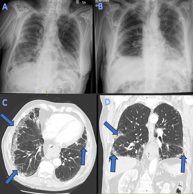
Initial investigations revealed neutrophilia with WCC 15.2 × 10⁹/L (normal ~ 2.0–8.0 × 10⁹/L) with an elevated C-reactive protein (CRP) measuring at 96 mg/L (normal < 3 mg/L) and erythrocyte sedimentation rate (ESR) at 41 (normal < 21 mm/h). Liver function testing demonstrated elevated transaminases (ALT 281 U/L, normal 4–36 U/L; AST 268 U/L, normal 8–33 U/L) with normal ALP and GGT. A chest X-ray found patchy opacification in bilateral lung spaces particularly involving the right mid to lower-zones, suggestive of atypical bronchopneumonia [Fig. 1A and B]. The patient was empirically treated for a post-COVID-19 pneumonia with intravenous antibiotics which included ceftriaxone 1 g daily and azithromycin 500 mg daily.
Fig. 1.
[1A] Chest X-ray demonstrating right middle to lower zone patchy heterogenous opacification. [1B] Repeat Chest X-ray demonstrating partially resolved changes in right lung with new ground glass opacity in the right lung in the subpleural location. [1C] CT-chest (axial view) demonstrating basal and lateral fibrotic changes consistent with a usual interstitial pneumonitis (UIP) pattern. [1D] CT-chest (coronal view) demonstrating basal and lateral fibrotic changes consistent with a usual interstitial pneumonitis (UIP) pattern
The patient had improved clinically following intravenous antibiotics before being transitioned to oral amoxicillin/clavulanate for a further three days. However he deteriorated two days later on oral antibiotics with a saturation of 86% on room air with a respiratory rate of 30 breaths per minute. A CT chest was performed subsequently.
The CT chest revealed changes suggestive of fibrotic lung disease, consistent with usual interstitial pneumonitis (UIP) pattern consistent with a drug reaction or connective tissue-related interstitial fibrosis [Fig. 1C and D].
Given the CT findings, auto-immune serology was sent. The extractable nuclear antigen (ENA), the anti-neutrophil cytoplasmic antibodies (ANCA), complement levels, the cyclic citrullinated peptide (CCP) antibodies and rheumatoid factor (RF) were all negative. Due to the deranged liver enzymes, other atypical causes of pneumonia were considered. Chlamydia pneumonia serology demonstrated a positive IgG and a negative IgM antibody consistent with a previous exposure, and mycoplasma serology wasnegative.
Despite two courses of broad-spectrum intravenous antibiotics, the patient had ongoing respiratory symptoms and had persistently elevated inflammatory markers.
After three weeks, the patient had a significant decline in function. He was notable to perform personal activities of daily living and was noted to have difficulty getting out of a chair as well as walking. As such, he was transferred to the rehabilitation unit where he remained for a further two weeks for ongoing care. Whilst in rehabilitation, the patient reported new oropharyngeal dysphagia, particularly coughing and spluttering when eating solid food. This was confirmed on a barium swallow, which found oesophageal dysmotility and diffuse oesophageal spasm.
A diagnosis of atypical COVID pneumonia with long COVID was made.
The patient represented the following day after a fall with a long lie of approximately 14 h. A physical examination noted no injuries however the patient was feeling lethargic and weak. He also demonstrated quite profound upper and lower limb weakness consistent with proximal myopathy. His cervical flexion power was also found to be weak. These constellation of symptoms were present five weeks from the time of diagnosis of his COVID illness.
Further investigations were performed; there was a mild neutrophilia (10.7 × 10⁹/L; normal ~ 2.0–8.0 × 10⁹/L), thrombocytosis (platelets 511 × 109/L, normal 150-450 × 109/L), persistently elevated C-reactive protein (52.4 mg/L, normal < 3 mg/L) and a significantly elevated creatinine kinase (CK) (4301 U/L, normal < 201 U/L).
A chest X-ray showed the right lung heterogenous opacification had partially resolved, however there are some new ground glass changes in the right subpleural space. There was also new development of patchy opacification in the left lower zone with background fibrotic changes in the lungs.
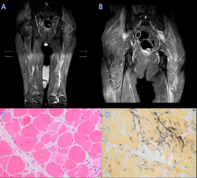
Due to the elevated CK, the dark-coloured urine and the fall with long lie, the patient was treated as rhabdomyolysis with fluid resuscitation. In spite of this, the patient’s CK remained elevated. Further testing revealed a strongly positive anti-OJ auto-antibody and a weakly positive Mi-2a auto-antibody on a myositis immunoblot assay [Table 1]. An MRI of his bilateral thighs were done [Fig. 2A and B] and subsequently a muscle biopsy was performed and showed numerous muscle fibres showing active degeneration with a tendency to perifascicular degeneration. There was no lymphocytic infiltration with a light histiocytic infiltrate in the perimysium and alkaline phosphatase was regulated in a perifascicular distribution [Fig. 2C and D]. A diagnosis was made of an active immune mediated perimysial muscle pathology (IMPP), in keeping with antisynthetase syndrome.
Table 1.
Patient’s auto-antibody panel demonstrating positive anti-OJ auto-antibody
| Autoantibody | Result |
|---|---|
| Anti (U1) RNP | Negative |
| Anti SM | Negative |
| Anti SSA (Ro) | Negative |
| Anti Ro52 | Negative |
| Anti SSB (La) | Negative |
| Anti Scl 70 | Negative |
| Anti PMScl | Negative |
| Anti Jo1 | Negative |
| Anti PCNA | Negative |
| Anti Ribosomal-P | Negative |
| C3 | 0.97 g/L |
| C4 | 0.30 g/L |
| OJ | Strong positive |
| Mi-2a | Weak positive |
| ANA titre | < 160 |
| C-ANCA | Negative |
| P-ANCA | Negative |
| CCP Antibodies | < 5 |
| Rheumatoid Factor | < 14 |
Fig. 2.
[2A, 2B] MRI shows high signals on STIR bilateral thighs demonstrating mild diffuse myositis of the gluteal muscles and the muscles of the posterior thigh with mild generalised fatty atrophy. MRI, magnetic resonance imaging; STIR, short T1 inversion recovery. [2C] High power image of the skeletal muscle showing peri fascicular degeneration of muscle fibres x 400 actual magnification, Haematoxylin and Eosin. [2D] Low power image showing upregulation of alkaline phosphatase (black) in a peri fascicular pattern x 100 actual magnification, Alkaline phosphatase
The patient received pulse methylprednisolone 1 g for 3 days with a long oral prednisolone wean. In addition, he was then commenced and escalated to a full dose of azathioprine given a normal purine metabolism. In view of multiple end-organ manifestations, loading immunoglobulin at 2 g/kg administered over two days was given. He was also given 1 g/kg immunoglobulin at a four-weekly frequency as maintenance. He has since then responded well to his immunosuppression and has not had any flares of his ASSD.
Discussion and conclusion
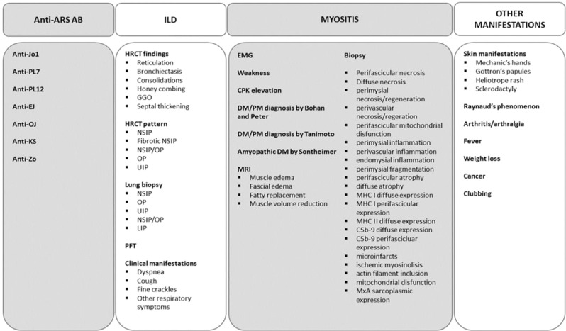
Anti-synthetase syndrome (ASSD) is a heterogenous subtype of autoimmune inflammatory myopathy [Table 2]. ASSD is characterized by the triad of myositis, interstitial lung disease (ILD) and arthritis [2]. This is serologically supported by the presence of anti-aminoacyl-tRNA synthetase antibodies with anti-Jo-1 being the most common. The proposed diagnostic criteria for ASSD includes presence of a serum anti-synthetase antibody and at least one of the following clinical manifestations: arthritis, ILD, Raynaud’s phenomenon, Mechanic’s hands or fever [3]. There have been reported cases of anti-synthetase syndrome following Sars-CoV-2 infection [4]. We here report the first anti-OJ antibody-positive ASSD following SARS-CoV-2 infection.
Table 2.
List of variables used to define ASSD. ARS-Ab: anti aminoacyl-RNA-synthetase autoantibodies; ILD: interstitial lung disease; HRCT: high resolution computed tomography; NSIP: non-specific idiopathic pneumonia; OP: organising pneumonia; UIP: usual interstitial pneumonia; GGO: ground glass opacities; LIP: lymphoid interstitial pneumonia; PFT: pulmonary function tests; EMG: electromyography; CPK: creatine phosphokinase; DM: dermatomyositis; PM: polymyositis; MRI: magnetic resonance imaging; MHC: major histocompatibility [9].
ASSD encompasses a subset of idiopathic inflammatory myopathies (IIM) where the presence of myositis specific autoantibodies (MSAs) and myositis-associated autoantibodies (MAAs) have become an essential aspect for classification and diagnosis of IIM [5]. Among the MSAs, autoantibody against anti-aminoacyl-tRNA synthetase were found in 25-35% with IIM [6]. Among the autoantibodies detected were anti-Jo-1(histidyl), PL-7 (threonyl), PL-12 (alanyl), OJ (isoleucyl), EJ (glycyl), KS (asparaginyl), Zo (phenylalanyl), and Ha (tyrosyl) [7]. Anti-nuclear antibody (ANA) has a poor sensitivity in ASSD and is found in less than half of the patients with ASSD, although 70–80% of patients will have positive cytoplasmic staining on indirect immunofluorescence [8].
The treatment of ASSD often requires multimodal immunosuppression in order to control muscle and extra-muscular manifestations, additionally managing immunosuppression complications and disease-related sequalae including interstitial lung disease requiring lung transplantation, pulmonary hypertension, malignancy as well as decreased survival [10].
SARS-CoV-2 is an RNA virus that can often lead to heterogeneous clinical presentations and complications. SARS-CoV-2 infection can cause pulmonary changes such as ground-glass opacities that are similar to that of patients with ASSD which can be difficult to differentiate between [11]. Similarly, the inflammatory process of SARS-CoV-2 infection involves macrophage activation and cytokine storm, which share similar clinical features as multiple rheumatological diseases [12]. We hypothesise that the absence of preceding myositis symptoms prior to the patient’s SARS-CoV2 infection and the presence of anti-OJ antibody makes this most likely a viral-induced idiopathic inflammatory myositis(IIM) [13]. There have been proposed mechanisms in which SARS-CoV-2 infection can trigger a molecular autoantigen transformation or modification [14]. Changes in adaptive immunity with dysregulation of neutrophil extracellular trap (NET) formation has been demonstrated to be involved in the pathophysiology following viral infections such as SARS-CoV-2 [15]. Other proposed mechanisms include molecular mimicry of viral antigen and damaged muscle antigen as well as an elevation of type 1 interferon (IFN) which plays a major role in many rheumatological diseases [16, 17]. Having reviewed the literature, the occurrence of ASSD following SARS-CoV-2 has not been commonly reported. However, the occurrence of dermatomyositis (DM) following SARS-CoV-2 infection is more frequent, especially anti-MDA-5 subtype. Shimizu et al. reported a case of anti-PL-7 ASSD following SARS-CoV-2 infection [18]. There have also been cases of interstitial lung disease (ILD) that was initially presumed to be COVID-19 related which later lead to the diagnosis of ASSD [19].
At least 267 cases of rheumatic diseases have been associated with SARS-CoV-2 infection as well as COVID-19 vaccination [20]. A literature search was made to determine the amount of case reports describing myositis associated with SARS-CoV-2 infection. Using PubMed database, we searched articles available anytime using the following search terms: “myositis” AND (“sars*” OR “covid*”), dermatomyositis AND (“sars*” OR “covid*”), “anti-synthetase*” AND (“sars*” OR “covid*”). 385 articles were screened. Among the articles, only case reports were chosen, duplicates were removed and articles other than English were excluded. Other variants of IIM besides ASSD were excluded as well. We found 3 case reports describing ASSD following SARS-CoV-2 infection [Table 3]. No article describing anti-OJ ASSD following SARS-CoV-2 infection was identified. Further literature searches on diagnostic approach and treatment of ASSD were done.
Table 3.
Cases of anti-synthetase syndrome (ASSD) following SARS-CoV-2 infection
| Author | Year | Sex | Age | CK(U/L) | Antibody | Biopsy | Biopsy results |
|---|---|---|---|---|---|---|---|
| Marmen et al. [4] | 2021 | Male | 62 | Anti-Jo1 | Yes | Perifascicular necrosis with perimysial fragmentation | |
| Peña et al. [20] | 2023 | Male | 36 | Anti-EJ | Yes | Perivascular and endomysial lymphocyte infiltration, myofiber necrosis and regeneration with perifascicular atrophy | |
| Shimizu et al. [17] | 2022 | Male | 47 | 3380 | Anti-PL7 | Yes | Inflammatory cells at the myofiber bundles with perivascular inflammatory cell infiltration and CD8- positive lymphocytes infiltration in atrophy of the myofibers |
In the first case described by Marmen et al., a sixty-two-year-old gentleman developed polyarthritis and interstitial lung changes a month after being treated for his COVID illness proven on a positive PCR test. Further tests including positive anti-Jo1 and muscle biopsy confirmed ASSD [4]. The second case described by Peña et al. describes a thirty-six-year-old gentleman with positive COVID PCR with simultaneous proximal muscle weakness, dysphagia and cutaneous changes on his digits. The suspicion of inflammatory myositis and further investigations demonstrate positive anti-EJ antibody and a positive muscle biopsy [21]. Lastly, a case by Shimizu et al. discusses a forty-seven-year-old gentleman who developed erythematous skin macules on the patient’s eyelid which extended to the anterior chest three weeks following a SARS-CoV-2 infection and subsequently dysphagia, odynophagia and severe proximal muscle weakness a further three weeks later despite prednisolone therapy. Investigations done demonstrated positive anti-PL7 antibody, HLADRB1 analysis with HLA-DR4 (DRB1*0405)) and HLA-DR15 (DRB1*15:01) and evidence of inflammation on muscle biopsy following MRI-STIR sequence [18]. A case of ASSD with ILD mimicking COVID pneumonitis with negative COVID PCR was also described by Elsayed at al. [19].
This case report found that the patient had features of myalgia, fatigue and muscle weakness which were initially presumed to be due to a viral illness. It highlighted that diagnosing anti-synthetase syndrome in a patient with SARS-CoV-2 infection can be difficult as approximately 33–46% of patients would demonstrate elevated CK levels and this depends on the duration of the COVID-19 illness [12].
During the COVID19 pandemic, the incidence of inflammatory disorders has increased and there may be a relationship between the virus precipitating loss of tolerance and leading to autoimmunity through various immunological pathways, although the mechanism of this remains unclear. Our patient represents a challenge in diagnosis as ASSD is strongly associated with ILD. Given that the patient tested positive for SARS-CoV-2 infection, it was questioned at the time of diagnosis whether the pulmonary changes were part of the spectrum of COVID-19 related lung disease rather than true ILD. Furthermore, the CK level was significantly elevated at the time of presentation which is not in keeping with ASSD.
Although SARS-CoV-2 infection itself can cause a directly mediated viral myositis, this case report highlights the possibility of developing virus-triggered inflammatory myositis through multiple aforementioned proposed mechanisms. Therefore, further studies are required to explore the relationship and pathophysiology of SARS-CoV-2 infection and the incidence of inflammatory myopathies. It is important that physicians are aware of the potential for the SARS-CoV-2 virus to induce exacerbations of pre-existing autoimmune diseases or even potentially unmask de novo autoimmune diseases in predisposed individuals.
Acknowledgements
Not applicable.
Author contributions
RS is the primary author who conducted the planning, write-up especially in regards to the case report and images, discussion and literature review, BM and NNK were involved in the initial patient care and collected materials for the write-up, BY was involved in editing the article, CM provided histopathology images, SC provided supervision over the entire process with final touch up to the article.
Funding
There is no funding involved in this case report and literature review.
Data availability
No datasets were generated or analysed during the current study.
Declarations
Ethical approval
Ethics has been approved by Northern Health Ethics Committee and consent has been taken from both employer and patient for publication.
Informed consent
Informed consent to participate was obtained for all involved in this article.
Consent for publication
Not applicable.
Competing interests
The authors declare no competing interests.
Footnotes
Publisher’s Note
Springer Nature remains neutral with regard to jurisdictional claims in published maps and institutional affiliations.
References
- 1.Gu JN, Yan W, Gao QL, Chen L. Anti-OJ antibody-positive anti-synthetase syndrome with repeated arthritis, fever, and recurrent liver cancer: a case report. J Gastrointest Oncol. 2022;13(5). [DOI] [PMC free article] [PubMed]
- 2.Monti S, Montecucco C, Cavagna L. Clinical spectrum of anti-jo-1-associated disease. 29, Current Opinion in Rheumatology. 2017. [DOI] [PubMed]
- 3.Connors GR, Christopher-Stine L, Oddis CV, Danoff SK. Interstitial lung disease associated with the idiopathic inflammatory myopathies: What progress has been made in the past 35 years? Vol. 138, Chest. 2010. [DOI] [PubMed]
- 4.Bouchard Marmen M, Ellezam B, Fritzler MJ, Troyanov Y, Gould PV, Satoh M et al. Anti-synthetase syndrome occurring after SARS-CoV-2 infection. Scand J Rheumatol. 2022;51(3). [DOI] [PubMed]
- 5.Ghirardello A, Bassi N, Palma L, Borella E, Domeneghetti M, Punzi L et al. Autoantibodies in polymyositis and dermatomyositis. Curr Rheumatol Rep. 2013;15(6). [DOI] [PubMed]
- 6.Koenig M, Fritzler MJ, Targoff IN, Troyanov Y, Senécal JL. Heterogeneity of autoantibodies in 100 patients with autoimmune myositis: insights into clinical features and outcomes. Arthritis Res Ther. 2007;9(4). [DOI] [PMC free article] [PubMed]
- 7.Huang K, Aggarwal R. Antisynthetase syndrome: a distinct disease spectrum. J Scleroderma Relat Disorders. 2020. [DOI] [PMC free article] [PubMed]
- 8.Aggarwal R, Dhillon N, Fertig N, Koontz D, Qi Z, Oddis CV. Negative antinuclear antibody does not indicate autoantibody negativity in myositis: role of anticytoplasmic antibody as a screening test for antisynthetase syndrome. J Rheumatol. 2017;44(2). [DOI] [PubMed]
- 9.Zanframundo G, Faghihi-Kashani S, Scirè CA, Bonella F, Corte TJ, Doyle TJ, Fiorentino D, Gonzalez-Gay MA, Hudson M, Kuwana M, Lundberg IE, Mammen A, McHugh N, Miller FW, Monteccucco C, Oddis CV, Rojas-Serrano J, Schmidt J, Selva-O’Callaghan A, Werth VP, Sakellariou G, Aggarwal R, Cavagna L. Defining anti-synthetase syndrome: a systematic literature review. Clin Exp Rheumatol. 2022;40(2):309–19. 10.55563/clinexprheumatol/8xj0b9 [DOI] [PMC free article] [PubMed] [Google Scholar]
- 10.W LJ, C JJ. M.E. S. The diagnosis and treatment of antisynthetase syndrome. Clin Pulm Med. 2016;23(5). [DOI] [PMC free article] [PubMed]
- 11.Alfraji N, Mazahir U, Chaudhri M, Miskoff J. Anti-synthetase syndrome: a rare and challenging diagnosis for bilateral ground-glass opacities—a case report with literature review. BMC Pulm Med. 2021;21(1). [DOI] [PMC free article] [PubMed]
- 12.Huang C, Wang Y, Li X, Ren L, Zhao J, Hu Y et al. Clinical features of patients infected with 2019 novel coronavirus in Wuhan, China. Lancet. 2020;395(10223). [DOI] [PMC free article] [PubMed]
- 13.Saud A, Naveen R, Aggarwal R, Gupta L. COVID-19 and myositis: what we know so far. 23, Curr Rheumatol Rep. 2021. [DOI] [PMC free article] [PubMed]
- 14.Chang SE, Feng A, Meng W, Apostolidis SA, Mack E, Artandi M et al. New-onset IgG autoantibodies in hospitalized patients with COVID-19. Nat Commun. 2021;12(1). [DOI] [PMC free article] [PubMed]
- 15.Papayannopoulos V. Neutrophil extracellular traps in immunity and disease. 18, Nat Rev Immunol. 2018. [DOI] [PubMed]
- 16.Cusick MF, Libbey JE, Fujinami RS. Molecular mimicry as a mechanism of autoimmune disease. Clin Rev Allergy Immunol. 2012;42(1). [DOI] [PMC free article] [PubMed]
- 17.Muskardin TLW, Niewold TB. Type i interferon in rheumatic diseases. 14, Nat Rev Rheumatol. 2018. [DOI] [PMC free article] [PubMed]
- 18.Shimizu H, Matsumoto H, Sasajima T, Suzuki T, Okubo Y, Fujita Y et al. New-onset dermatomyositis following COVID-19: a case report. Front Immunol. 2022;13. [DOI] [PMC free article] [PubMed]
- 19.Elsayed M, Abdelgabar A, Karmani J, Majid M. A case of antisynthetase syndrome initially presented with interstitial lung Disease Mimicking COVID-19. J Med Cases. 2023;14(1). [DOI] [PMC free article] [PubMed]
- 20.Ursini F, Ruscitti P, Addimanda O, Foti R, Raimondo V, Murdaca G et al. Inflammatory rheumatic diseases with onset after SARS-CoV-2 infection or COVID-19 vaccination: a report of 267 cases from the COVID-19 and ASD group. RMD Open. 2023;9(2). [DOI] [PMC free article] [PubMed]
- 21.Peña C, Kalara N, Velagapudi P. A case of antisynthetase syndrome in the setting of SARS-Cov-2 infection. Cureus. 2023. [DOI] [PMC free article] [PubMed]
Associated Data
This section collects any data citations, data availability statements, or supplementary materials included in this article.
Data Availability Statement
No datasets were generated or analysed during the current study.


