Abstract
BACKGROUND
Recent advancements in biliary tract cancer (BTC) treatment have expanded beyond surgery to include adjuvant therapy, yet the prognosis remains poor. Identifying prognostic biomarkers could enhance the assessment of patients who have undergone radical resection for BTC.
AIM
To determine transmembrane serine protease 4 (TMPRSS4) utility as a prognostic biomarker of radical resection for BTC.
METHODS
Medical records of patients who underwent radical resection for BTC, excluding intrahepatic cholangiocarcinoma, were retrospectively reviewed. The associations between TMPRSS4 expression and clinicopathological factors, overall survival, and recurrence-free survival were analyzed.
RESULTS
Among the 85 patients undergoing radical resection for BTC, 46 (54%) were TMPRSS4-positive. The TMPRSS4-positive group exhibited significantly higher preoperative carbohydrate antigen 19-9 (CA19-9) values and greater lymphatic invasion than the TMPRSS4-negative group (P = 0.019 and 0.039, respectively). Postoperative overall survival and recurrence-free survival were significantly worse in the TMPRSS4-positive group (median survival time: 25.3 months vs not reached, P < 0.001; median survival time: 28.7 months vs not reached, P = 0.043, respectively). Multivariate overall survival analysis indicated TMPRSS4 positivity, pT3/T4, and resection status R1 were independently associated with poor prognosis (P = 0.032, 0.035 and 0.030, respectively). TMPRSS4 positivity correlated with preoperative CA19-9 values ≥ 37 U/mL and pathological tumor size ≥ 30 mm (P = 0.016 and 0.038, respectively).
CONCLUSION
TMPRSS4 is a potential prognostic biomarker of radical resection for BTC.
Keywords: Biliary tract cancer, Biomarker, Prognosis, Radical resection, Transmembrane serine protease 4
Core Tip: Transmembrane serine protease 4 (TMPRSS4) expression correlates with poor prognosis in patients with biliary tract cancer post-radical resection, indicating its potential as a prognostic biomarker. TMPRSS4 positivity is linked to higher preoperative carbohydrate antigen 19-9 levels, lymphatic invasion, and larger tumor size. This study underscores the importance of TMPRSS4 in enhancing prognostic assessment and guiding treatment strategies for patients with biliary tract cancer undergoing radical resection.
INTRODUCTION
Surgical resection has been the only curative treatment option for biliary tract cancer (BTC). However, in recent years, multidisciplinary treatment, including adjuvant therapy, has become part of the standard treatment for BTC[1-3]. Nonetheless, even with curative resection followed by adjuvant chemotherapy, the three-year overall survival (OS) rate is limited to 77.1%[4]; therefore, the prognosis of BTC remains poor. A positive surgical margin, lymph node metastasis, perineural invasion, histological differentiation, invasion to major vessels, and pancreatic infiltration have been identified as prognostic factors after radical resection of BTC[5-10]. It is essential to identify candidate biomarkers for the prognosis of BTC.
Serine proteases facilitate the degradation of the basement membrane and extracellular matrix, promoting tumor cell invasion into the surrounding tissue. Serine proteases also play a role in the proliferation, migration, invasion, and angiogenesis of cancer cells[11]. A novel subfamily of serine proteases called type II transmembrane serine proteases (TTSPs) has been identified, which contain a large extracellular domain that mediates catalytic activity and a short cytoplasmic domain that can interact with cytoskeletal and cellular signaling molecules[12]. Several TTSPs are overexpressed in a variety of tumors and are potential novel markers of tumor development and progression. Transmembrane protease serine 4 (TMPRSS4), a member of the TTSP family, is reportedly upregulated in pancreatic, colorectal, gastric, lung, thyroid, prostate, and several other cancers. Elevated expression of TMPRSS4 often correlates with poor prognosis[13]. However, few studies have reported the effects of TMPRSS4 on the prognosis of patients who have undergone radical resection for BTC[14]. Therefore, we aimed to determine the potential of TMPRSS4 as a prognostic biomarker of radical resection for BTC.
MATERIALS AND METHODS
Study design
We retrospectively reviewed the medical records of patients who underwent radical resection for BTC (perihilar cholangiocarcinoma, distal cholangiocarcinoma, gallbladder cancer, and ampulla of Vater cancer) at the Department of Surgery, National Hospital Organization Kure Medical Center, in Hiroshima, Japan, between March 2012 and July 2023. The present study was approved by the Institutional Review Board of the National Hospital Organization Kure Medical Center (2023-45) and conducted in accordance with the principles of the Declaration of Helsinki. In addition, all patients provided written informed consent.
Data collection
We collected the demographic and clinicopathological data of patients, including age, sex, preoperative carbohydrate antigen 19-9 (CA19-9) level, diseases according to general rules for clinical and pathological studies on cancer of the biliary tract 7th edition[15], operating time, operative blood loss, histology, pathological tumor stage according to tumor node metastasis classification of malignant tumors 8th edition[16], pathological tumor size, lymphatic invasion, venous invasion, neural invasion, tumor infiltrative type, lymph node metastasis, resection status, and adjuvant chemotherapy.
Adjuvant therapy and surveillance
Adjuvant chemotherapy was recommended to patients (excluding those with pathological T1N0M0) and was administered to those who were tolerant and who consented after radical resection. Regular surveillance was performed by blood testing, which included the detection of tumor markers and multidetector computed tomography at intervals of 3–6 months. When two or more modalities, such as magnetic resonance imaging or positron emission tomography-computed tomography, indicated the presence of recurrence or a recurrent lesion at two different time points, recurrence was confirmed and recorded. The survival time after surgery and the cause of death were recorded for patients who died, whereas the OS time and recurrence status were recorded for those who survived.
Immunohistochemical assessment
Immunohistochemical assessment was performed as described previously[11]. The slides were incubated with rabbit anti-TMPRSS4 antibody (1:200) (abl188816; Abcam, Cambridge, United Kingdom). TMPRSS4 expression was assessed as either positive or negative across all slides. The scoring criteria for the percentage of positively stained tumor cells were as follows: TMPRSS4-positive, ≥ 50% positive cells; TMPRSS4-negative, < 50% positive cells. A surgical pathologist applied this classification system and reviewed the immunoreactivity of each specimen.
Statistical analysis
Data are expressed as medians with ranges or absolute values with percentages. Categorical variables were analyzed using the χ2 or Fisher’s exact test, while continuous variables were compared using the Wilcoxon two-sample test in the univariate analysis. Survival curves were generated using the Kaplan–Meier method, with distribution comparisons conducted using the log-rank test. The proportional hazard regression model (Cox regression) was used to perform multivariate OS analyses based on a P value ≤ 0.1 in univariate analysis. Hazard ratios (HRs) and 95% confidence intervals (CIs) were calculated. Cut-off values were defined using receiver operating characteristic curve analysis for three-year OS after surgery. Logistic regression analysis was used to perform multivariate analyses of factors associated with TMPRSS4 positivity. All tests were two-sided, with statistical significance set at P < 0.05. All statistical analyses were performed using JMP statistical software (version 16.0; SAS Institute, Cary, NC, United States).
RESULTS
Patients
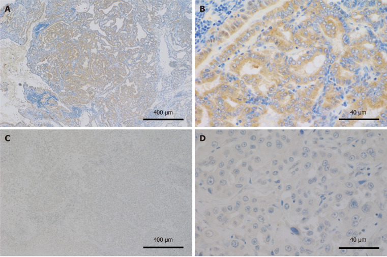
In total, 85 patients underwent radical resection for BTC. TMPRSS4-positive staining was mainly observed in the cytoplasm of BTC cells, as evidenced by immunohistochemical analysis (Figure 1).
Figure 1.
Immunohistochemical staining of transmembrane serine protease 4 in biliary tract cancer. A: Transmembrane serine protease 4 (TMPRSS4)-positive biliary tract cancer (BTC) tissues; B: TMPRSS4-positive BTC tissues; C: TMPRSS4-negative BTC tissues; D: TMPRSS4-negative BTC tissues. Original magnification: 40 × (A and C); 400 × (B and D).
The clinicopathological characteristics of patients are shown in Table 1. The TMPRSS4 expression rate was 54% (46/85) in all patients with BTC. Neoadjuvant chemotherapy was administered to three (3%) patients. Considering clinicopathological factors, the TMPRSS4-positive group had a significantly higher preoperative CA19-9 value and greater lymphatic invasion than the TMPRSS4-negative group (P = 0.019 and 0.039, respectively), exhibiting no other significant differences. Of the 85 patients, 14 had perihilar cholangiocarcinoma, 23 had distal cholangiocarcinoma, 37 had gallbladder cancer, and 11 had ampulla of Vater cancer. Of these, 40 (47%) patients had lymph node metastasis, and adjuvant chemotherapy was administered to 49 (58%) patients.
Table 1.
Patients’ characteristics (n = 85)
|
|
TMPRSS4 expression
|
P value
|
|
|
Positivity (n = 46)
|
Negativity (n = 39)
|
||
| Age, median (range), years | 75 (47-86) | 75 (57-87) | 0.612 |
| Sex, male, n (%) | 23 (50) | 22 (56) | 0.555 |
| Neoadjuvant chemotherapy, n (%) | 2 (4) | 1 (2) | 0.653 |
| Preoperative CA19-9 level, median (range), U/mL | 37 (0-27146) | 13 (0-1246) | 0.019 |
| Diseases, n (%) | |||
| Perihilar cholangiocarcinoma | 8 (17) | 6 (15) | 0.176 |
| Distal cholangiocarcinoma | 10 (22) | 13 (34) | |
| Gallbladder cancer | 19 (41) | 18 (46) | |
| Ampulla of vater cancer | 9 (20) | 2 (5) | |
| Operating time, median (range), min | 465 (82-962) | 425 (239-1052) | 0.853 |
| Operative blood loss, median (range), mL | 310 (5-3950) | 410 (5-1600) | 0.609 |
| Histology, tub1/tub2, n (%) | 37 (80) | 31 (79) | 0.913 |
| Tumor stage, pT3/T4, n (%) | 14 (30) | 10 (26) | 0.624 |
| Pathological tumor size, median (range), mm | 35 (0-110) | 25 (1-88) | 0.081 |
| Lymphatic invasion (ly), n (%) | 16 (35) | 6 (15) | 0.039 |
| Venous invasion (v), n (%) | 20 (43) | 14 (36) | 0.477 |
| Neural invasion (ne), n (%) | 22 (48) | 17 (44) | 0.696 |
| Tumor infiltrative type c (IFN c), n (%) | 8 (17) | 5 (13) | 0.558 |
| Lymph node metastasis, n (%) | 24 (52) | 16 (41) | 0.304 |
| Resection status, R0, n (%) | 36 (78) | 31 (80) | 0.890 |
| Adjuvant chemotherapy, n (%) | 27 (59) | 22 (56) | 0.832 |
CA19-9: Carbohydrate antigen 19-9; tub1: Well-differentiated tubular adenocarcinoma; tub2: Moderately differentiated tubular adenocarcinoma.
Survival analysis
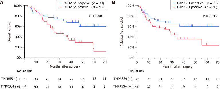
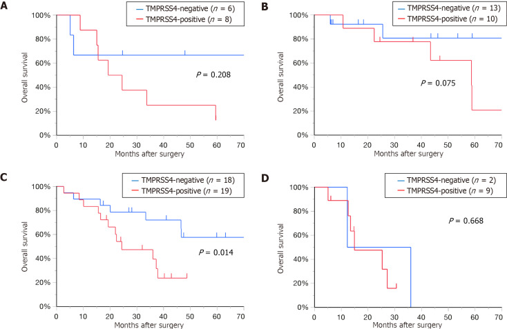
The median follow-up duration was 25.6 (range, 1.9-123.8) months. The TMPRSS4-positive group had a significantly worse OS than the TMPRSS4-negative group [median survival time (MST): 25.3 months vs not reached, P < 0.001; Figure 2A]. Postoperative one-, three-, and five-year survival rates in the TMPRSS4-positive and-negative groups were 86% vs 88%, 40% vs 68%, and 12% vs 60%, respectively. Postoperative recurrence-free survival (RFS) differed significantly between the two groups (MST: 28.7 months vs not reached, P = 0.043; Figure 2B). Postoperative one-, three-, and five-year RFS rates in the TMPRSS4-positive and -negative groups were 67% vs 75%, 37% vs 63%, and 25% vs 59%, respectively. Based on post-surgical OS for each disease classified by TMPRSS4 expression, patients with gallbladder cancer in the TMPRSS4-positive group exhibited a significantly worse OS than those in the TMPRSS4-negative group (MST: 24.4 months vs 75.3 months; P = 0.014). Considering other patients with cancer, the TMPRSS4-positive group tended to exhibit poor OS without significant differences (perihilar cholangiocarcinoma, MST: 21.9 months vs not reached, P = 0.208, distal cholangiocarcinoma, MST: 58.7 months vs not reached, P = 0.075, ampulla of Vater cancer, MST: 14.9 months vs 24.1 months, P = 0.668; Figure 3).
Figure 2.
Kaplan–Meier curves for all patients with biliary tract cancer classified based on the expression of transmembrane serine protease 4. A: Overall survival after surgery for all patients with biliary tract cancer (BTC), classified according to the expression of transmembrane serine protease 4 (TMPRSS4); B: Recurrence-free survival after surgery for all patients with BTC, classified according to the expression of TMPRSS4. TMPRSS4: Transmembrane serine protease 4.
Figure 3.
Overall survival after surgery for each disease, classified according to transmembrane serine protease 4 expression. A: Perihilar cholangiocarcinoma; B: Distal cholangiocarcinoma; C: Gallbladder cancer; D: Ampulla of vater cancer. TMPRSS4: Transmembrane serine protease 4.
Univariate and multivariate OS analyses of poor prognostic factors are described in Table 2. The univariate OS analysis of poor prognostic factors revealed a significant association with OS for TMPRSS4 positivity (P < 0.001), pT3/T4 (P = 0.002), pathological tumor size ≥ 30 mm (P = 0.017), lymphatic and venous invasions (all P < 0.001), lymph node metastasis (P = 0.004), and resection status R1 (P = 0.022). Furthermore, multivariate analysis identified TMPRSS4 positivity (HR: 2.33; 95%CI: 1.08-5.08; P = 0.032), pT3/T4 (HR: 2.26; 95%CI: 1.06-4.81; P = 0.035), and resection status R1 (HR: 2.21; 95%CI: 1.08-4.52; P = 0.030), as independent poor prognostic factors. Multivariate analysis showed that TMPRSS4 positivity was significantly associated with a preoperative CA 19-9 value of ≥ 37 U/mL [odds ratio (OR): 4.02; 95%CI: 1.30-12.47; P = 0.016] and pathological tumor size of ≥ 30 mm (OR: 2.78; 95%CI: 1.05-7.30; P = 0.038; Table 3).
Table 2.
Univariate and multivariate overall survival analyses of poor prognostic factors (n = 85)
| Factors |
Univariate
|
Multivariate
|
||||
|
Total patients, (n = 85), n(%)
|
MST (months)
|
P value
|
HR
|
95%CI
|
P value
|
|
| Age, years | ||||||
| < 80 | 62 (73) | 37.8 | 0.718 | |||
| ≥ 80 | 23 (27) | 58.7 | ||||
| Sex | ||||||
| Male | 45 (53) | 37.2 | 0.378 | |||
| Female | 40 (47) | 46.5 | ||||
| Neoadjuvant chemotherapy | ||||||
| Yes | 3 (4) | 36.0 | 0.956 | |||
| No | 82 (96) | 46.5 | ||||
| Preoperative CA 19-9 level, U/mL | ||||||
| < 37 | 55 (65) | 46.5 | 0.504 | |||
| ≥ 37 | 30 (35) | 43.4 | ||||
| TMPRSS4 | ||||||
| Positivity | 46 (54) | 25.3 | < 0.001 | 2.33 | 1.08-5.08 | 0.032 |
| Negativity | 39 (46) | 1.0 | ||||
| Histology, tub1/tub2 | ||||||
| Yes | 68 (80) | 46.5 | 0.752 | |||
| No | 17 (20) | 36.0 | ||||
| Tumor stage, pT3/T4 | ||||||
| Yes | 24 (28) | 22.0 | 0.002 | 2.26 | 1.06-4.81 | 0.035 |
| No | 61 (72) | 59.4 | 1.0 | |||
| Pathological tumor size, mm | ||||||
| < 30 | 43 (51) | 0.017 | 1.0 | |||
| ≥ 30 | 42 (49) | 33.7 | 1.32 | 0.65-2.65 | 0.439 | |
| Lymphatic invasion (ly) | ||||||
| Yes | 22 (26) | 16.3 | < 0.001 | 1.33 | 0.62-2.84 | 0.461 |
| No | 63 (74) | 75.3 | 1.0 | |||
| Venous invasion (v) | ||||||
| Yes | 34 (40) | 22.0 | < 0.001 | 1.93 | 0.91-4.07 | 0.086 |
| No | 51 (60) | 59.4 | 1.0 | |||
| Neural invasion (ne) | ||||||
| Yes | 39 (46) | 33.3 | 0.058 | 0.60 | 0.26-1.40 | 0.237 |
| No | 46 (54) | 58.7 | 1.0 | |||
| Tumor infiltrative type c (IFN c) | ||||||
| Yes | 13 (15) | 33.7 | 0.081 | 1.18 | 0.58-2.42 | 0.648 |
| No | 72 (85) | 58.7 | 1.0 | |||
| Lymph node metastasis | ||||||
| Yes | 40 (47) | 25.6 | 0.004 | 1.44 | 0.60-3.46 | 0.412 |
| No | 45 (53) | 75.3 | 1.0 | |||
| Resection status, | ||||||
| R0 | 67 (79) | 46.6 | 0.022 | 1.0 | ||
| R1 | 18 (21) | 24.5 | 2.21 | 1.08-4.52 | 0.030 | |
| Adjuvant chemotherapy | ||||||
| Yes | 49 (58) | 36.0 | 0.229 | |||
| No | 36 (42) | 75.3 | ||||
CA19-9: Carbohydrate antigen 19-9; CI: Confidence interval; HR: Hazard ratio; MST: Median survival time; tub1: Well-differentiated tubular adenocarcinoma; tub2: moderately differentiated tubular adenocarcinoma; TMPRSS4: Transmembrane serine protease 4.
Table 3.
Multivariate analyses of factors associated with transmembrane serine protease 4 positivity
|
Factors
|
Odds ratio (95%CI)
|
P value
|
| Preoperative CA19-9 ≥ 37 U/mL | 4.02 (1.30-12.47) | 0.016 |
| Pathological tumor size ≥ 30 mm | 2.78 (1.05-7.30) | 0.038 |
| Lymphatic invasion (ly) | 2.67 (0.84-8.53) | 0.097 |
| Neural invasion (ne) | 0.45 (0.15-1.35) | 0.153 |
CA19-9: Carbohydrate antigen 19-9; CI: Confidence interval; TMPRSS4: Transmembrane serine protease 4.
DISCUSSION
BTC remains a highly fatal disease with a poor prognosis. Thus, finding novel and effective biomarkers associated with advanced tumor progression is crucial for early diagnosis and discovery of a promising therapeutic target for BTC. The findings of the current study indicate that the five-year OS rates of patients with BTC who exhibit TMPRSS4-negativity were significantly higher than their TMPRSS4-positive counterparts. Our multivariate analysis identified TMPRSS4 as an independent poor prognostic factor for patients with BTC who underwent radical surgery, indicating its potential as a useful prognostic biomarker for BTC.
TMPRSS4 is highly expressed on the cell surface of the esophagus, stomach, small intestine, colon, bladder, and kidney, although the physiological roles of TMPRSS4 remain unclear[13]. TMPRSS4 induces epithelial-mesenchymal transition and promotes invasion, migration, and metastasis of human tumor cells[17]. Katopodis et al[18] reported substantial upregulation of TMPRSS4 in 11 types of cancer, including lung adenocarcinoma, lung squamous cell carcinoma, cervical squamous cell carcinoma, thyroid carcinoma, ovarian cancer, cancer of the rectum, pancreatic cancer, colon and stomach adenocarcinoma, uterine carcinosarcoma, and uterine corpus endometrial carcinoma, compared with normal control tissues; conversely, TMPRSS4 expression was downregulated in six types of cancer, including kidney carcinomas, acute myeloid leukemia, skin cutaneous melanoma, and testicular germ cell tumor. Elevated TMPRSS4 expression correlates with poor prognosis in patients with various cancers, including gastric cancer[19,20], colorectal cancer[21-23], hepatocellular carcinoma[24], and pancreatic ductal adenocarcinoma[25], suggesting its implication in the progression of noninvasive tumors to invasive malignancies. Additionally, Wu et al[14] highlighted that high TMPRSS4 expression levels are potentially associated with markedly poor prognosis in patients with gallbladder cancer. However, studies exploring the effects of TMPRSS4 on the prognosis of patients who underwent radical resection for BTC are limited. Herein, we found that TMPRSS4-positive patients with BTC, including perihilar cholangiocarcinoma, distal cholangiocarcinoma, gallbladder cancer, and ampulla of Vater cancer, who underwent radical surgery had a poor prognosis. In our study, the median OS time (25.3 months) for the TMPRSS4-positive group was shorter than the median RFS time (28.7 months). This difference can be attributed to mortality from unrelated causes of the seven patients in the TMPRSS4-positive group who did not experience recurrence. In patients with perihilar cholangiocarcinoma, distal cholangiocarcinoma, and ampulla of Vater cancer, the TMPRSS4-positive group showed a tendency for poor prognosis, although the difference was not statistically significant.
Patients with non-small cell lung cancer that lacked TMPRSS4 expression were found to be substantially more sensitive to cisplatin than the controls[26]. In gastric cancer, downregulated TMPRSS4 expression reportedly increases susceptibility to 5-fluorouracil[11]. This suggests that TMPRSS4-negative expression might enhance sensitivity to chemotherapy, leading to reduced recurrence in patients exhibiting TMPRSS4-negativity. Collectively, these findings highlight TMPRSS4 as a potential novel target and that the inhibiting TMPRSS4 expression increases chemosensitivity.
We excluded patients who underwent radical resection for intrahepatic cholangiocarcinoma (ICC). We enrolled patients with BTC according to the guidelines outlined in the 7th edition of the general rules for clinical and pathological studies on cancer of the biliary tract[15]. Moreover, the prevalence of ICC tends to be high in east and southeast asia[27]. Patients with HBV-associated ICC reportedly display substantially different clinicopathological characteristics and survival outcomes[28-30]. ICC has the greatest variety of mutations, which contributes to its high resistance to pharmacotherapy[31,32]. The marked heterogeneity of ICC may lead to insensitivity to TMPRSS4 expression.
In the current study, TMPRSS4 positivity was significantly associated with preoperative CA 19-9 levels ≥ 37 U/mL. Kondo et al[33] reported that preoperative CA 19-9 levels can predict the survival of patients with resectable cholangiocarcinoma. Accordingly, TMPRSS4 positivity could indicate a malignancy contributing to poor prognosis. TMPRSS4 positivity was also significantly associated with pathological tumor size ≥ 30 mm. Similar results have been reported previously. For example, Gu et al[25] identified TMPRSS4 as an independent prognostic factor in patients with pancreatic ductal adenocarcinoma, with TMPRSS4 levels closely associated with age, tumor size, and differentiation status. Wang et al[24] reported that TMPRSS4 levels in patients with hepatocellular carcinoma are closely correlated with tumor size and vascular invasion. In patients with colorectal cancer, TMPRSS4 levels reportedly correlate with tumor size, depth of tumor invasion, and lymph node metastasis[23].
Nevertheless, this study had some limitations. This study involved a single institution with a relatively small number of patients who underwent radical resection for BTC. Hence, further studies utilizing multicenter data or a nationwide database with a larger number of patients are warranted.
CONCLUSION
High TMPRSS4 expression is associated with a poor prognosis of radical resection for BTC. TMPRSS4 is a potential prognostic biomarker of radical resection for BTC.
Footnotes
Institutional review board statement: The protocol for this research project has been approved by a suitably constituted Ethics Committee of the institution, and it conforms to the provisions of the Declaration of Helsinki. The Institutional Review Board of National Hospital Organization Kure Medical Center approved this study (Approval No. 2023-45 on October 4, 2023).
Informed consent statement: All patients provided written informed consent.
Conflict-of-interest statement: The authors have no financial relationships to disclose.
Provenance and peer review: Unsolicited article; Externally peer reviewed.
Peer-review model: Single blind
Corresponding Author's Membership in Professional Societies: The Japanese Society of Gastroenterological surgery, G0379458.
Specialty type: Gastroenterology and hepatology
Country of origin: Japan
Peer-review report’s classification
Scientific Quality: Grade B, Grade C
Novelty: Grade B, Grade B
Creativity or Innovation: Grade B, Grade B
Scientific Significance: Grade B, Grade B
P-Reviewer: Song T S-Editor: Fan M L-Editor: A P-Editor: Zhang L
Contributor Information
Yoshiyuki Shibata, Department of Surgery, National Hospital Organization Kure Medical Center and Chugoku Cancer Center, Hiroshima 737-0023, Japan. yshibata.hiroshima@gmail.com.
Takeshi Sudo, Department of Surgery, National Hospital Organization Kure Medical Center and Chugoku Cancer Center, Hiroshima 737-0023, Japan.
Sho Tazuma, Department of Surgery, National Hospital Organization Kure Medical Center and Chugoku Cancer Center, Hiroshima 737-0023, Japan.
Naoki Tanimine, Department of Surgery, National Hospital Organization Kure Medical Center and Chugoku Cancer Center, Hiroshima 737-0023, Japan.
Takashi Onoe, Department of Surgery, National Hospital Organization Kure Medical Center and Chugoku Cancer Center, Hiroshima 737-0023, Japan.
Yosuke Shimizu, Department of Surgery, National Hospital Organization Kure Medical Center and Chugoku Cancer Center, Hiroshima 737-0023, Japan.
Atsushi Yamaguchi, Department of Gastroenterology, National Hospital Organization Kure Medical Center and Chugoku Cancer Center, Hiroshima 737-0023, Japan.
Kazuya Kuraoka, Department of Diagnostic Pathology, National Hospital Organization Kure Medical Center, and Chugoku Cancer Center, Hiroshima 737-0023, Japan.
Shinya Takahashi, Department of Surgery, Graduate School of Biochemical and Health Science, Hiroshima University, Hiroshima 734-8551, Japan.
Hirotaka Tashiro, Department of Surgery, National Hospital Organization Kure Medical Center and Chugoku Cancer Center, Hiroshima 737-0023, Japan.
Data sharing statement
The datasets used and/or analyzed during the current study are available from the corresponding author upon reasonable request.
References
- 1.Murakami Y, Uemura K, Sudo T, Hayashidani Y, Hashimoto Y, Nakamura H, Nakashima A, Sueda T. Adjuvant gemcitabine plus S-1 chemotherapy improves survival after aggressive surgical resection for advanced biliary carcinoma. Ann Surg. 2009;250:950–956. doi: 10.1097/sla.0b013e3181b0fc8b. [DOI] [PubMed] [Google Scholar]
- 2.Primrose JN, Fox RP, Palmer DH, Malik HZ, Prasad R, Mirza D, Anthony A, Corrie P, Falk S, Finch-Jones M, Wasan H, Ross P, Wall L, Wadsley J, Evans JTR, Stocken D, Praseedom R, Ma YT, Davidson B, Neoptolemos JP, Iveson T, Raftery J, Zhu S, Cunningham D, Garden OJ, Stubbs C, Valle JW, Bridgewater J BILCAP study group. Capecitabine compared with observation in resected biliary tract cancer (BILCAP): a randomised, controlled, multicentre, phase 3 study. Lancet Oncol. 2019;20:663–673. doi: 10.1016/S1470-2045(18)30915-X. [DOI] [PubMed] [Google Scholar]
- 3.Cheng Q, Luo X, Zhang B, Jiang X, Yi B, Wu M. Predictive factors for prognosis of hilar cholangiocarcinoma: postresection radiotherapy improves survival. Eur J Surg Oncol. 2007;33:202–207. doi: 10.1016/j.ejso.2006.09.033. [DOI] [PubMed] [Google Scholar]
- 4.Nakachi K, Ikeda M, Konishi M, Nomura S, Katayama H, Kataoka T, Todaka A, Yanagimoto H, Morinaga S, Kobayashi S, Shimada K, Takahashi Y, Nakagohri T, Gotoh K, Kamata K, Shimizu Y, Ueno M, Ishii H, Okusaka T, Furuse J Hepatobiliary and Pancreatic Oncology Group of the Japan Clinical Oncology Group (JCOG-HBPOG) Adjuvant S-1 compared with observation in resected biliary tract cancer (JCOG1202, ASCOT): a multicentre, open-label, randomised, controlled, phase 3 trial. Lancet. 2023;401:195–203. doi: 10.1016/S0140-6736(22)02038-4. [DOI] [PubMed] [Google Scholar]
- 5.Su CH, Tsay SH, Wu CC, Shyr YM, King KL, Lee CH, Lui WY, Liu TJ, P'eng FK. Factors influencing postoperative morbidity, mortality, and survival after resection for hilar cholangiocarcinoma. Ann Surg. 1996;223:384–394. doi: 10.1097/00000658-199604000-00007. [DOI] [PMC free article] [PubMed] [Google Scholar]
- 6.Neuhaus P, Jonas S, Bechstein WO, Lohmann R, Radke C, Kling N, Wex C, Lobeck H, Hintze R. Extended resections for hilar cholangiocarcinoma. Ann Surg. 1999;230:808–18; discussion 819. doi: 10.1097/00000658-199912000-00010. [DOI] [PMC free article] [PubMed] [Google Scholar]
- 7.Iwatsuki S, Todo S, Marsh JW, Madariaga JR, Lee RG, Dvorchik I, Fung JJ, Starzl TE. Treatment of hilar cholangiocarcinoma (Klatskin tumors) with hepatic resection or transplantation. J Am Coll Surg. 1998;187:358–364. doi: 10.1016/s1072-7515(98)00207-5. [DOI] [PMC free article] [PubMed] [Google Scholar]
- 8.Todoroki T, Kawamoto T, Koike N, Takahashi H, Yoshida S, Kashiwagi H, Takada Y, Otsuka M, Fukao K. Radical resection of hilar bile duct carcinoma and predictors of survival. Br J Surg. 2000;87:306–313. doi: 10.1046/j.1365-2168.2000.01343.x. [DOI] [PubMed] [Google Scholar]
- 9.Miyazaki M, Ito H, Nakagawa K, Ambiru S, Shimizu H, Okaya T, Shinmura K, Nakajima N. Parenchyma-preserving hepatectomy in the surgical treatment of hilar cholangiocarcinoma. J Am Coll Surg. 1999;189:575–583. doi: 10.1016/s1072-7515(99)00219-7. [DOI] [PubMed] [Google Scholar]
- 10.Chan C, Herrera MF, de la Garza L, Quintanilla-Martinez L, Vargas-Vorackova F, Richaud-Patín Y, Llorente L, Uscanga L, Robles-Diaz G, Leon E. Clinical behavior and prognostic factors of periampullary adenocarcinoma. Ann Surg. 1995;222:632–637. doi: 10.1097/00000658-199511000-00005. [DOI] [PMC free article] [PubMed] [Google Scholar]
- 11.Tazawa H, Suzuki T, Saito A, Ishikawa A, Komo T, Sada H, Shimada N, Hadano N, Onoe T, Sudo T, Shimizu Y, Kuraoka K, Tashiro H. Utility of TMPRSS4 as a Prognostic Biomarker and Potential Therapeutic Target in Patients with Gastric Cancer. J Gastrointest Surg. 2022;26:305–313. doi: 10.1007/s11605-021-05101-2. [DOI] [PMC free article] [PubMed] [Google Scholar]
- 12.Bugge TH, Antalis TM, Wu Q. Type II transmembrane serine proteases. J Biol Chem. 2009;284:23177–23181. doi: 10.1074/jbc.R109.021006. [DOI] [PMC free article] [PubMed] [Google Scholar]
- 13.Kim S. TMPRSS4, a type II transmembrane serine protease, as a potential therapeutic target in cancer. Exp Mol Med. 2023;55:716–724. doi: 10.1038/s12276-023-00975-5. [DOI] [PMC free article] [PubMed] [Google Scholar]
- 14.Wu XY, Zhang L, Zhang KM, Zhang MH, Ruan TY, Liu CY, Xu JY. Clinical implication of TMPRSS4 expression in human gallbladder cancer. Tumour Biol. 2014;35:5481–5486. doi: 10.1007/s13277-014-1716-4. [DOI] [PubMed] [Google Scholar]
- 15.Japanese Society of Hepato-Biliary-Pancreatic Surgery. General Rules for Clinical and Pathological Studies on Cancer of the Biliary Tract. 7th ed. Kanehara & Co., Ltd., 2021: 3-78. [Google Scholar]
- 16.James DB, Mary KG, Christian W. TNM Classification of Malignant Tumors. 8th ed. John Wiley & Sons, Ltd., 2017: 85-92. [Google Scholar]
- 17.Kim S, Kang HY, Nam EH, Choi MS, Zhao XF, Hong CS, Lee JW, Lee JH, Park YK. TMPRSS4 induces invasion and epithelial-mesenchymal transition through upregulation of integrin alpha5 and its signaling pathways. Carcinogenesis. 2010;31:597–606. doi: 10.1093/carcin/bgq024. [DOI] [PubMed] [Google Scholar]
- 18.Katopodis P, Kerslake R, Davies J, Randeva HS, Chatha K, Hall M, Spandidos DA, Anikin V, Polychronis A, Robertus JL, Kyrou I, Karteris E. COVID-19 and SARS-CoV-2 host cell entry mediators: Expression profiling of TMRSS4 in health and disease. Int J Mol Med. 2021;47 doi: 10.3892/ijmm.2021.4897. [DOI] [PMC free article] [PubMed] [Google Scholar]
- 19.Luo ZY, Wang YY, Zhao ZS, Li B, Chen JF. The expression of TMPRSS4 and Erk1 correlates with metastasis and poor prognosis in Chinese patients with gastric cancer. PLoS One. 2013;8:e70311. doi: 10.1371/journal.pone.0070311. [DOI] [PMC free article] [PubMed] [Google Scholar]
- 20.Sheng H, Shen W, Zeng J, Xi L, Deng L. Prognostic significance of TMPRSS4 in gastric cancer. Neoplasma. 2014;61:213–217. doi: 10.4149/neo_2014_027. [DOI] [PubMed] [Google Scholar]
- 21.Huang A, Zhou H, Zhao H, Quan Y, Feng B, Zheng M. TMPRSS4 correlates with colorectal cancer pathological stage and regulates cell proliferation and self-renewal ability. Cancer Biol Ther. 2014;15:297–304. doi: 10.4161/cbt.27308. [DOI] [PMC free article] [PubMed] [Google Scholar]
- 22.Zhao XF, Yang YS, Gao DZ, Park YK. TMPRSS4 overexpression promotes the metastasis of colorectal cancer and predicts poor prognosis of stage III-IV colorectal cancer. Int J Biol Markers. 2021;36:23–32. doi: 10.1177/17246008211046368. [DOI] [PubMed] [Google Scholar]
- 23.Huang A, Zhou H, Zhao H, Quan Y, Feng B, Zheng M. High expression level of TMPRSS4 predicts adverse outcomes of colorectal cancer patients. Med Oncol. 2013;30:712. doi: 10.1007/s12032-013-0712-7. [DOI] [PubMed] [Google Scholar]
- 24.Wang CH, Guo ZY, Chen ZT, Zhi XT, Li DK, Dong ZR, Chen ZQ, Hu SY, Li T. TMPRSS4 facilitates epithelial-mesenchymal transition of hepatocellular carcinoma and is a predictive marker for poor prognosis of patients after curative resection. Sci Rep. 2015;5:12366. doi: 10.1038/srep12366. [DOI] [PMC free article] [PubMed] [Google Scholar]
- 25.Gu J, Huang W, Zhang J, Wang X, Tao T, Yang L, Zheng Y, Liu S, Yang J, Zhu L, Wang H, Fan Y. TMPRSS4 Promotes Cell Proliferation and Inhibits Apoptosis in Pancreatic Ductal Adenocarcinoma by Activating ERK1/2 Signaling Pathway. Front Oncol. 2021;11:628353. doi: 10.3389/fonc.2021.628353. [DOI] [PMC free article] [PubMed] [Google Scholar]
- 26.Exposito F, Villalba M, Redrado M, de Aberasturi AL, Cirauqui C, Redin E, Guruceaga E, de Andrea C, Vicent S, Ajona D, Montuenga LM, Pio R, Calvo A. Targeting of TMPRSS4 sensitizes lung cancer cells to chemotherapy by impairing the proliferation machinery. Cancer Lett 2019; 453: 21-33. [DOI] [PubMed] [Google Scholar]
- 27.Bridgewater J, Galle PR, Khan SA, Llovet JM, Park JW, Patel T, Pawlik TM, Gores GJ. Guidelines for the diagnosis and management of intrahepatic cholangiocarcinoma. J Hepatol. 2014;60:1268–1289. doi: 10.1016/j.jhep.2014.01.021. [DOI] [PubMed] [Google Scholar]
- 28.Fujita T. An unusual risk factor for intrahepatic cholangiocarcinoma. J Hepatol. 2012;57:1396–1397. doi: 10.1016/j.jhep.2012.07.021. [DOI] [PubMed] [Google Scholar]
- 29.Palmer WC, Patel T. Are common factors involved in the pathogenesis of primary liver cancers? A meta-analysis of risk factors for intrahepatic cholangiocarcinoma. J Hepatol. 2012;57:69–76. doi: 10.1016/j.jhep.2012.02.022. [DOI] [PMC free article] [PubMed] [Google Scholar]
- 30.Ahn CS, Hwang S, Lee YJ, Kim KH, Moon DB, Ha TY, Song GW, Lee SG. Prognostic impact of hepatitis B virus infection in patients with intrahepatic cholangiocarcinoma. ANZ J Surg. 2018;88:212–217. doi: 10.1111/ans.13753. [DOI] [PubMed] [Google Scholar]
- 31.Sirica AE, Strazzabosco M, Cadamuro M. Intrahepatic cholangiocarcinoma: Morpho-molecular pathology, tumor reactive microenvironment, and malignant progression. Adv Cancer Res. 2021;149:321–387. doi: 10.1016/bs.acr.2020.10.005. [DOI] [PMC free article] [PubMed] [Google Scholar]
- 32.Ravichandra A, Bhattacharjee S, Affò S. Cancer-associated fibroblasts in intrahepatic cholangiocarcinoma progression and therapeutic resistance. Adv Cancer Res. 2022;156:201–226. doi: 10.1016/bs.acr.2022.01.009. [DOI] [PubMed] [Google Scholar]
- 33.Kondo N, Murakami Y, Uemura K, Sudo T, Hashimoto Y, Sasaki H, Sueda T. Elevated perioperative serum CA 19-9 levels are independent predictors of poor survival in patients with resectable cholangiocarcinoma. J Surg Oncol. 2014;110:422–429. doi: 10.1002/jso.23666. [DOI] [PubMed] [Google Scholar]
Associated Data
This section collects any data citations, data availability statements, or supplementary materials included in this article.
Data Availability Statement
The datasets used and/or analyzed during the current study are available from the corresponding author upon reasonable request.


