Abstract
Background/Aim
Skin and soft tissue infections (SSTIs) can be life-threatening, but the conventional bacterial cultures have low sensitivity and are time-consuming. Metagenomic next-generation sequencing (mNGS) is widely used as a diagnostic tool for detecting pathogens from infection sites. However, the use of mNGS for pathogen detection in SSTIs and related research is still relatively limited.
Patients and Methods
From January 2020 to October 2021, 19 SSTI samples from 16 patients were collected in a single center (Taichung Veterans General Hospital, Taichung, Taiwan). The clinical samples were simultaneously subjected to mNGS and conventional bacterial culture methods to detect pathogens. Clinical characteristics were prospectively collected through electronic chart review. The microbiological findings from conventional bacterial culture and mNGS were analyzed and compared.
Results
The mNGS method detected a higher proportion of multiple pathogens in SSTIs compared to conventional bacterial culture methods. Pseudomonas spp. was among the most commonly identified Gram-negative bacilli using mNGS. Additionally, the mNGS method identified several rare pathogens in patients with SSTIs, including Granulicatella adiacens, Bacillus thuringiensis, and Bacteroides fragilis. Antimicrobial resistance genes were detected in 10 samples (52.6%) using the mNGS method, including genes for extended-spectrum beta-lactamase, Ambler class C β-lactamases, and carbapenemase.
Conclusion
mNGS not only plays an important role in the detection of pathogens in soft tissue infections, but also informs clinical professionals about the presence of additional microbes that may be important for treatment decisions. Further studies comparing conventional pathogen culture with the mNGS method in SSTIs are required.
Keywords: Clinical metagenomics, next-generation sequencing, skin and soft tissue infection
Skin and soft tissue infections (SSTIs) range from mild to serious life-threatening infections, such as necrotizing skin and soft-tissue infections (nSSTIs), which are not common, but often require intensive unit care (1). SSTIs are one of the most common infectious diseases and their incidence is 2-fold that of urinary tract infection and 10-fold that of pneumonia (2). Additionally, due to the diversity of pathogens, clinical doctors prefer to choose broad-spectrum antibiotics as empirical treatment. However, this will increase the prevalence rate of resistant bacteria (3). Currently, we rely on pathogen culture report to offer appropriate antibiotic treatment, but the conventional bacterial cultures have low sensitivity and are time consuming, which has great impact on physician’s decisions when treatment is not sufficient.
There are several advanced methods for pathogen detection in SSTIs. Broad-range PCR of the 16S rRNA/rDNA gene is a widely used method for pathogen detection. However, due to the inability of identifying polymicrobial infections, the sensitivity was not high, approximately 70% (4). In addition, multiplex PCR can detect several specific pathogens in one assay, but the number of targets is limited (5).
Metagenomic next-generation sequencing (mNGS) is an approach for nearly all infectious agents with DNA or RNA genomes (6). It is a powerful method for identifying and sequencing nucleic acids from a mixed population of microorganisms (7). Compared to conventional methods such as culture, mNGS has high precision and efficacy, and it is thought that it will become an important clinical diagnostic tool in the current era (8). However, etiological diagnosis of SSTIs by clinical application of mNGS is relatively less studied. There have been 96 SSTIs cases studies showing that the sensitivity of mNGS to detect pathogens was superior to traditional culture methods, and that the use of mNGS testing could guide antibiotic treatment strategies and improve clinical outcomes (9). In our study, we evaluated the performance of mNGS in universally detecting pathogens from various types of soft tissue infection samples in a tertiary hospital in Taiwan. We compared the positive rate and pathogen identification between conventional and mNGS methods. Furthermore, we focused on the resistance genes detected using mNGS and compared the relationship between antibiotic susceptibility test and genotype.
Patients and Methods
Study population. The present study initially included 19 soft tissue infection samples from 16 patients who underwent conservative or surgical therapy for SSTIs. The institutional review board of the Taichung Veterans General Hospital granted ethical approval for this study (CE24012B). Data were prospectively collected and analyzed at a single institution (Taichung Veterans General Hospital, Taichung, Taiwan, ROC), over a 22-month period, from January 2020 to October 2021. The patients included met one of the diagnoses below: cutaneous abscess, cellulitis, acute lymphangitis, necrotizing fasciitis, superficial or deep surgical site infection or pressure sore. The physicians and surgeons diagnosed patients with infectious diseases according to clinical manifestation, laboratory tests, imaging, and culture examinations. The tissues of the wounds were debrided from mainly pressure sore, surgical wound and abscess. Tissue swabs were collected in deep wounds. The samples were delivered to the microbiology laboratory for simultaneous bacterial culture and mNGS.
Initial species identification was conducted using Matrix-assisted laser desorption ionization–time of flight mass spectrometry (MALDI-TOF MS) (bioMérieux, Lyon, France) according to the manufacturer’s instructions. The minimum inhibitory concentration (MIC) values were determined by VITEK®2 (bioMérieux) and interpreted using the Clinical and Laboratory Standards Institute (CLSI) guidelines. If the results of mNGS encompassed all pathogens identified by bacterial culture at the species level or genus level, we considered them to be concordant. If the results of mNGS included only a portion of the pathogens identified by bacterial culture, we considered the results to be partially concordant. If the pathogens identified by mNGS were entirely different from those identified by bacterial culture at the genus level, we considered the results to be discordant.
Clinical sample preparation and sequencing. The tissue samples were firstly grounded and mixed with 1 g of 0.5-mm diameter glass beads and then placed on a vortex mixer for 30 min at 3,000 rpm. DNeasy Blood and Tissue Kit (Qiagen, Valencia, CA, USA) was used for DNA extraction from 300 μl of the sample according to manufacturer’s instructions. We used an enzymatic method to fragment the DNA to 150-200 bp long. The DNA library was built using end-repaired adapter and polymerase chain reaction amplification. The DNA Qubit Assay (Thermo Fisher, Waltham, MA, USA) was used to determine DNA concentrations. DNA quality was assessed electrophoretically using the Agilent 2100 system (Agilent Technologies, Santa Clara, CA, USA). The DNA library was built using end-repaired adapters and polymerase chain reaction amplification using the MGIEasy FS DNA Library Prep Kit (MGI, ShenZhen, PR China). We then transformed the single-strand circularized DNA library into DNA nanoballs (DNB) and sequenced using DNBSeq-G50 (MGI) with an average read length of 50 bp.
Bioinformatics analysis. The sequencing reads were preprocessed by removing low-quality (i.e., reads <80% phred score Q30), duplicated reads, and reads shorter than 35 bp in length. The remaining high-quality reads were aligned to the human genome (hg38) using the Burrows-Wheeler Aligner (BWA) to remove reads derived from human sequences (10). The non-human reads were BWA-aligned to the NCBI microbial reference genomes (RefSeq) for taxonomic classification. The species of lower read counts were considered as reagent/environmental contamination or alignment errors due to short-read mapping ambiguity.
Results
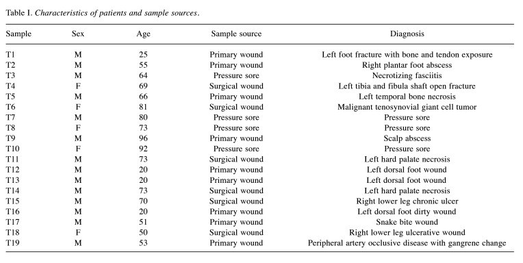
Characteristics of the study participants. In total, 16 patients diagnosed with SSTIs were included in this study; 11 were male and 5 were female. A total of 19 SSTI samples were collected from 16 patients, two patients subjected to surgery at least 2 times and therefore offered more than one sample. Samples were classified into three types: pressure sore (four cases, 21.0%), surgical wound (six cases, 31.5.0%), and wound infection (nine cases, 47.3%) (Figure 1). Details of the demographic characteristics of the patients enrolled in this study are provided in Table I. Sample 11 and Sample 14 were collected from the same patient with diagnosis of left tonsil squamous cell carcinoma, who was subjected to surgery twice. Samples 12, 13, and 16 were collected separately from the same patient with left dorsal foot open wound who was subjected to debridement thrice.
Figure 1. Among the 19 samples from 16 patients with soft tissue infection, four cases were pressure sore, six cases were surgical wounds, and nine cases were wound infection tissues.
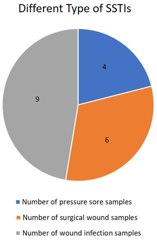
Table I. Characteristics of patients and sample sources.
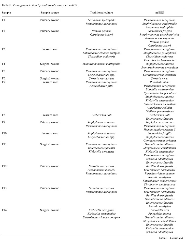
Pathogen detection using mNGS and traditional bacterial culture methods. All pathogens detected in different specimens using traditional bacterial cultures and mNGS method are listed in Table II. The microbiological cultures were positive in 19 out of 19 samples and mNGS showed positive results in 18 out of 19 samples.
Table II. Pathogen detection by traditional culture vs. mNGS.
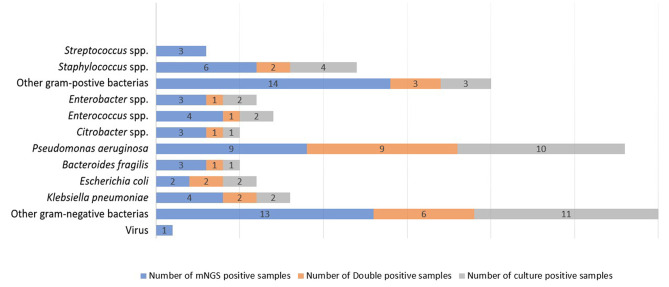
The numbers of different pathogens detected by mNGS and traditional bacterial cultures are shown in Figure 2. Among the 47 pathogens detected using mNGS or traditional bacterial cultures, gram-negative bacillus was predominant, especially Pseudomonas spp. (n=10), followed by Staphylococcus aureus (n=6), and Streptococci (n=3). The mNGS method showed higher positive detection rates than the culture in Staphylococcus aureus, Streptococci spp., Enterococcus spp., Enterobacter spp. and Bacteroids spp.
Figure 2. Comparison the pathogen detection between mNGS and culture in SSTI cases. Higher positive rate for Streptococcus, Staphylococcus, other gram-positive bacteria and other gram negative bacterial was demonstrated by mNGS when compared to culture.
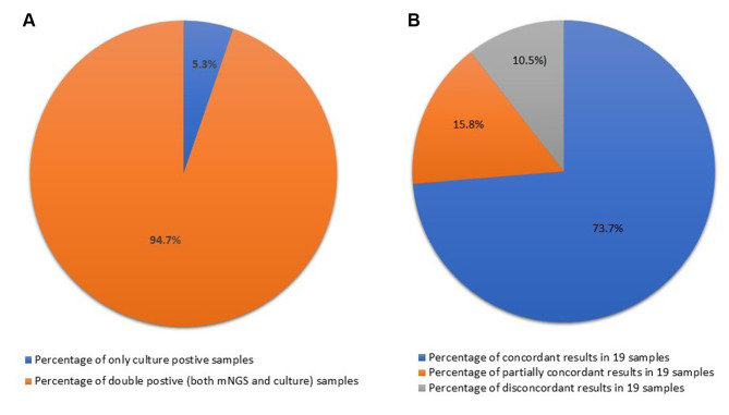
The results in seven of 19 samples (36.8%) obtained using mNGS and conventional bacterial culture were concordant at the species level; seven of 18 (36.8%) were concordant at the genus level. Among 19 samples, three (15.8%) were regarded partially concordant, and two (10.5%) were discordant (Figure 3). Sample T17 obtained from a wound of a snake bite (Table II) was negative in mNGS but positive for Staphylococcus epidermis in conventional culture.
Figure 3. Composite pie chart showing consistency between mNGS and conventional bacterial culture results. There was a 95% rate of double-positive results. Most results were concordant between the two methods (14 cases, 73.7%), with only two cases (10.5%) being discordant.
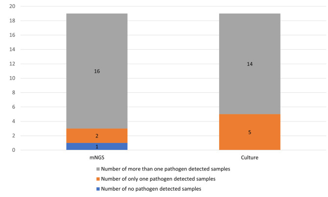
The mNGS also showed a higher proportion of polymicrobial pathogen detection in soft tissue infections (Figure 4). Multiple pathogens were detected in 73% of the 19 samples by the traditional bacterial culture method, while mNGS detected multiple pathogens in 84% of the 19 samples.
Figure 4. Bar graph showing the proportions of multiple pathogens in SSTI cases as detected using mNGS and culture. mNGS identified multiple pathogen infections in 16 cases, which was more than those detected using conventional bacterial culture (14 cases).
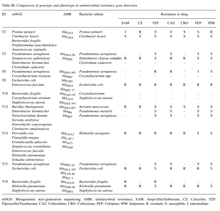
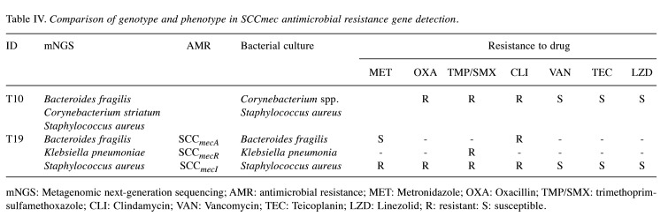
Antimicrobial resistance gene detection using mNGS. In this study, several types of antimicrobial resistance genes were detected (Table III and Table IV), including carbapenemase gene, broad spectrum and extended spectrum beta-lactamase gene (blaTEM, blaSHV, blaOXA), Ambler class C (AmpC) beta-lactamase, Bacteroides spp. related beta-lactamase gene (blaCfXA and blaCepA), Bacillus spp. related resistant gene (blaBCI and blaBla), SCCmec and quinolone resistance gene.
Table III. Comparison of genotype and phenotype in antimicrobial resistance gene detection.
mNGS: Metagenomic next-generation sequencing; AMR: antimicrobial resistance; SAM: Ampicillin/Sulbactam; CZ: Cefazolin; TZP: Piperacillin/Tazobactam; CAZ: Ceftazidime; CRO: Ceftriaxone; FEP: Cefepime; IPM: Imipenem; R: resistant; S: susceptible; I: intermediate.
Table IV. Comparison of genotype and phenotype in SCCmec antimicrobial resistance gene detection.
mNGS: Metagenomic next-generation sequencing; AMR: antimicrobial resistance; MET: Metronidazole; OXA: Oxacillin; TMP/SMX: trimethoprim-sulfamethoxazole; CLI: Clindamycin; VAN: Vancomycin; TEC: Teicoplanin; LZD: Linezolid; R: resistant: S: susceptible.
Discussion
Metagenomic sequencing is a promising and novel tool for the etiological diagnosis of SSTIs, particularly for identifying viruses, anaerobes, fastidious, and multi-pathogen infections (9). In this study, the clinical performance of mNGS was assessed in patients with soft tissue infection, in comparison with conventional microbiology testing. This study demonstrated the capability of mNGS as the first-line pathogen test. The mNGS method could detect more pathogens compared to conventional culture. Furthermore, mNGS was shown to overcome limitations of current diagnostic tests, such as 16S rRNA PCR, allowing universal pathogen detection without prior knowledge of specific microorganisms.
In our study, most of microbiological findings in each sample were concordant and partially concordant between traditional bacterial cultures and mNGS (17 of 19). For discordant and partially concordant samples, the discrepancy was primary on anaerobes, fastidious pathogens, and viral pathogens.
The detection of anaerobes, especially obligate anaerobes, requires a strict anaerobic environment during specimen collection and transport. In our study, mNGS revealed the presence of Bacteroides fragilis in three samples (T2, T10, T19), but traditional bacterial culture showed its presence in only one sample (T19). Bacteroides spp. are small, pleomorphic gram-negative bacilli. Among the more than 50 known Bacteroides spp. (11), Bacteroides fragilis is the most important in human disease. It is the most common anaerobe isolated from intra-abdominal infections, and the most frequent anaerobic isolate in patients with bacteremia (12). This highlights the importance of quick detection of anaerobes using mNGS, which can provide novel and fast turnaround time in the detection of Bacteroides fragilis.
In addition, mNGS identified several fastidious pathogens in patients with SSTI, such as Granulicatella spp. (formerly known as nutritionally variant streptococci). It is an important etiologic species in blood culture negative endocarditis (BCNE) (13). Besides BCNE, Granulicatella adiacens are also involved in prosthetic joint infection, and are often being missed due to difficult culture, especially in the cases of polymicrobial infection. In our study, G. adiacens was found in three samples (T11, T13, T14) using mNGS, but was not detected using conventional bacterial culture. A study showed that prolonged antimicrobial treatment (≥8 weeks) should be considered for prosthetic joint infection involving G. adiacens (14). The ability of the mNGS method to detect fastidious pathogens in SSTI helps guiding antimicrobial agent selection and determining duration of treatment.
Additionally, due to viral pathogens usually not being taken seriously in SSTI condition, mNGS provides an effective way to detect viral pathogens. In the T9 sample, left occipital carbuncle, the bacterial culture detected Staphylococcus aureus, Pseudomonas aeruginosa and mNGS Staphylococcus aureus, Pseudomonas aeruginosa and Human betaherpesvirus 5. Although Human betaherpesvirus 5 (also known as Human cytomegalovirus) infection is typically asymptomatic in the immunocompetent population, this infection can still result in conditions, such as mononucleosis and certain cancers (15). In nowadays, mNGS is also used in central nervous system infections due to the inefficiency of the current diagnostic workflows. mNGS offers the chance to circumvent these challenges by using unbiased laboratory and computational methods (16).
The major advantage of mNGS in soft tissue infection compared to conventional bacterial culture is not only the much shorter turnaround time but also the acquisition of drug resistance data that can guide optimal antibiotic therapy at an early stage of infection. In this study, several types of antimicrobial resistance genes were detected (Table III and Table IV), including carbapenemase, broad- and extended spectrum beta-lactamase (blaTEM, blaSHV, blaOXA), AmpC beta-lactamase, Bacteroides spp.-related beta-lactamase (blaCfXA and blaCepA), and Bacillus spp. related resistant gene (blaBCI and blaBla), SCCmec.
One of the problems with the detected antimicrobial resistance genes is that it is difficult to attribute a resistant gene to a specific species. However, because certain resistant genes tend to be present in certain bacterial species, there are still hints that can be used for identification. For example, in the T8 sample, Escherichia coli and Enterococcus faecium were detected using mNGS. The resistant genes blaCMY, blaOXA, and blaKPC-17 were more likely to belong to E. coli. In the T19 sample, SCCmecA, SCCmecR, and SCCmecI were more recognized as belonging to S. aureus.
Specific resistance genes mainly appear in specific bacterial species. When using mNGS to simultaneously detect multiple pathogens and multiple resistance genes in a sample, if a specific resistance gene is identified, it can be linked to a specific bacterial species. The blaCfxA is known to be present in Bacteroides spp. and oral anaerobic bacteria, such as Prevotella spp. and Capnocytophaga spp. (17). Insertional activation of blacepA leads to high-level beta-lactamase expression in clinical isolates of B. fragilis (18). In our study, mNGS showed the presence of B. fragilis reads in the T2, T10, T19 samples. Beta-lactamase genes, such as blaCfxA, blacepA, and blaCfiA, which are often found in Bacteroides spp., were also detected in T2, T10, and T19 samples. Although other pathogens were also detected in the T2, T10, and T19 samples, based on the characteristics of blaCfxA, blacepA, and blaCfiA, these genes are likely to belong to Bacteroides spp. in these samples. In addition, blaBCI and blaBla1 were detected in Bacillus spp. Expression of the bla1 and bla2 genes in certain strains of Bacillus anthracis is associated with penicillin resistance (19). In sample T12, Bacillus thuringiensis and blaBCI and blaBla resistant genes was detected using mNGS.
In Pseudomonas spp., the main resistance mechanism against β-lactams is the expression of the class C blaPDC (Pseudomonas-derived cephalosporinase) resistant gene (20). In our study, P. aeruginosa was detected using mNGS and conventional bacterial culture in T3, T6, T15. The blaPDC gene was also detected in those samples using mNGS.
There are still some limitations to the use of mNGS for pathogen and resistant gene detection. First, in our study, mNGS could detect 24 more potential pathogens from samples positive based on mNGS and culture. However, culturable pathogens were also missed by mNGS because of the low read numbers or low relative abundance. For example, T3 culture revealed Enterobacter cloacae complex, but mNGS did not detect Enterobacter hormaechei due to low reads. T7 conventional bacterial culture (a bedsore wound) yielded Acinetobacter pittii but not mNGS. Second, resistant gene detection can’t always correlate to specific pathogens precisely. For example, there are several samples in our study where there was simultaneous detection of P. aeruginosa and other non-Pseudomonas Enterobacterales using mNGS. However, if OXA beta-lactamase was detected, we cannot determine which pathogen carried the resistant gene until a phenotype report is issued. However, there are also some specific resistance genes, such as blaPDC, which can be more confidently interpreted as originating from P. aeruginosa. This implies that interpretation plays a more and more significant role when mNGS is implemented in clinical conditions.
Conclusion
The mNGS plays a significant role in pathogen detection in soft tissue infection. It identifies more pathogens and can allow clinical practitioners to make precise patient treatment decisions. However, careful interpretation of the mNGS report is needed. We need further studies comparing conventional bacterial cultures with mNGS in SSTI.
Conflicts of Interest
The Authors declare no competing interests in relation to this study.
Authors’ Contributions
Conceptualization: Kuan-Pei Lin, Po-Yu Liu, Yan-Chiao Mao, Chih-Sheng Lai, Kuo-Lung Lai, Ting-Kuang Yeh; Methodology: Kuan-Pei Lin, Yao-Ting Huang, Po-Yu Liu, Ting-Kuang Yeh; Formal analysis: Yao-Ting Huang, Po-Yu Liu; Investigation: Kuan-Pei Lin, Yao-Ting Huang, Po-Yu Liu, Chien-Hao Tseng, Ting-Kuang Yeh; Data Curation: Kuan-Pei Lin, Yao-Ting Huang, Po-Yu Liu, Chia-Wei Liu; Writing - Original Draft: Kuan-Pei Lin, Yao-Ting Huang, Po-Yu Liu, Yan-Chiao Mao, Chih-Sheng Lai, Kuo-Lung Lai, Chien-Hao Tseng, Chia-Wei Liu, Wei-Hsuan Huang, Hsien-Po Huang, Ting-Kuang Yeh; Writing - Review & Editing: Kuan-Pei Lin, Po-Yu Liu, Ting-Kuang Yeh; All Authors had final approval of the version to be published and agreed to be accountable for all aspects of the work in ensuring that questions related to the accuracy or integrity of any part of the work are appropriately investigated and resolved.
Funding
Yao-Ting Huang was supported by National Science and Technology Council grant: 111-2221-E-194-031-MY3, 112-2628-E-194-001-MY3. Po-Yu Liu was supported by Taichung Veterans General Hospital: TCVGH-1133901C, TCVGH-1133901D; National Science and Technology Council: 112-2314-B-075A-006.
References
- 1.Peetermans M, de Prost N, Eckmann C, Norrby-Teglund A, Skrede S, De Waele JJ. Necrotizing skin and soft-tissue infections in the intensive care unit. Clin Microbiol Infect. 2020;26(1):8–17. doi: 10.1016/j.cmi.2019.06.031. [DOI] [PubMed] [Google Scholar]
- 2.Miller LG, Eisenberg DF, Liu H, Chang CL, Wang Y, Luthra R, Wallace A, Fang C, Singer J, Suaya JA. Incidence of skin and soft tissue infections in ambulatory and inpatient settings, 2005-2010. BMC Infect Dis. 2015;15:362. doi: 10.1186/s12879-015-1071-0. [DOI] [PMC free article] [PubMed] [Google Scholar]
- 3.Moffarah AS, Al Mohajer M, Hurwitz BL, Armstrong DG. Skin and soft tissue infections. Microbiol Spectr. 2016;4(4) doi: 10.1128/microbiolspec.DMIH2-0014-2015. [DOI] [PubMed] [Google Scholar]
- 4.Huang Z, Wu Q, Fang X, Li W, Zhang C, Zeng H, Wang Q, Lin J, Zhang W. Comparison of culture and broad-range polymerase chain reaction methods for diagnosing periprosthetic joint infection: analysis of joint fluid, periprosthetic tissue, and sonicated fluid. Int Orthop. 2018;42(9):2035–2040. doi: 10.1007/s00264-018-3827-9. [DOI] [PubMed] [Google Scholar]
- 5.Malandain D, Bémer P, Leroy AG, Léger J, Plouzeau C, Valentin AS, Jolivet-Gougeon A, Tandé D, Héry-Arnaud G, Lemarié C, Kempf M, Bret L, Burucoa C, Corvec S, Centre de Référence des Infections Ostéo-articulaires du Grand Ouest (CRIOGO) Study Team Assessment of the automated multiplex-PCR Unyvero i60 ITI® cartridge system to diagnose prosthetic joint infection: a multicentre study. Clin Microbiol Infect. 2018;24(1):83.e1–83.e6. doi: 10.1016/j.cmi.2017.05.017. [DOI] [PubMed] [Google Scholar]
- 6.Gu W, Miller S, Chiu CY. Clinical metagenomic next-generation sequencing for pathogen detection. Annu Rev Pathol. 2019;14:319–338. doi: 10.1146/annurev-pathmechdis-012418-012751. [DOI] [PMC free article] [PubMed] [Google Scholar]
- 7.Shi Y, Wang G, Lau HC, Yu J. Metagenomic sequencing for microbial DNA in human samples: emerging technological advances. Int J Mol Sci. 2022;23(4):2181. doi: 10.3390/ijms23042181. [DOI] [PMC free article] [PubMed] [Google Scholar]
- 8.Zeng X, Wu J, Li X, Xiong W, Tang L, Li X, Zhuang J, Yu R, Chen J, Jian X, Lei L. Application of metagenomic next-generation sequencing in the etiological diagnosis of infective endocarditis during the perioperative period of cardiac surgery: a prospective cohort study. Front Cardiovasc Med. 2022;9:811492. doi: 10.3389/fcvm.2022.811492. [DOI] [PMC free article] [PubMed] [Google Scholar]
- 9.Wang Q, Miao Q, Pan J, Jin W, Ma Y, Zhang Y, Yao Y, Su Y, Huang Y, Li B, Wang M, Li N, Cai S, Luo Y, Zhou C, Wu H, Hu B. The clinical value of metagenomic next-generation sequencing in the microbiological diagnosis of skin and soft tissue infections. Int J Infect Dis. 2020;100:414–420. doi: 10.1016/j.ijid.2020.09.007. [DOI] [PubMed] [Google Scholar]
- 10.Li H. Aligning sequence reads, clone sequences and assembly contigs with BWA-MEM. arXiv. 2013:1303.3997. doi: 10.48550/arXiv.1303.3997. [DOI] [Google Scholar]
- 11.Wexler HM. Bacteroides: the good, the bad, and the nitty-gritty. Clin Microbiol Rev. 2007;20(4):593–621. doi: 10.1128/CMR.00008-07. [DOI] [PMC free article] [PubMed] [Google Scholar]
- 12.Polk BF, Kasper DL. Bacteroides fragilis subspecies in clinical isolates. Ann Intern Med. 1977;86(5):569. doi: 10.7326/0003-4819-86-5-569. [DOI] [PubMed] [Google Scholar]
- 13.Reyes C, Barthel ME. [Granulicatella spp] Rev Chilena Infectol. 2015;32:359–360. doi: 10.4067/S0716-10182015000400017. [DOI] [PubMed] [Google Scholar]
- 14.Quénard F, Seng P, Lagier JC, Fenollar F, Stein A. Prosthetic joint infection caused by Granulicatella adiacens: a case series and review of literature. BMC Musculoskelet Disord. 2017;18(1):276. doi: 10.1186/s12891-017-1630-1. [DOI] [PMC free article] [PubMed] [Google Scholar]
- 15.Fulkerson HL, Nogalski MT, Collins-McMillen D, Yurochko AD. Overview of human cytomegalovirus pathogenesis. Methods Mol Biol. 2021;2244:1–18. doi: 10.1007/978-1-0716-1111-1_1. [DOI] [PubMed] [Google Scholar]
- 16.Piantadosi A, Mukerji SS, Ye S, Leone MJ, Freimark LM, Park D, Adams G, Lemieux J, Kanjilal S, Solomon IH, Ahmed AA, Goldstein R, Ganesh V, Ostrem B, Cummins KC, Thon JM, Kinsella CM, Rosenberg E, Frosch MP, Goldberg MB, Cho TA, Sabeti P. Enhanced virus detection and metagenomic sequencing in patients with meningitis and encephalitis. mBio. 2021;12(4):e0114321. doi: 10.1128/mBio.01143-21. [DOI] [PMC free article] [PubMed] [Google Scholar]
- 17.Binta B, Patel M. Detection of cfxA2, cfxA3, and cfxA6 genes in beta-lactamase producing oral anaerobes. J Appl Oral Sci. 2016;24(2):142–147. doi: 10.1590/1678-775720150469. [DOI] [PMC free article] [PubMed] [Google Scholar]
- 18.Rogers MB, Bennett TK, Payne CM, Smith CJ. Insertional activation of cepA leads to high-level beta-lactamase expression in Bacteroides fragilis clinical isolates. J Bacteriol. 1994;176(14):4376–4384. doi: 10.1128/jb.176.14.4376-4384.1994. [DOI] [PMC free article] [PubMed] [Google Scholar]
- 19.Chen Y, Tenover FC, Koehler TM. Beta-lactamase gene expression in a penicillin-resistant Bacillus anthracis strain. Antimicrob Agents Chemother. 2004;48(12):4873–4877. doi: 10.1128/AAC.48.12.4873-4877.2004. [DOI] [PMC free article] [PubMed] [Google Scholar]
- 20.Colque CA, Albarracín Orio AG, Tomatis PE, Dotta G, Moreno DM, Hedemann LG, Hickman RA, Sommer LM, Feliziani S, Moyano AJ, Bonomo RA, K Johansen H, Molin S, Vila AJ, Smania AM. Longitudinal evolution of the pseudomonas-derived cephalosporinase (PDC) structure and activity in a cystic fibrosis patient treated with β-lactams. mBio. 2022;13(5):e0166322. doi: 10.1128/mbio.01663-22. [DOI] [PMC free article] [PubMed] [Google Scholar]


