Abstract
Interfacial ferroelectricity, prevalent in various parallel-stacked layered materials, allows switching of out-of-plane ferroelectric order by in-plane sliding of adjacent layers. Its resilience against doping potentially enables next-generation storage and logic devices. However, studies have been limited to indirect sensing or visualization of ferroelectricity. For transition metal dichalcogenides, there is little knowledge about the influence of ferroelectric order on their intrinsic valley and excitonic properties. Here, we report direct probing of ferroelectricity in few-layer 3R-MoS2 using reflectance contrast spectroscopy. Contrary to a simple electrostatic perception, layer-hybridized excitons with out-of-plane electric dipole moment remain decoupled from ferroelectric ordering, while intralayer excitons with in-plane dipole orientation are sensitive to it. Ab initio calculations identify stacking-specific interlayer hybridization leading to this asymmetric response. Exploiting this sensitivity, we demonstrate optical readout and control of multi-state polarization with hysteretic switching in a field-effect device. Time-resolved Kerr ellipticity reveals direct correspondence between spin-valley dynamics and stacking order.
Subject terms: Ferroelectrics and multiferroics, Two-dimensional materials, Electronic and spintronic devices
In parallel-stacked materials, ferroelectric order is switched by layer sliding. In 3R-MoS2, excitons react to ferroelectricity via stacking-specific hybridization. Authors show field effect control of ferroelectricity and its effect on spin dynamics.
Introduction
The basic building block for any interfacial ferroelectrics is two layers of certain van der Waals materials aligned parallel to each other1–11. In the case of transition metal dichalcogenides (TMDs) like MoS2, layer-asymmetric atomic registry12 along the out-of-plane direction leads to interlayer hybridization between the valence band of one layer and the conduction band of the other but not vice versa13. Such a preferential coupling induces unidirectional charge transfer, leading to the spontaneous emergence of an electric dipole bound at the interface. A bilayer unit of parallelly-stacked (rhombohedral or so-called 3R)-TMDs thus possesses two equivalent yet opposite ferroelectric orders viz. MX and XM, marking the stacking of metal (M) and chalcogen (X) atoms at the eclipsed sites (Fig. 1).
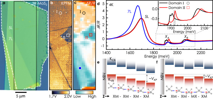
Fig. 1. Reflection contrast imaging of ferroelectric domains in few-layer 3R-MoS2.
a Optical micrograph of a 3R-MoS2 flake on Si/SiO2/hBN. Topographical steps and edges of the bottom hBN have been marked by white dotted lines. b Surface potential map within the area enclosed by the black rectangle in (a). Two domains, marked by ∘ and □, can be identified by the difference in contrast. c. Integrated intensity map of ΔR/R at the XA spectral region, i.e., from 1906 to 1918 meV (gray bar in inset of d). d Low-temperature reflectance contrast spectra from various spatial locations marked by symbols of corresponding color in c (black - 5L domain I, red - 5L domain II, and blue - 7L). Inset shows high-resolution spectra collected from domain I and II. e, f Two crystal configurations of 5L 3R-MoS2 overlayed on layer-projected KV B and KCB band-edges. To first order, the band edge variation along Z and the degeneracies, highlighted for the XM-XM-MX-XM stacking by dotted lines, result from the ferroelectricity-induced electrostatic considerations (SI Figs. S12, 13 for other configurations). Arrows represent polarization vectors at the interfaces.
Besides hosting ferroelectricity, parallel stacking of TMDs preserves the spin-valley locking, regarded predominantly as a property of monolayers14, even in the multilayer limit due to broken inversion and mirror symmetries15–18. Therefore, the 3R-polymorph of TMDs provide means for the bottom-up construction of three-dimensional spin-valleytronic devices on a ferroelectric platform. A visionary goal would be to engineer the optical response in TMDs by exploiting the ferroelectricity-induced interaction, which can be highly localized, non-volatile, and reconfigurable. Despite a tremendous interest in this emerging phenomenon19,20, so far the field has been limited to indirect sensing or visualizing the ferroelectricity employing surface-sensitive probes, such as atomic force microscopy2,4,6,7,11, scanning electron microscopy21, or sensing-layer-based approaches2,5 and, therefore, could only provide limited physical insight. In the case of TMDs, this leaves a void of in-depth knowledge about the influence of ferroelectric order on their intrinsic valley and excitonic properties. To fill this gap, we exploit hyperspectral reflectivity imaging to discern the correspondence between ferroelectric stacking and optical response in these structures. We demonstrate ferroelectricity-induced control of spin-valley dynamics by systematically studying the ultrafast response of excitons using transient reflection and Kerr ellipticity. As-grown samples are chosen over artificially stacked parallel structures, which provides a moiré-free crystal, enabling us to explore excitonic phenomena of purely ferroelectric origin and to employ far-field optical spectroscopy on mesoscopic domains with well-defined stacking. Clear signatures of stacking-dependent K-valley excitonic response and a striking contrast in valley relaxation dynamics are observed. This presents the prospect of using spin and valley as useful degrees of freedom in a robust multilayer platform. It is worth mentioning that recent experimental efforts22 also underscore the possibility of utilizing multilayer 2H-TMDs for valleytronic applications despite their centro-symmetric structure23.
Results
Ferroelectric domains in 3R single crystals
We begin by probing the electric surface potential of deterministically stamped 3R-MoS2 flakes on non-polar hexagonal boron nitride (hBN) exfoliated on Si/SiO2(-285 nm) substrate (SI. S1). An atomic force microscope operated in side-band Kelvin probe mode (KPFM) (SI. S2) is used to map the surface potential, VKP. Figure 1a shows an optical microscopy image of a representative flake (Sample 1) composed of five and seven layers of MoS2. In a multilayer flake, each of the interfaces may have either XM (polarization pointing upward) or MX (downward) stacking order creating a potential ladder either going upwards or downwards, respectively7,13. The difference in the total number of upward- and downward-pointing polarized interfaces decides the cumulative polarization state, hence the surface potential. Therefore, different ferroelectric domains in a given thickness would be visible in a VKP map. Based on this understanding, the existence of two ferroelectric domains, viz. domain I and II in the 5L region of the sample, becomes evident from Fig. 1b. The measured surface potential variation of ~ 127 mV between these domains (SI Fig. S1) corresponds to the sum of effectively two ferroelectric interfaces, consistent with earlier results7,24.
The appearance of different domains in a single crystal flake can be attributed either to inherent stacking faults in the bulk 3R crystal or to the sliding by slip-avalanche of constituting layers from XM to MX stacking order or vice versa. Since the energies of all possible stacking configurations are similar (SI Fig. S19), the latter occurs as a result of inadvertent shear strain during exfoliation25 or deterministic stamping. To further illustrate the mechanism, we fabricated a sample by intentionally applying an in-plane perturbation, achieved by laterally tapping the substrate holder during the dry transfer of 3R-MoS2 from PDMS to the substrate. This horizontal force introduces a shear stress between the layers that remain anchored to PDMS and the layers attached to the substrate. This deliberate horizontal force, which is significantly stronger than the ubiquitously present shear forces during standard operations, generates smaller and densely packed ferroelectric domains, as identified by KPFM (SI Fig. S2). The ferroelectric domains, in this case, are of similar dimension as in the artificially stacked moiré interfacial ferroelectrics. However, achieving precise control over domain formation and their structural organization remains a challenge.
Correspondence of surface potential and exciton fine structure
To check for the ferroelectric-field-induced changes in optical response, we map the exciton transition energies over the entire flake (i.e. Sample 1) using confocal differential reflectance spectroscopy (SI. S3). Typical reflectance contrast (RC) spectra, ΔR/R = (RRef − RFlake)/RFlake26 obtained at different spatial locations of the flake at liquid helium temperature are presented in Fig. 1d. Here RFlake is the reflection from MoS2 and RRef is the same from hBN (SI Fig. S3). In the energy range of interest, the reflectance contrast of the few-layer MoS2 is dominated by three excitonic features, viz. the momentum-indirect transition at ~1700 meV and the well-known A and B excitons at XA ~1900 and XB ~2050 meV27–29. Notably, the lowest energy feature despite being a momentum-indirect transition has a prominent appearance. This can be attributed to the favorable thin-film interference condition in the Fabry-Pérot cavity formed by Si/SiO2/hBN/MoS2 in the given energy range (SI Figs. S4, S5 for additional examples and further discussion). We note that RC spectra, ΔR/R can also be calculated using (RRef − RFlake)/RRef30. We found that both representations, ΔR/RRef and ΔR/RFlake share similar characteristic viz. the presence of three distinct features and their energetic positions (SI Figs. S4, S5, S6, S15). However, the choice of definition modifies the line shape of each transition and therefore, the overall appearance of the spectra. Evidently, the lowest energy peak appears vividly in ΔR/RFlake (SI Figs. S4–S5, S10). Therefore, in the following we refer to ΔR/RFlake as RC spectra unless mentioned otherwise.
We proceed by constructing RC maps corresponding to different transitions by integrating the intensity over selected spectral ranges. The map of the momentum-indirect transition brings out a clear contrast between the five- and seven-layer regions (SI Figs. S7–S8) due to the red-shift in its spectral position with increasing thickness. On the other hand, the XA intensity map exhibits signatures of two distinct domains within the topographically smooth 5L area, similar to the KPFM map. The contrast in the intensity map (Fig. 1c) is due to the appearance of higher- and lower-energy sub-features in the spectral region from 1880 to 1930 meV. The energy positions and relative oscillator strengths of these fine features distinguishably mark the ferroelectric stacking configuration, as revealed by the high-resolution spectra shown in Fig. 1d-inset. In contrast to previous studies on artificial R-stacked TMDs31, these subfeatures are not related to nanoscale reconstruction of moiré domains (as confirmed by the complementary KPFM measurements). A similar spatial map is obtained from room temperature spectroscopy measurements (SI Fig. S9); however, due to thermal broadening, spectral features can not be fully resolved. Correspondence between RC maps and KPFM images has been observed in several samples of different thicknesses, see Fig. S10 for example. Upon closer inspection, it becomes evident that XB exhibits a similar trait (SI Figs. S7, S8). To improve the signal-to-noise ratio of the RC spectra around the XB transition, we exploit the cavity effect by carefully choosing the bottom hBN of our stack. SI Fig. S11 shows an RC spectrum of a trilayer-MoS2 flake on a 95 nm-hBN/285 nm-SiO2/Si substrate, where the splitting in the XB becomes apparent. In a nutshell, momentum-direct excitons at the K points in the Brillouin zone reveal the stacking order, while the spectral feature related to the momentum-indirect transition remains insensitive to it. Based on the trend of red-shift32 with increasing layer number, as shown in Fig. S4 and previous studied on similar systems29,33, one can tentatively assign the low-energy peak either to the Γ-Q or to K-Q hybrid-excitonic34 transition.
To explain the asymmetric response of different exciton species to ferroelectric ordering, we consider the band structure of 3R-MoS2. It has been shown that in bilayer 3R-TMDs, states at the Γ point of the valence band (VB) or Q point of conduction band (CB) are fully delocalized due to strong interlayer hybridization7,13,16. In contrast, the wavefunctions at the K points are almost entirely layer-localized. The ferroelectricity-induced built-in potential manifests as a local electrostatic perturbation and introduces a shift between the layer-projected energy levels at K points, leading to a type-II band alignment at the interfaces16, with the energies decreasing from layer X to layer M. By the same token, in a multilayer sample, the spatial profile of K-point band extrema along the out-of-plane direction follows the underlying ferroelectric potential and is, therefore, susceptible to the stacking order, as depicted schematically in Fig. 1e–f and SI Figs. S12, S13.
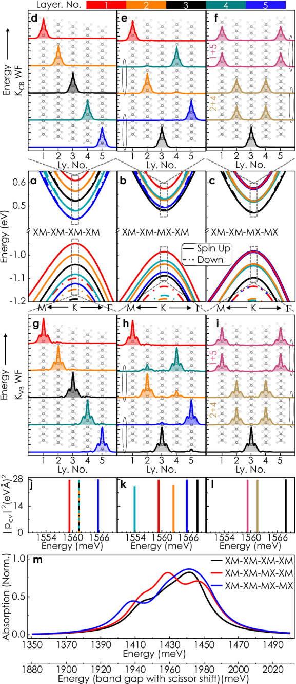
Neglecting interlayer coupling through hybridization, a built-in electric field should shift the conduction and valence bands by the same amount, leaving the transition energies unchanged. Clearly, this alone is insufficient for interpreting the experimentally observed emerging multiplicity of transition energies. Therefore, to this end, we inspect the stacking-dependent strength of K states delocalization based on our band structure calculations depicted in Fig. 2a–c. Figure 2d–f, g–i presents the five lowest energy conduction-band and highest-energy valence-band spin-up electron wavefunctions at the K point, sorted according to their energy eigenvalues (for the following sections, we have dropped the term ‘K point’ while referring to bands and wavefunctions unless stated otherwise). For the fully polarized, XM-XM-XM-XM case (Fig. 2a, d, g), the individual wave functions both at the valence and conduction band remain layer-localized with their energy levels corresponding to the ferroelectric potential ascending with each additional layer. However, for other variants, interlayer hybridization is evident (e.g., Fig. 2b, e, h, c, f, i). This dependence of hybridization on ferroelectric ordering is the underlying mechanism for the observed contrast in optical spectra. Before proceeding further, we note that a five-layer 3R-MoS2 crystal can have sixteen (2N−1, N = 5) stacking configurations, out of which KPFM can distinguish among five combinations based on their surface potential (SI Fig. S12). At this stage, therefore, instead of attempting a one-to-one quantitative description of the stacking-dependent excitonic response, we focus on the qualitative generic trend with three specific cases as examples (all the studied systems are shown in the SI Figs. S20–S27).
Fig. 2. Ab initio calculation of stacking-dependent band structure, wavefunctions and optical response.

a–c Band structure for different stacking orders. Spin orientation (∥ Z) of the individual bands is indicated by solid (spin up) and dashed (spin down) lines, respectively. The bands are colored corresponding to the dominant contribution of specific layers. The stacking of the band extrema at K follows the underlying ferroelectric potential. Due to mirror symmetry for the XM-XM-MX-MX case, some bands are (nearly) degenerate and cannot be attributed to an individual layer (indicated by different colors in f, i). d–i Absolute value of the lowest-energy spin-up conduction band (d–f) and highest-energy valence band (g–i) wavefunctions sorted according to their energies superimposed over the crystal structure. Colors indicate the dominant layer(s) for each wavefunction. Ellipses are used to indicate (near-)degenerate states. The wave functions are layer-localized for the fully polarized case (d, g). For XM-XM-MX-MX (f, i), the lowest-energy wave function is localized in the center layer, while the other wave functions have equal weight in the mirror-symmetric layers. j–l Oscillator strength for transitions from all spin-up KV B to all KCB with s+ polarization. The magnitude of these intralayer transitions is in good agreement with values for pristine monolayer MoS256,57. m Calculated absorption spectra in the A excitonic region based on intralayer dipole matrix elements and layer-specific exciton binding energy, including a phenomenological Lorentzian line width of 20 meV.
In the fully co-polarized XM-XM-XM-XM stacking, the potential varies monotonically from layer to layer7. Thus, the three central layers experience a similar ferroelectric field - preserving the energy degeneracy of their intralayer excitonic transitions, appearing as a single vertical line in the dipole matrix element plot (Fig. 2j). However, the top and bottom layers show energetically distinct transitions due to broken translational symmetry at the surfaces. The scenario changes in partially polarized stacking due to selective interlayer coupling between specific layers. The mechanism described below is similar to the quantum correction that leads to level anti-crossing by lifting the energy degeneracy of coupled quantum systems. For instance, in the XM-XM-MX-XM configuration, electrostatic degeneracy (cf. Fig. 1f) leads to layer-specific resonant hybridization35 of KVB states, which is particularly strong between the second and fourth layers. Pronounced hybridization also occurs in the XM-XM-MX-MX case between the mirror-symmetric layers, namely the first and fifth, as well as the second and fourth. In contrast to the valence band states, the KCB states largely retain their layer-localized nature in almost all cases except mirror-symmetric stacking orders. This layer- and band-specific strength of interlayer hybridization leads to unequal shifts of layer-projected conduction and valence bands, lifting the intralayer transition degeneracy even for the center layers and increasing the maximum spread of energies, clearly visible in Fig. 2j–l. These findings are of a general nature for any parallel-stacked multilayer TMDs. This stacking-specific resonant hybridization is not relevant for the Γ and Q states, which have a significant degree of hybridization irrespective of stacking.
In addition to the corrections to the electronic band structure arising from hybridization, changes in the exciton binding energy due to asymmetric dielectric surroundings also play a role in determining the energetic position of individual transitions36. In order to provide a qualitative comparison to the experimental reflectance contrast spectra, we calculate the absorption spectra based on the intralayer dipole matrix elements and exciton binding energy (SI. S5, S6), considering a phenomenological Lorentzian broadening with a full-width-half-maximum of 20 meV. These results are depicted in Fig. 2m. Evidently, the general shape with multiple sub-features, which strongly depend on the individual stacking, closely mimics the experimental observations. However, it is important to note that DFT typically underestimates the band gap of semiconductors due to the unaccounted derivative discontinuity of the typical exchange-correlation functionals37 and, therefore, the calculated absorption spectra are red-shifted by about 480 meV in comparison with the experimental results. By applying a rigid energy shift to the band gap, estimated from GW calculations, one can achieve transition energies that are more consistent with experimental data. For MoS2, this estimate yields a band gap increase of about 530 meV38, which nearly matches the experimentally observed energies.
These observations provide an alternative experimental approach to identify local changes in ferroelectric ordering in 3R-TMD flakes without resorting to surface-sensitive5–7,39 measurements, which become a challenge for heterostructures. In contrast to other optical measurement schemes relying on a sensing layer40, the TMD itself reveals its ferroelectric order in optical spectra. This allows for identifying local domains, even in ‘buried’ layers, as required for integrating more complex, functional heterostructures based on ferroelectric TMDs.
Non-volatile electrical control and optical readout of multi-state polarization
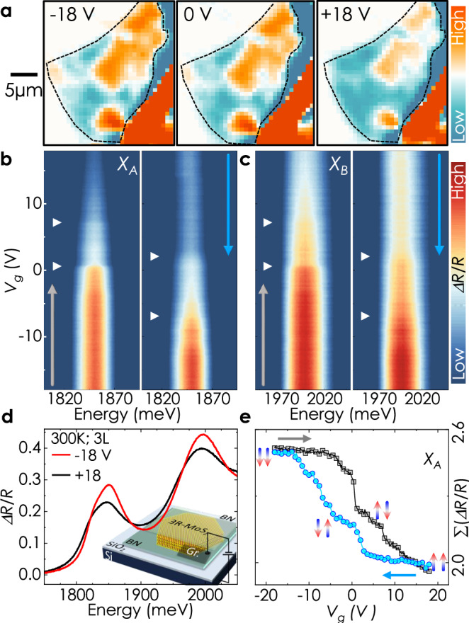
Our findings, therefore, present an unprecedented opportunity of exploiting ferroelectric fields to gain non-volatile control over excitonic transitions in a field effect transistor architecture- a crucial step forward for applications. We prepare devices on a conducting Si/SiO2(-90 nm) substrate, which acts as the gate electrode, and use few-layer graphite flakes to make electrical contact with 3R-MoS2 (viz. Sample 4). We use fully hBN-encapsulated structures for these measurements (therefore, RFlake is the reflection from hBN/MoS2/hBN heterostructure and RRef is the same from hBN/hBN). Here, we present results obtained on a trilayer sample at room temperature. First, we map the ferroelectric domains at a few different gate voltages. Two different domains are visible in the false color map as blue and orange regions. Application of positive gate voltage results in an expansion of the blue domains at the cost of the orange domains. An opposite change is observed for negative gate voltage. Next, we measure the reflection contrast close to a domain boundary as a function of gate voltage. The XA and XB transitions becomes weaker as the gate voltage is swept forward from -18 V to 18 V, corresponding to increasing electron doping. This reduction is due to a combined effect of Pauli blocking and reduced oscillator strength with state filling - typical of n-type TMDs41–43. Notably, the reduction of intensity from the highest to lowest occurs through two distinct steps during the sweeping process (Fig. 3e). Furthermore, a prominent hysteretic behavior in the intensity of XA and XB transitions is observed as the gate voltage decreases during a backward sweep, for which the intensity rises again and saturates (Fig. 3b–e and SI Fig. S16). We also examine the standard deviation, which correlates with the full-width half maximum of XA and XB. Evidently, the resulting plots exhibit the same features (SI Fig. S17).
Fig. 3. Hysteretic reflectance contrast as a function of gate voltage.

a Integrated amplitude false color map of the numerically calculated first derivative of RC spectra at different gate voltages to visualize domain switching. All measurements were performed at room temperature. Evidently, the out-of-plane electric field drives the blue-colored region to grow in area coverage with increasing gate voltage at the expense of the orange-colored region. b, c False color map of A and B exciton intensity as a function of gate voltage during forward and backward sweep. They share the same y-axis, given on the left. d Two representative room temperature reflectance contrast spectra at opposite gate voltages. Inset- schematic illustration of the field effect device. e Integrated intensity profile ( ≡ vertical line cuts from b) at 1852 ± 2 meV. The vertical arrows represent the ferroelectric ordering of each interface.
The step-like features indicate discrete changes in optical response due to ferroelectric switching through a mechanism known as slidetronics, where the out-of-plane electric field induces an in-plane sliding of adjacent layers4,21,44–46. At high electric fields, all the interfaces align to achieve an energetically favorable configuration, viz. MX-MX or XM-XM, as indicated in Fig. 3e. Therefore, the hysteretic occurrence of the discrete switching events in response to the external field directly results from a finite coercive field - a hallmark of ferroelectric materials. The two distinctive steps observed for the trilayer sample reveal that switching between the multiple ferroelectric states of a multilayer is feasible and controllable via external voltages - a pivotal insight for scalability.
Our study yields an average coercive field of 0.03–0.035 Vnm−1 for domain wall motion. Moreover, the hysteretic behavior of the monotonous intensity variation with gate voltage indicates a stacking-dependent change in average carrier concentration, observed earlier in transport measurements6,47. We note that a complete ferroelectric switching is not achieved throughout the whole crystal within the experimentally accessible gate voltage. This can be attributed to the increasing domain boundary deformation energy48 and domain wall pinning at localized stacking faults as well as surface contaminants21.
Domain-resolved exciton population and spin-valley dynamics
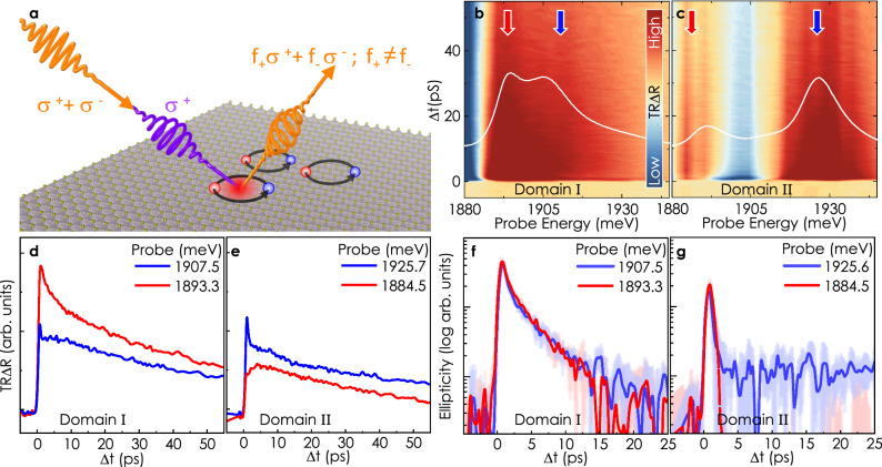
Besides gaining control over the excitonic oscillator strength, a meticulous understanding of excitonic lifetime is essential in the pursuit of realizing exciton-based circuits. To this end, we focus on probing the stacking-order-dominated exciton dynamics and spin relaxation using two-color pump-probe Kerr microscopy. Figure 4a illustrates the measurement scheme. We use a circularly polarized pump pulse, slightly blue detuned to XA, to induce a spin polarization of chosen helicity. The photo-generated excitons lead to photo-bleaching of a given valley, resulting in helicity-dependent reflectivity. Therefore, the transient exciton population and its spin-valley polarization can be probed by recording the intensity and induced ellipticity of a time-delayed linearly polarized probe pulse after reflection49,50. A series of simultaneous transient reflectivity (TRΔR) and transient ellipticity (TRKE) measurements are performed in which the probe laser wavelength is tuned across the energy range of XA with the pump energy set at 1958 meV. Figure 4b, c are false-color representations of TRΔR obtained from domain I and II of Sample 1 (cf. Fig. 1), respectively, as a function of pump-probe delay. The energy-dependent temporal evolution of reflectivity in individual domains clearly corresponds to their respective steady-state response (superimposed white lines). For both the domains, TRΔR traces at the higher resonance (marked by blue arrows) evolve similarly viz. an ultrafast decay component followed by relatively slower dynamics, as depicted by the blue colored traces in Fig. 4d, e. The response at the lower resonance energy has different characters for different domains. For instance, in domain I, we observe a monotonous decrease in the signal from t=0. Remarkably, for domain II, during the initial five ps, the reflectivity of the probe pulse increases with time, followed by a slower decay. The initial increase likely stems from slow state filling at the probe energy during relaxation, possibly suggesting interlayer charge transfer prompted by interlayer hybridization. These observations, therefore, further substantiate the theoretical understanding developed in the context of steady-state optical response.
Fig. 4. Exciton population and spin dynamics.
a Schematic of pump-probe Kerr measurement. A blue detuned (to XA) circularly polarized light pulse for the pump and linearly polarized pulse for the probe were used. Upon reflection, the linearly polarized pulses turn elliptically polarized. By projecting the reflected elliptic pulse on a quarter waveplate and a Wollaston prism, we can separate out the relative strength of right and left circular components. The total probe intensity change is equivalent to net exciton density, and the intensity difference between left and right circular components gives the net spin-valley polarization. All the pump-probe measurements were done at 4 K. b, c False color map of transient reflection as a function of probe energy from domain I and II. White contours (without y-scale) are copied from Fig. 1d-inset for comparison. d, e Transient reflection at selected energies, marked by colored arrows in (b, c). The opposite trend in the amplitude of the TRΔR signal at the lower and higher energy states from the two domains is a direct consequence of opposite relative strength of these sub-features in the steady-state spectroscopic signal. f, g Traces of transient ellipticity at resonant energies in respective domains. The spin-valley lifetime in domain I is on the order of 10-15 ps, while in domain II the signal decays down to the detection noise-floor on sub-ps timescales.
Unlike the transient reflection, the ellipticity signal manifests no prominent energy dependence. Instead, it is remarkably sensitive to the ferroelectric stacking configuration and exhibits entirely different traits for the two domains investigated. Figure 4f and g portrays the TRKE results for domain I and II probed at higher and lower resonance energies (SI Fig. S28 for the complete spectrum). In domain I, the TRKE decay is relatively slow, albeit faster than the decay of the transient reflectivity traces, indicating that it is not predominantly driven by exciton recombination but by a spin dephasing mechanism on a similar timescale as observed in MoS2 monolayers51,52. By contrast, in domain II, the excitons lose their spin polarization almost instantly. The microscopic mechanism for this striking dependence of exciton lifetimes and spin-valley dynamics on ferroelectric order remains an open question. However, recent calculations53,54 show that in parallel-stacked TMDs, dependent on the symmetry of the stacking order, in-plane spin-orbit coupling terms can arise that lead to spin mixing of K states. In this way, specific stacking orders may facilitate spin dephasing, while others protect spin orientation, yielding prolonged spin-valley lifetimes comparable to TMD monolayers. This opens up the use of spin and valley as useful degrees of freedom in 3R-stacked multilayers, which have so far been limited predominantly to monolayer TMDs.
To conclude, we demonstrate the direct correspondence between ferroelectric order and intralayer excitons in 3R-stacked MoS2 multilayers, allowing us to map ferroelectric domains directly using far-field optical spectroscopy. Ab initio theory provides a comprehensive explanation of the microscopic mechanism. In a field-effect device, we are able to control and map domain wall motion at room temperature and observe clear hysteretic features with a coercive field of roughly 0.03–0.035 V nm−1. The relatively small switching field of ferroelectricity by domain wall sliding could facilitate efficient electrical control of various optical properties of 2D materials55. Addressing individual domains in time-resolved experiments, we find that depending on ferroelectric order, 3R-stacked MoS2 multilayers can show spin-valley dynamics on timescales comparable to TMD monolayers or near-instantaneous loss of valley polarization. Our study paves the way for utilizing ferroelectric order in few-layer TMDs embedded within functional van der Waals heterostructures as a local, nonvolatile control lever for excitonic and valleytronic properties. Theses findings introduce a distinctive perspective to the field of quantum optoelectronics with van der Waals materials and will enable novel optoelectronic device architectures that rely on efficient electrical manipulation of excitons or the optical response in general.
Methods
Device fabrication
3R-MoS2, obtained from HQ Graphene, were exfoliated onto polydimethylsiloxane (PDMS). MoS2 flakes were selected according to their optical contrast. Chosen TMD flakes were stamped on pre-exfoliated hBN flakes. We use a polycarbonate (PC)/PDMS-based hot pickup method to transfer hBN flakes from bare Si/SiO2 substrate to encapsulate hBN/TMD stack. A few layer thick graphite flakes (procured from NGS Naturgraphit), exfoliated on PDMS, are deterministically stamped to make external contact to the sample.
KPFM measurements
KPFM measurements were performed using Park System NX20. The electrostatic signal was measured at side-band frequencies using two built-in lock-in amplifiers. We used PointProbe Plus Electrostatic Force Microscopy (PPP-EFM) n-doped tips. For the unencapsulated sample (Sample 1 c.f. Fig. 1) we used non-contact mode of operation. The average height above the surface was controlled via a two-pass measurement. The first pass records the topography, whereas, in the second pass, the tip follows the same scan line with a predefined lift (typically 4–5 nm) and measures the KPFM signal. In case of Sample 4 c.f. Fig. 3 where we used a ~ 85 nm hBN for top encapsulation, we operated the AFM in tapping mode and the KPFM measurements were done in a single pass.
Reflectance measurements and Spatial filtering
For reflectance measurements, the samples were illuminated with a quartz tungsten halogen lamp. The collimated beam was focused on the sample using an 80X objective (NA=0.50, f=200). The reflected light from the sample was collected using the same objective and spectrally resolved using a spectrometer and a charge-coupled detector. In the detection path, before the spectrograph, we introduce a home-built Spatial-Filtering module to enhance the spatial resolution. The input side of the spatial filter consists of an aspheric lens mounted on a Z-translator. It focuses the reflected beam onto a pinhole. The pinhole was carefully aligned to the beam path with the help of an XY translator. A fraction of the focused light passes through the pinhole aperture. Another aspheric lens was used to collect the beam from the pinhole and collimate along the detection path.
Time resolved reflectivity and Kerr ellipticity
In the pump-probe setup, two separately tunable pulsed lasers (Toptica: femtoFiberPro) were used. Each system emitted with a pulse repetition rate of 80 MHz, a spectral width of 6 meV, a pulse duration of about ~ 200 fs. Probe and pump pulses were electronically time-synchronized with aa auto- and cross-correlation width of ~ 300 and 700 fs, respectively. The cross-correlation time of ~ 700 fs marks temporal resolution of our measurement. The pump beam was circularly polarized through an achromatic λ/4 plate. The pump beam was set to a power of 30 μW and the probe beam was set to 20 μW. Both the beams were focused with an 80X (NA=0.50, f=200) objective onto the sample. Therefore, the pump and probe fluence roughly correspond to a maximum of 1.7 and 1.1 KW/cm2, respectively. After reflection the pump beam was filtered out by a long-pass filter. The polarization of the probe beam was analyzed for its ellipticity by a combination of a λ/4 plate, a Wollaston prism, and two photodiodes (ThorLabs PDB210A Si Photodetector). The difference signal of the two diodes was fed into a lock-in amplifier, yielding a TRKE signal. The sum signal of the diodes was fed into a second lock-in amplifier, yielding the TRΔR signal.
Supplementary information
Acknowledgements
The authors would like to thank I. Barke for technical assistance. S.D. acknowledges financial support by the Humboldt foundation and a startup funding grant provided by the DFG via SPP2244. The authors also acknowledge financial support by the DFG via the following grants: SFB1277 (project No. 314695032), SFB1477 (project No. 441234705), SPP2244 projects FA791/8-1 and KO3512/6-1 (project No. 443361515), INST 264/181-1 FUGG (project No. 441219355) and KO3612/7-1 (project No. 467549803). K.W. and T.T. acknowledge support from the JSPS KAKENHI (grant numbers 21H05233 and 23H02052) and World Premier International Research Center Initiative (WPI), MEXT, Japan.
Author contributions
S.D. and T.K. conceived the idea for the study. S.D. performed the experiments, prepared the samples together with J.K., analyzed the data together with T.K. and P.F.J. and wrote the manuscript in close interaction with all other authors. M.K., R.S., and T.K. designed, built, and tested the setup for the time-resolved Kerr experiments. S.D. upgraded the setup for simultaneous time-resolved reflectivity measurements. K.W. and T.T. provided high-quality hBN crystals for sample preparation. P.F.J. and J.F. performed the DFT calculations and analyzed the results together with S.D. and T.K.
Peer review
Peer review information
Nature Communications thanks the anonymous reviewer(s) for their contribution to the peer review of this work. A peer review file is available.
Funding
Open Access funding enabled and organized by Projekt DEAL.
Data availability
The data generated in this study have been deposited in the FigShare database under accession code https://figshare.com/projects/ExcitonicSignaturesOfFerroelectricOrderInParallel-stackedMoS2/217963.
Competing interests
The authors declare no competing interests.
Footnotes
Publisher’s note Springer Nature remains neutral with regard to jurisdictional claims in published maps and institutional affiliations.
Contributor Information
Swarup Deb, Email: swarupdeb2580@gmail.com.
Tobias Korn, Email: tobias.korn@uni-rostock.de.
Supplementary information
The online version contains supplementary material available at 10.1038/s41467-024-52011-3.
References
- 1.Li, L. & Wu, M. Binary compound bilayer and multilayer with vertical polarizations: two-dimensional ferroelectrics, multiferroics, and nanogenerators. ACS Nano11, 6382–6388 (2017). 10.1021/acsnano.7b02756 [DOI] [PubMed] [Google Scholar]
- 2.Woods, C. R. et al. Charge-polarized interfacial superlattices in marginally twisted hexagonal boron nitride. Nat. Commun.12, 1–7 (2021). 10.1038/s41467-020-20667-2 [DOI] [PMC free article] [PubMed] [Google Scholar]
- 3.Yasuda, K., Wang, X., Watanabe, K., Taniguchi, T. & Jarillo-Herrero, P. Stacking-engineered ferroelectricity in bilayer boron nitride. Science372, 1458–1462 (2021). 10.1126/science.abd3230 [DOI] [PubMed] [Google Scholar]
- 4.Vizner Stern, M. et al. Interfacial ferroelectricity by van der waals sliding. Science372, 1462–1466 (2021). 10.1126/science.abe8177 [DOI] [PubMed] [Google Scholar]
- 5.Wang, X. et al. Interfacial ferroelectricity in rhombohedral-stacked bilayer transition metal dichalcogenides. Nat. Nanotechnol.17, 367–371 (2022). 10.1038/s41565-021-01059-z [DOI] [PubMed] [Google Scholar]
- 6.Weston, A. et al. Interfacial ferroelectricity in marginally twisted 2d semiconductors. Nat. Nanotechnol.17, 390–395 (2022). 10.1038/s41565-022-01072-w [DOI] [PMC free article] [PubMed] [Google Scholar]
- 7.Deb, S. et al. Cumulative polarization in conductive interfacial ferroelectrics. Nature612, 465–469 (2022). 10.1038/s41586-022-05341-5 [DOI] [PubMed] [Google Scholar]
- 8.Rogée, L. et al. Ferroelectricity in untwisted heterobilayers of transition metal dichalcogenides. Science376, 973–978 (2022). 10.1126/science.abm5734 [DOI] [PubMed] [Google Scholar]
- 9.Garcia-Ruiz, A., Enaldiev, V., McEllistrim, A. & Fal’ko, V. I. Mixed-stacking few-layer graphene as an elemental weak ferroelectric material. Nano Lett.23, 4120−4125 (2023). [DOI] [PMC free article] [PubMed]
- 10.Yang, L., Ding, S., Gao, J. & Wu, M. Atypical sliding and moiré ferroelectricity in pure multilayer graphene. Phys. Rev. Lett.131, 096801 (2023). 10.1103/PhysRevLett.131.096801 [DOI] [PubMed] [Google Scholar]
- 11.Atri, S. S. et al. Spontaneous electric polarization in graphene polytypes. Adv. Phys. Res.3, 2300095 (2023).
- 12.Cao, W., Hod, O. & Urbakh, M. Interlayer registry dictates interfacial 2d material ferroelectricity. ACS Appl. Mater. Interfaces14, 57492–57499 (2022). 10.1021/acsami.2c20411 [DOI] [PubMed] [Google Scholar]
- 13.Ferreira, F., Enaldiev, V. V., Fal’ko, V. I. & Magorrian, S. J. Weak ferroelectric charge transfer in layer-asymmetric bilayers of 2d semiconductors. Sci. Rep.11, 13422 (2021). 10.1038/s41598-021-92710-1 [DOI] [PMC free article] [PubMed] [Google Scholar]
- 14.Xiao, D., Liu, G.-B., Feng, W., Xu, X. & Yao, W. Coupled spin and valley physics in monolayers of mos2 and other group-vi dichalcogenides. Phys. Rev. Lett.108, 196802 (2012). 10.1103/PhysRevLett.108.196802 [DOI] [PubMed] [Google Scholar]
- 15.Suzuki, R. et al. Valley-dependent spin polarization in bulk MoS2 with broken inversion symmetry. Nat. Nanotechnol.9, 611–617 (2014). 10.1038/nnano.2014.148 [DOI] [PubMed] [Google Scholar]
- 16.Sung, J. et al. Broken mirror symmetry in excitonic response of reconstructed domains in twisted MoSe2/MoSe2 bilayers. Nat. Nanotechnol.15, 750–754 (2020). 10.1038/s41565-020-0728-z [DOI] [PubMed] [Google Scholar]
- 17.Xu, X. et al. Towards compact phase-matched and waveguided nonlinear optics in atomically layered semiconductors. Nat. Photonics16, 698–706 (2022). 10.1038/s41566-022-01053-4 [DOI] [Google Scholar]
- 18.Dong, Y. et al. Giant bulk piezophotovoltaic effect in 3R-MoS2. Nat. Nanotechnol.18, 36–41 (2023). 10.1038/s41565-022-01252-8 [DOI] [PubMed] [Google Scholar]
- 19.Fox, C., Mao, Y., Zhang, X., Wang, Y. & Xiao, J. Stacking order engineering of two-dimensional materials and device applications. Chem. Rev.124, 1862 (2024). 10.1021/acs.chemrev.3c00618 [DOI] [PubMed] [Google Scholar]
- 20.Naumis, G. G., Herrera, S. A., Poudel, S. P., Nakamura, H. & Barraza-Lopez, S. Mechanical, electronic, optical, piezoelectric and ferroic properties of strained graphene and other strained monolayers and multilayers: an update. Rep. Prog. Phys.87, 016502 (2023). 10.1088/1361-6633/ad06db [DOI] [PubMed] [Google Scholar]
- 21.Ko, K. et al. Operando electron microscopy investigation of polar domain dynamics in twisted van der waals homobilayers. Nat. Mater.22, 992–998 (2023). 10.1038/s41563-023-01595-0 [DOI] [PubMed] [Google Scholar]
- 22.Tyulnev, I. et al. Valleytronics in bulk mos2 with a topologic optical field. Nature628, 746–751 (2024). 10.1038/s41586-024-07156-y [DOI] [PubMed] [Google Scholar]
- 23.Paradisanos, I. et al. Controlling interlayer excitons in MoS2 layers grown by chemical vapor deposition. Nat. Commun.11, 2391 (2020). 10.1038/s41467-020-16023-z [DOI] [PMC free article] [PubMed] [Google Scholar]
- 24.Cao, W. et al. Polarization saturation in multi-layered interfacial ferroelectrics. Advanced Materials36, 2400750 (2024). [DOI] [PubMed]
- 25.Liang, J. et al. Shear strain-induced two-dimensional slip avalanches in rhombohedral MoS2. Nano Letters, 23, (2023). [DOI] [PubMed]
- 26.Frisenda, R. et al. Micro-reflectance and transmittance spectroscopy: a versatile and powerful tool to characterize 2d materials. J. Phys. D: Appl. Phys.50, 074002 (2017). 10.1088/1361-6463/aa5256 [DOI] [Google Scholar]
- 27.Wang, G. et al. Colloquium: Excitons in atomically thin transition metal dichalcogenides. Rev. Mod. Phys.90, 021001 (2018). 10.1103/RevModPhys.90.021001 [DOI] [Google Scholar]
- 28.Li, Z. et al. Stacking-dependent exciton multiplicity in WSe2 bilayers. Phys. Rev. B106, 045411 (2022). 10.1103/PhysRevB.106.045411 [DOI] [Google Scholar]
- 29.Ullah, F. et al. Selective growth and robust valley polarization of bilayer 3R-MoS2. ACS Appl. Mater. Interfaces13, 57588–57596 (2021). 10.1021/acsami.1c16889 [DOI] [PubMed] [Google Scholar]
- 30.McIntyre, J. D. E. & Aspnes, D. E. Differential reflection spectroscopy of very thin surface films. Surf. Sci.24, 417–434 (1971). 10.1016/0039-6028(71)90272-X [DOI] [Google Scholar]
- 31.Zhao, S. et al. Excitons in mesoscopically reconstructed moiré heterostructures. Nat. Nanotechnol.18, 572–579 (2023). 10.1038/s41565-023-01356-9 [DOI] [PMC free article] [PubMed] [Google Scholar]
- 32.Splendiani, A. et al. Emerging photoluminescence in monolayer mos2. Nano Lett.10, 1271–1275 (2010). 10.1021/nl903868w [DOI] [PubMed] [Google Scholar]
- 33.Khestanova, E. et al. Robustness of momentum-indirect interlayer excitons in MoS2/WSe2 heterostructure against charge carrier doping. ACS Photonics10, 1159–1168 (2023). [Google Scholar]
- 34.Tagarelli, F. et al. Electrical control of hybrid exciton transport in a van der waals heterostructure. Nat. Photonics17, 615–621 (2023). 10.1038/s41566-023-01198-w [DOI] [PMC free article] [PubMed] [Google Scholar]
- 35.Fox, A. M. et al. Excitons in resonant coupling of quantum wells. Phys. Rev. B42, 1841–1844 (1990). 10.1103/PhysRevB.42.1841 [DOI] [PubMed] [Google Scholar]
- 36.Liang, J. et al. Optically probing the asymmetric interlayer coupling in rhombohedral-stacked MoS2 bilayer. Phys. Rev. X12, 041005 (2022). [Google Scholar]
- 37.Godby, R. W., Schlüter, M. & Sham, L. J. Self-energy operators and exchange-correlation potentials in semiconductors. Phys. Rev. B37, 10159–10175 (1988). 10.1103/PhysRevB.37.10159 [DOI] [PubMed] [Google Scholar]
- 38.Kim, H.-G. & Choi, H. J. Thickness dependence of work function, ionization energy, and electron affinity of Mo and W dichalcogenides from DFT and GW calculations. Phys. Rev. B103, 085404 (2021). 10.1103/PhysRevB.103.085404 [DOI] [Google Scholar]
- 39.Zhang, S. et al. Visualizing moiré ferroelectricity via plasmons and nano-photocurrent in graphene/twisted-wse2 structures. Nat. Commun.14, 6200 (2023). 10.1038/s41467-023-41773-x [DOI] [PMC free article] [PubMed] [Google Scholar]
- 40.Fraunié, J. et al. Electron and hole doping of monolayer WSe2 induced by twisted ferroelectric hexagonal boron nitride. Phys. Rev. Mater.7, L121002 (2023). 10.1103/PhysRevMaterials.7.L121002 [DOI] [Google Scholar]
- 41.Newaz, A. K. M. et al. Electrical control of optical properties of monolayer MoS2. Solid State Commun.155, 49–52 (2013). 10.1016/j.ssc.2012.11.010 [DOI] [Google Scholar]
- 42.Wang, Z., Shan, J. & Mak, K. F. Valley-and spin-polarized landau levels in monolayer WSe2. Nat. Nanotechnol.12, 144–149 (2017). 10.1038/nnano.2016.213 [DOI] [PubMed] [Google Scholar]
- 43.Li, M., Biswas, S., Hail, C. U. & Atwater, H. A. Refractive index modulation in monolayer molybdenum diselenide. Nano Lett.21, 7602–7608 (2021). 10.1021/acs.nanolett.1c02199 [DOI] [PubMed] [Google Scholar]
- 44.Meng, P. et al. Sliding induced multiple polarization states in two-dimensional ferroelectrics. Nat. Commun.13, 7696 (2022). 10.1038/s41467-022-35339-6 [DOI] [PMC free article] [PubMed] [Google Scholar]
- 45.Van Winkle, M. et al. Engineering interfacial polarization switching in van der waals multilayers. Nat. Nanotechnol.19, 751–757 (2024). [DOI] [PubMed]
- 46.Sui, F. et al. Atomic-level polarization reversal in sliding ferroelectric semiconductors. Nat. Commun.15, 3799 (2024). 10.1038/s41467-024-48218-z [DOI] [PMC free article] [PubMed] [Google Scholar]
- 47.Lv, M. et al. Multiresistance states in ferro- and antiferroelectric trilayer boron nitride. Nat. Commun.15, 295 (2024). 10.1038/s41467-023-44617-w [DOI] [PMC free article] [PubMed] [Google Scholar]
- 48.Enaldiev, V. V., Ferreira, F. & Fal’ko, V. I. A scalable network model for electrically tunable ferroelectric domain structure in twistronic bilayers of two-dimensional semiconductors. Nano Lett.22, 1534–1540 (2022). 10.1021/acs.nanolett.1c04210 [DOI] [PMC free article] [PubMed] [Google Scholar]
- 49.Kempf, M., Schubert, A., Schwartz, R. & Korn, T. Two-color kerr microscopy of two-dimensional materials with sub-picosecond time resolution. Rev. Sci. Instrum.92, 113904 (2021). 10.1063/5.0058110 [DOI] [PubMed] [Google Scholar]
- 50.Kempf, M. A. et al. Rapid spin depolarization in the layered 2d ruddlesden–popper perovskite (BA)(MA) PbI. ACS nano17, 25459–25467 (2023). 10.1021/acsnano.3c09001 [DOI] [PubMed] [Google Scholar]
- 51.Lagarde, D. et al. Carrier and polarization dynamics in monolayer MoS2. Phys. Rev. Lett.112, 047401 (2014). 10.1103/PhysRevLett.112.047401 [DOI] [PubMed] [Google Scholar]
- 52.Glazov, M. M. et al. Exciton fine structure and spin decoherence in monolayers of transition metal dichalcogenides. Phys. Rev. B89, 201302 (2014). 10.1103/PhysRevB.89.201302 [DOI] [Google Scholar]
- 53.Jafari, H. et al. Robust Zeeman-type band splitting in sliding ferroelectrics. Phys. Rev. Mater.8, 024005 (2024). 10.1103/PhysRevMaterials.8.024005 [DOI] [Google Scholar]
- 54.Frank, T., Faria Junior, P. E., Zollner, K. & Fabian, J. Emergence of radial Rashba spin-orbit fields in twisted van der waals heterostructures. Phys. Rev. B109, L241403 (2024). 10.1103/PhysRevB.109.L241403 [DOI] [Google Scholar]
- 55.Zhong, T.-T., Gao, Y., Ren, Y. & Wu, M. Theoretical designs of low-barrier ferroelectricity. WIREs Computational Mol. Sci.13, e1682 (2023). 10.1002/wcms.1682 [DOI] [Google Scholar]
- 56.Woźniak, T., Faria Junior, P. E., Seifert, G., Chaves, A. & Kunstmann, J. Exciton g factors of van der waals heterostructures from first-principles calculations. Phys. Rev. B101, 235408 (2020). 10.1103/PhysRevB.101.235408 [DOI] [Google Scholar]
- 57.Faria Junior, P. E. et al. First-principles insights into the spin-valley physics of strained transition metal dichalcogenides monolayers. N. J. Phys.24, 083004 (2022). 10.1088/1367-2630/ac7e21 [DOI] [Google Scholar]
Associated Data
This section collects any data citations, data availability statements, or supplementary materials included in this article.
Supplementary Materials
Data Availability Statement
The data generated in this study have been deposited in the FigShare database under accession code https://figshare.com/projects/ExcitonicSignaturesOfFerroelectricOrderInParallel-stackedMoS2/217963.


