Abstract
This case report presents a 72-year-old female with a unique anatomical variation of the median nerve recurrent motor branch that has not been described in the literature. During her open carpal tunnel release, the recurrent motor branch was found to divide from the median nerve within the carpal tunnel, pierce the proximal aspect of the transverse carpal ligament in a transligamentous fashion, and then immediately divide into one branch that pierced the thenar muscles and another branch that traveled superficial to the transverse carpal ligament before piercing the thenar muscles more distal. This variation in anatomy stresses the importance of thoughtful incision design and direct visualization of all structures during carpal tunnel release.
Keywords: Carpal Tunnel Syndrome, Median Nerve, Recurrent Motor Branch, Carpal Tunnel Release
Introduction
The median nerve is a mixed motor and sensory nerve that enters the hand through the carpal tunnel along with the tendons of the flexor digitorum superficialis, flexor digitorum profundus, and flexor pollicis longus muscles.1 After exiting the carpal tunnel, the median nerve gives off the recurrent motor branch to the thenar musculature and the proper and common digital branches, which supply sensation to the thumb, index, middle, and radial aspect of the ring finger.1 There exist several anatomical variations of the median nerve thenar motor branch that have been well described in the literature.2,3 It is important to understand these anatomical variants, as the recurrent motor branch is at risk of injury during open and endoscopic carpal tunnel release and has been aptly nicknamed the “million-dollar nerve” due to legal costs incurred in the setting of iatrogenic injury.4 This case report describes a unique anatomical variant that has not been described in the literature.
Case Presentation
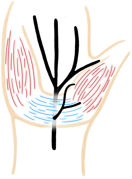
A 72-year-old female with a history of well-controlled type 2 diabetes presented to clinic with right hand parasthesias consistent with carpal tunnel syndrome and right ring trigger finger. The diagnosis was supported on EMG/NCS, and she ultimately underwent right open carpal tunnel release and ring finger trigger release. During the open carpal tunnel release, an incision was made over the ulnar aspect of the carpal tunnel in the palm and the dissection was carried to the palmar aponeurosis. A palmaris brevis muscle was notable during the dissection, and after incising the palmar fascia, the transverse carpal ligament (TCL) was exposed. Care was taken to look for an aberrant nerve branch due to the presence of the palmaris brevis muscle. The recurrent motor branch was identified as a transligamentous variant, exiting through the proximal aspect of the transverse carpal ligament, and dividing immediately into a proximal and distal branch that coursed toward the thenar muscles (Figure 1). The recurrent motor branch was protected as the ligament was released. The patient did well postoperatively with resolution of her parasthesias and no complications.
Figure 1.

Median nerve diagram.
Discussion
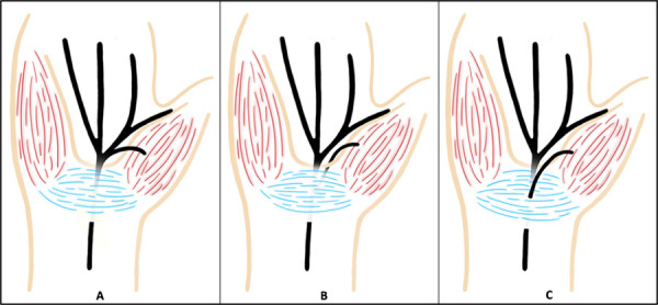
Most studies utilize the Poisel classification to describe the course of the recurrent motor branch: extraligamentous (type 1), subligamentous (type 2), and transligamentous (type 3) (Figure 2). In the extraligamentous type, the recurrent motor branch divides from the median nerve distal to the TCL and takes a retrograde course to the thenar muscles. In the subligamentous type, the recurrent motor branch divides within the carpal tunnel and remains deep to the TCL as it courses distally to the thenar muscles. In the transligamentous variant, the recurrent motor branch takes off within the carpal tunnel and pierces through the TCL on its course to the thenar muscles.3 A supraligamentous course and a preligamentous course have also been described in the literature.2,5,6 In the supraligamentous course, the recurrent motor branch exits the carpal tunnel and runs retrograde over the TCL to reach the thenar muscles.2 In the preligamentous course, the recurrent motor branch arises proximal to the TCL and takes a superficial course over the ligament to reach the thenar muscles.5,6 The preligamentous and transligamentous courses are often associated with a hypertrophic thenar muscle origin over the TCL and the branch is within this muscle6.
Figure 2.

Common branch patterns of the recurrent motor branch: (A) extraligamentous, (B) subligamentous, (C) transligamentous.
The recurrent motor branch typically arises from the radial or palmar aspect of the median nerve, though some studies have noted an ulnar-directed branch pattern.5,7,8 The current literature supports that the extraligamentous type is the most common and the ulnar origin of the thenar motor branch is the least common.1,8,10
There exist several case reports in the literature of anatomic variants. Linburg et al noted a proximal transligamentous division with a separate accessory recurrent branch at the distal aspect of the TCL.11 Graham et al encountered a recurrent motor branch that exited the ulnar aspect of the median nerve within the carpal tunnel, crossed the median nerve volarly, and pierced the TCL to enter the thenar muscles.12 Other case reports describe a patient with 3 distinct recurrent motor branches, a motor branch that divided proximal to the TCL in the forearm and traveled through the carpal tunnel, and a motor branch that began proximally and ran with a persistent median artery to separate into 3 transligamentous divisions.13,15 This case presents a patient with a transligamentous recurrent motor branch that divided from the median nerve within the carpal tunnel, pierced the proximal aspect of the TCL, and immediately divided into 2 branches that ran across the ligament and entered the thenar musculature proximally and distally at 2 separate locations. This variant has not been described in the literature but again stresses the importance of direct visualization of the transverse carpal ligament during release to avoid inadvertent injury to the recurrent motor branch. It also reinforces the need to take extra time and care to look for an aberrant nerve variant with the presence of a palmaris brevis muscle.
Acknowledgments
Ethics: All persons involved in this case report provided informed consent. The research was conducted in accordance with ethical standards of national and international committees on human experimentation.
Disclosures: The authors disclose no relevant financial or nonfinancial interests.
References
- 1.Henry BM, Zwinczewska H, Roy J, et al. The prevalence of anatomical variations of the median nerve in the carpal tunnel: A systematic review and meta-analysis [published correction appears in PLoS One. 2015 Sep 11;10(9):e0138300. doi: 10.1371/journal.pone.0138300]. PLoS One. 2015;10(8):e0136477. Published 2015 Aug 25. doi:10.1371/journal.pone.0136477 10.1371/journal.pone.0136477 [DOI] [PMC free article] [PubMed] [Google Scholar]
- 2.Lanz U. Anatomical variations of the median nerve in the carpal tunnel. J Hand Surg Am. 1977;2(1):44-53. doi:10.1016/s0363-5023(77)80009-9 10.1016/S0363-5023(77)80009-9 [DOI] [PubMed] [Google Scholar]
- 3.Poisel S. Ursprung und Verlauf des R. muscularis des Nervus digitalis palmaris communis I (N. medianus), Chir Praxis. 1974;18:471-474. [Google Scholar]
- 4.Krishnan P, Mishra R, Jena M, Das A. Transligamentous thenar branch of the median nerve: the million dollar nerve. Neurol India. 2013;61(3):311-312. doi:10.4103/0028-3886.115078 10.4103/0028-3886.115078 [DOI] [PubMed] [Google Scholar]
- 5.Ahn DS, Yoon ES, Koo SH, Park SH. A prospective study of the anatomic variations of the median nerve in the carpal tunnel in Asians. Ann Plast Surg. 2000;44(3):282-287. doi:10.1097/00000637-200044030-00006 10.1097/00000637-200044030-00006 [DOI] [PubMed] [Google Scholar]
- 6.Al-Qattan MM. Variations in the course of the thenar motor branch of the median nerve and their relationship to the hypertrophic muscle overlying the transverse carpal ligament. J Hand Surg Am. 2010;35(11):1820-1824. doi:10.1016/j.jhsa.2010.08.011 10.1016/j.jhsa.2010.08.011 [DOI] [PubMed] [Google Scholar]
- 7.Hurwitz PJ. Variations in the course of the thenar motor branch of the median nerve. J Hand Surg Br. 1996;21(3):344-346. doi:10.1016/s0266-7681(05)80198-6 10.1016/S0266-7681(05)80198-6 [DOI] [PubMed] [Google Scholar]
- 8.Mizia E, Tomaszewski KA, Goncerz G, Kurzydło W, Walocha J. Median nerve thenar motor branch anatomical variations. Folia Morphol (Warsz). 2012;71(3):183-186. [PubMed] [Google Scholar]
- 9.Kozin SH. The anatomy of the recurrent branch of the median nerve. J Hand Surg Am. 1998;23(5):852-858. doi:10.1016/S0363-5023(98)80162-7 10.1016/S0363-5023(98)80162-7 [DOI] [PubMed] [Google Scholar]
- 10.Biswas Kanta K, Baishya RJ, Talukdar K. A study on variations of branching patterns of median nerve in the carpal tunnel. Nat J Clin Anat. 2017. Jul-Sep;6(3):193-197. 10.1055/s-0039-1700746 [DOI] [Google Scholar]
- 11.Linburg RM, Albright JA. An anomalous branch of the median nerve. A case report. J Bone Joint Surg Am. 1970;52:182-183. 10.2106/00004623-197052010-00022 [DOI] [PubMed] [Google Scholar]
- 12.Graham WP 3rd. Variations of the motor branch of the median nerve at the wrist. Case report. Plast Reconstr Surg. 1973;51(1):90-92. doi:10.1097/00006534-197301000-00025 10.1097/00006534-197301000-00025 [DOI] [PubMed] [Google Scholar]
- 13.Rockwell WB, Stone B, Zakhireh M. Three thenar motor branches of the median nerve. Ann Plast Surg. 2001;46(6):661-662. doi:10.1097/00000637-200106000-00025 10.1097/00000637-200106000-00025 [DOI] [PubMed] [Google Scholar]
- 14.Kuvat SV, Ozçakar L, Yazar M. A foregoing thenar muscular branch of the median nerve. Indian J Plast Surg. 2010;43(1):106-107. doi:10.4103/0970-0358.63963 10.4103/0970-0358.63963 [DOI] [PMC free article] [PubMed] [Google Scholar]
- 15.Kornberg M, Aulicino PL, DuPuy TE. Bifid median nerve with three thenar branches–case report. J Hand Surg Am. 1983;8(5 Pt 1):583-584. doi:10.1016/s0363-5023(83)80131-2 10.1016/S0363-5023(83)80131-2 [DOI] [PubMed] [Google Scholar]


