Abstract
Juvenile myelomonocytic leukemia (JMML) is an aggressive pediatric myeloproliferative neoplasm requiring hematopoietic stem cell transplantation (HSCT) in most cases. We retrospectively analyzed 119 JMML patients who underwent first allogeneic HSCT between 2002 and 2021. The majority (97%) carried a RAS-pathway mutation, and 62% exhibited karyotypic alterations or additional mutations in SETBP1, ASXL1, JAK3 and/or the RAS pathway. Relapse was the primary cause of death, with a 5-year cumulative incidence of 24.6% (95% CI: 17.1-32.9). Toxic deaths occurred in 12 patients, resulting in treatment-related mortality (TRM) of 9.0% (95% CI: 4.6-15.3). The 5-year overall (OS) and event-free survival were 73.6% (95% CI: 65.7-82.4) and 66.4% (95% CI: 58.2-75.8), respectively. Four independent adverse prognostic factors for OS were identified: age at diagnosis >2 years, time from diagnosis to HSCT ≥6 months, monocyte count at diagnosis >7.2x10⁹/L, and the presence of additional genetic alterations. Based on these factors, we proposed a predictive classifier. Patients with 3 or more predictors (21% of the cohort) had a 5-year OS of 34.2%, whereas those with none (7%) had a 5-year OS of 100%. Our study demonstrates improved transplant outcomes compared to prior published data, which can be attributed to the synergistic impacts of a low TRM and a reduced, yet still substantial, relapse incidence. By integrating genetic information with clinical and hematologic features, we have devised a predictive classifier. This classifier effectively identifies a subgroup of patients who are at a heightened risk of unfavorable post-transplant outcomes who would benefit from novel therapeutic agents and post-transplant strategies.
Introduction
Juvenile myelomonocytic leukemia (JMML) is an aggressive hematologic malignancy of early childhood. It results from the hyperactivation of the RAS signal transduction pathway mainly caused by mutations of PTPN11, KRAS, NRAS, RRAS, RRAS2, CBL, SH2B3 and NF1.1-3 Except for a small subset of patients who exhibit spontaneous remissions, the disease usually progresses and leads to death within months of diagnosis. Unlike acute leukemia, intensive chemotherapy is insufficient to eradicate the disease and hematopoietic stem cell transplantation (HSCT) is the only curative treatment for most patients.4,5 However, the rarity of the disease has resulted in only a few large cohorts of HSCT being reported over the last 20 years.6-10 These studies showed that JMML is associated with a poor outcome, with a 5-year overall survival (OS) ranging from 52% to 72% due to high-risk post-transplant relapse combined with high treatment-related mortality (TRM). Predictive variables of poor outcome identified across these trials encompass age at diagnosis and at transplant, abnormal karyotype and HLA disparities. Previously published studies including non-transplanted patients also identified other predictive factors of death and relapse, such as low platelet count and high fetal hemoglobin level (HbF).11,12 Since then, progress has been made in deciphering the genomic landscape of JMML, contributing to a deeper understanding of the marked heterogeneity that characterizes this disease.13,14 Indeed, certain initiating mutations, such as NF1 or PTPN11, have been shown to correlate with disease aggressiveness. Furthermore, the occurrence of additional genetic mutations, including double RAS mutations, ASXL1, SETBP1 or JAK3, while uncommon in JMML, further worsens the overall prognosis of patients.13-16
However, it is worth noting that most previously published studies on HSCT in JMML have provided either incomplete or no genetic information. Consequently, the recent molecular insights associated with aggressive disease have not been challenged within a cohort of transplanted JMML patients. In this study, we report the outcome of 119 children with JMML who underwent HSCT over the past 20 years and were genetically characterized. We evaluated the impact of previously described parameters, as well as the role of the initiating RAS mutation and additional ASXL1, SETBP1 and JAK3 mutations on prognosis.
Methods
Patients and data collection
This study investigated 119 consecutive children diagnosed with JMML who received a first allogeneic HSCT between June 2002 and August 2021 in France (Table 1). All patients met the World Health Organization (WHO) consensus criteria for JMML.2 Patients’ data were collected retrospectively using the PROMISE database of the European Bone Marrow Transplant group (EBMT) through the Société Francophone de Greffe de Moelle et de Thérapie Cellulaire (SFGM-TC). JMML patient samples, bone marrow (BM) and/or peripheral blood (PB), were collected on EDTA at diagnosis. Genomic DNA was extracted from mononucleated cells. Mutational screening using bi-directional Sanger and/or next generation sequencing (NGS) of exons and their flanking intron-exon boundaries was performed on genomic DNA as part of the classic diagnostic workup for JMML, and included NRAS, KRAS, PTPN11, CBL, NF1, SH2B3, RRAS, RRAS2, ASXL1, SETBP1 and JAK3, as previously described.13,17 Written informed consent for the study was provided by the patients or their guardians in accordance with the Declaration of Helsinki (IRB: 00006477).
Definitions and endpoints
Elevated HbF levels at the time of JMML diagnosis were defined as follows: ≥10% for children aged ≥6 to <12 months, and ≥1% for children aged ≥12 months. HbF levels were considered not interpretable for children under six months old. Relapse was defined as the recurrence of JMML, clinically and morphologically on PB and/or BM analysis, after HSCT. HLA matching and engraftment definitions are provided in the Online Supplementary Methods. Acute and chronic graft-versus-host disease (GvHD) were diagnosed and graded by each transplantation center according to conventional criteria.18,19 Treatment of GvHD was based on the protocols used in each center.
Treatment-related mortality was defined as any death occurring from any cause but disease relapse. One patient died of Pneumocystis jirovecii lung infection after JMML relapse while waiting for a second transplant. We considered the death of this patient as related to the HSCT. Event-free survival (EFS) was defined as a composite outcome, including relapse and death, whichever occurred first. In exploratory analyses, we also considered secondary allograft and secondary malignancy as additional events defining a ‘stringent EFS’.
Statistical analysis
Time-to-event outcomes were measured from the date of transplant to the date of event or date of last follow-up, with a cut-off date of December 30, 2021. TRM and relapse were considered as mutually competing risk events, while death was considered a competing risk for engraftment and GvHD. Engraftment and acute GvHD were arbitrarily censored at 100 days.
The OS and EFS were estimated using Kaplan-Meier estimator. For competing risk analyses of TRM, relapse and GvHD, cumulative incidence functions were estimated.20 Factors associated with outcomes were analyzed using the Fine and Gray model for GvHD, proportional hazards models for the cause-specific hazard for relapse and TRM, and Cox proportional hazards models for EFS and OS.21,22
The proportional hazards assumption was checked by examination of Schoenfeld residuals and Grambsch and Therneau lack-of-fit test.23
All tests were two-sided. P≤0.05 was considered statistically significant. The analyses were performed using the R statistical software version 4.1.1.
Results
Patients’ characteristics at juvenile myelomonocytic leukemia diagnosis and pre-transplant treatment
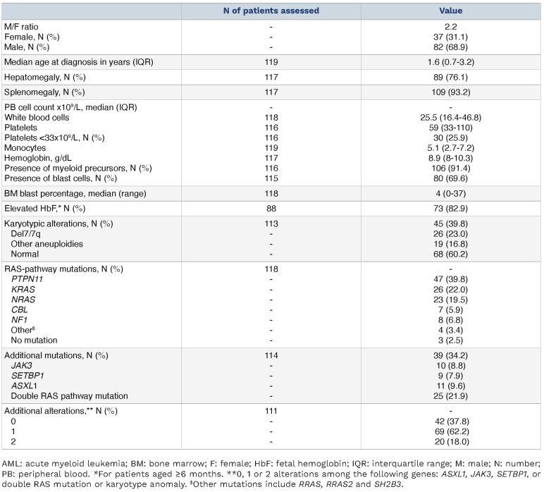
Table 1 provides a summary of patients’ characteristics (N=119). At diagnosis, 64 patients (54%) were <2 years of age. JMML occurred in the setting of a germline predisposing condition in 16 patients (14%): type 1 neurofibromatosis (N=8), Casitas B-lineage lymphoma (CBL) syndrome (N=7), and SH2B3 germline biallelic mutation (N=1). A RAS-pathway mutation was observed in 115 out of 118 patients (97%). The most commonly mutated gene was PTPN11, observed in 40% of patients, followed by KRAS (22%), NRAS (19%), NF1 (7%), CBL (6%), and other less frequent mutations (3%; including RRAS, RRAS2 and SH2B3).
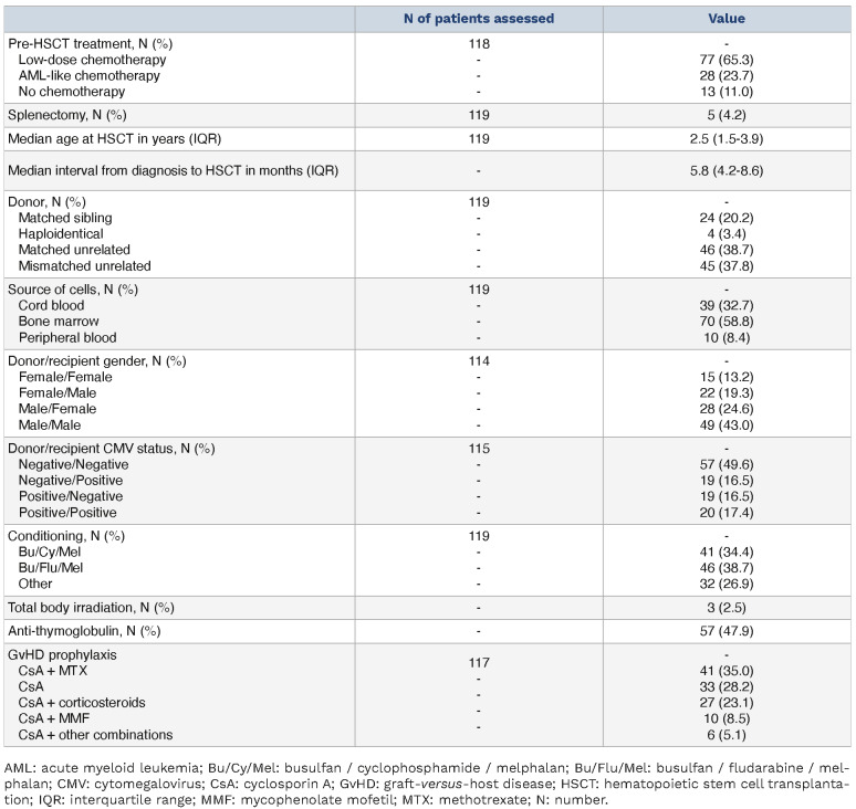
The time from diagnosis to HSCT varied among patients, ranging from 1.8 to 44.3 months, but remained consistent throughout the study period and across the genetic sub-groups (Table 2, Online Supplementary Figure S1). Prior to conditioning regimen and transplant, patients received either no treatment (N=13, 11%), low-dose chemotherapy (N=77, 65%; including 6-mercaptopurine, azacytidine [N=8] and low-dose cytarabine), or acute myeloid leukemia-type (AML-type) chemotherapy (N=28, 23.7%) at the physician’s discretion (Table 2). The pre-HSCT strategies were uniformly distributed among the NF1, PTPN11, KRAS, and NRAS groups, while CBL patients exclusively received low-intensity treatment and those without mutation, only intensive chemotherapy (Online Supplementary Figure S1).
Table 1.
Patients’ characteristics at diagnosis.
Transplant, engraftment and graft-versus-host disease occurrence
Transplant characteristics are described in Table 2. All patients underwent myeloablative conditioning regimens, with the majority receiving busulfan / fludarabine / melphalan (Bu/Flu/Mel, N=46) or busulfan / cyclophosphamide / melphalan (Bu/Cy/Mel, N=41) (Table 2, Online Supplementary Table S1). The Bu/Cy/Mel and Bu/Flu/Mel conditioning regimens were administered at the median year of 2007 (range, 2002-2019) and 2016 (range, 2010-2021), respectively. GvHD prophylaxis according to donor type is provided in Online Supplementary Table S2.
Table 2.
Pre-transplant treatment and transplant characteristics.
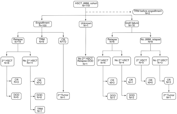
Of the 116 patients assessable for engraftment, 100 showed sustained engraftment (Figure 1). The median time to neutrophil recovery was 23 days (range, 12-56), and the median time to a self-sustained platelet count higher than 50x109/L was 43 days (range, 14-160). Sixteen patients experienced either primary (N=9) or secondary (N=7) graft failure after a median time of four months (range, 2-10). Complete loss of chimerism was concomitant with relapse in 4/16 patients (1 patient with primary and 3 patients with secondary graft failure) (Online Supplementary Figure S2). Patients who encountered graft failure exhibited a higher prevalence of CBL mutations, HLA disparities, cord blood source, and alternative conditioning regimens (other than Bu/Cy/Mel or Bu/Flu/Mel) compared to the rest of the cohort (Online Supplementary Table S3).
Acute GvHD grade 2-4 was observed in 63 patients (100-day cumulative incidence 53.8%, 95% CI: 44.4-62.3) and acute GvHD grade 3-4 in 38 patients (100-day cumulative incidence 31.9%, 95% CI: 23.7-40.4) (Online Supplementary Figure S3). Univariate analyses identified cytomegalovirus (CMV) status (donor positive / recipient negative) and the absence of elevated HbF as risk factors for developing grade 2-4 acute GvHD, and the NRAS mutation as a risk factor for grade 3-4 acute GvHD (Online Supplementary Table S4). Chronic GvHD was observed in 42 patients (with a 36-month cumulative incidence of 36.0%, 95% CI: 27.2-44.9) (Online Supplementary Figure S3). Twenty-four had extensive disease and 17 had limited disease (unknown for 1). Only the presence of a mismatched relative HLA status of the donor was significantly associated with the onset of chronic GvHD while having NF1, KRAS, and no/ other mutation appeared to be protective factors (Online Supplementary Table S4). The occurrence of acute GvHD grade 3-4 did not significantly impact the 5-year EFS, which was 69.0% (95% CI: 56.8-84.0) with acute GvHD, compared to 65.4% without (95% CI: 54.5-78.6) (P=0.72). In contrast, the occurrence of chronic GvHD led to a reduction in the incidence of relapse or death, although not statistically significant, with a 5-year EFS of 78.8% (95% CI: 64.7-96.1) with chronic GvHD versus 68.3% without (95% CI: 59.5-78.4) (P=0.09) (Online Supplementary Figure S3).
Patient outcome
The median follow-up after transplant was 59.5 months (IQR: 21.7-118.6). The 5-year OS was 73.6% (95% CI: 65.7-82.4), and the 10-year OS 72.2% (95% CI: 64.1-81.4) (Figure 2). Twenty-eight patients relapsed after a median time of 4.6 months (range: 0.5-43.6) from HSCT, resulting in a 5-year cumulative incidence of relapse (CIR) of 24.6% (95% CI: 17.1-32.8). Twelve of them (43%) received a second allogeneic transplant, including 6 with the same donor. Six patients remained disease-free over a prolonged post-transplant follow up (median 7 years, range 5-17) while 6 patients relapsed once again (Figure 1). Five died of disease; none from TRM. Time to relapse, time to second transplant, type of donor and conditioning regimen did not differ between the 2 groups. Of note, 4/6 who did not relapse developed grade 2-4 acute GvHD while it occurred in 1/6 patients in the relapse group. Among the 16 patients who relapsed but did not receive a 2nd transplant, 3 are still alive at three, eight and nine years after HSCT. Patient #192, carrying a PTPN11 mutation, received 7 cycles of azacitidine. Patient #44, without any identified mutation, received weekly etoposide injections for three months, followed by rapamycine until the 6th-month post HSCT. Finally, patient #55, with a CBL mutation, achieved CR within a few months of a course of 6-mercaptopurine.
Figure 1.
Flow chart of the transplanted juvenile myelomonocytic leukemia cohort. CR: complete remission; DOD: dead of disease; HSCT: hematopoietic stem cell transplant; JMML: juvenile myelomonocytic leukemia; N: number; TRM: treatment-related mortality.
Twelve patients died from transplant toxicity with a median time of 2.9 months (range, 23 days-67.2 months) resulting in an estimated TRM of 9.0% (95% CI: 4.6-15.3). Toxic causes of deaths included severe GvHD + associated with disseminated viral or bacterial infections (N=5), infections (N=4), acute hepatitis of unknown origin (N=1), sinusoidal obstruction syndrome (SOS) (N=1), and thrombotic micro-angiopathy (N=1). SOS was observed in 32 patients (26.9%). Of the 16 patients who did not engraft, 9 underwent a second transplant within a year, in the absence of relapse (N=5) or after JMML relapse (N=4) (Figure 1, Online Supplementary Figure S2). Among the 7 patients who did not receive a subsequent transplant, 3 died of disease recurrence. The remaining 4 patients, comprising one CBL patient and 3 KRAS patients, maintained sustained JMML remission with autologous reconstitution. None had undergone splenectomy before HSCT. The KRAS patients have been followed up for five, eight, and ten years.
Two patients developed secondary malignancies. One patient with a KRAS mutation, who experienced graft failure, developed T-cell precursor acute lymphoblastic leukemia (ALL) seven years post HSCT while still in autologous remission. Remarkably, the same KRAS p.Gly13Cys mutation detected in the patient’s JMML cells was also identified in the acute lymphoblastic leukemia (ALL) blast cells. Another patient, who carried a PTPN11 mutation, developed AML with minimal differentiation four years after transplant. The PTPN11 p.Ala72Val mutation initially detected in JMML was also found in the AML blasts.
Overall, 39 patients experienced an event, resulting in a 5-year EFS at 66.4% (95% CI: 58.2-75.8), and a 10-year EFS at 65.0% (95% CI: 56.5-74.7) (Figure 2). Also considering second transplants for graft failure without relapse and secondary malignancies as events, the 5-year ‘stringent EFS’ was estimated at 63.6% (95% CI: 55.3-73.3) (Online Supplementary Figure S4).
Prognostic factors for overall survival, event-free survival, relapse and treatment-related mortality
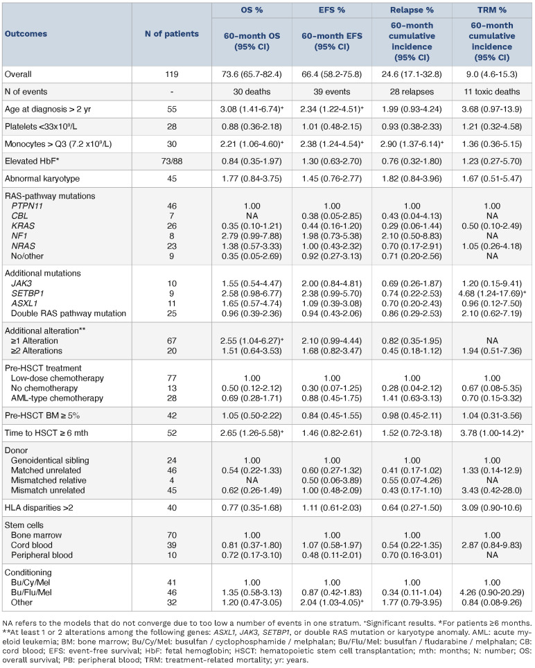
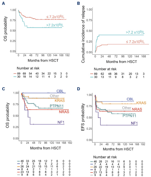
Table 3 presents the univariate analysis of the patients’ characteristics that influence the outcomes. Age at diagnosis or age at transplant >2 years, as well as a monocyte count at diagnosis >3rd quartile (>7.2x10⁹/L), were associated with a lower rate of OS, EFS, and ‘stringent EFS’ (Table 3, Figure 3, Online Supplementary Table S5). The negative effect of monocyte count on survival was linked to a higher incidence of relapse (Figure 3). Monocytes >7.2x10⁹/L were associated with higher white blood cell, neutrophil, and lymphocyte counts, as well as a higher BM blast percentage but were not correlated with platelet count, HbF levels, cytogenetic features, or molecular features (Online Supplementary Table S6). Although patients with NF1 mutations tended to show worse outcomes, no significant association was found between RAS initiating variants and OS, EFS, or cumulative incidence of relapse (CIR) (Figure 3). Considered individually, abnormal karyotype, pathogenic variants of SETBP1, ASXL1, JAK3 or additional RAS mutation had no impact on outcome. However, when analyzed collectively, the presence of any of them had a negative impact on OS (Table 3). Conditioning regimens other than Bu/Cy/Mel or Bu/Flu/Mel were associated with lower EFS. Finally, time to HSCT >6 months had a negative impact on OS related to a higher TRM.
Figure 2.
Estimated outcomes of the 119 transplanted patients. Overall survival (OS) (A) and event-free survival (EFS) (B) were analyzed using Kaplan-Meier methodology. Cumulative incidence function for relapse and treatment-related mortality (TRM) (C).
Table 3.
Univariable predictive analyses of the outcomes based on Cox models.
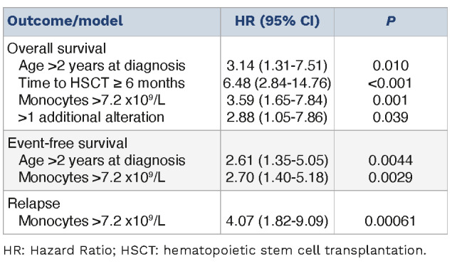
The multivariable model confirmed the prognostic impact of monocyte count on OS, EFS and relapse. Age at diagnosis remained significant for OS and EFS. Additionally, time to HSCT and any additional alteration were found significant for OS (Table 4).
Finally, we derived a prognostic classifier based on the 4 predictors of death in the multivariate analysis (age at diagnosis, time to transplant, monocyte count and any additional alteration). This classifier defined prognostic groups of patients with 5-year OS ranging from 34.2% for patients with at least 3 predictors (N=23, 20.7%) to 100% for patients with none of the four predictors (N=8, 7%) (Figure 4).
Discussion
We present here a comprehensive analysis of the outcome of a large cohort of transplanted JMML patients. All patients met the JMML diagnostic criteria, recently revised in the WHO classification, including genetic characterization, which enables us to formally establish the JMML diagnosis.24,25
With a 5-year EFS of 66% and OS of 74%, our results compare favorably with previous published studies5,7-10,26-30 (Online Supplementary Table S7). The improvement in OS among JMML patients over time has been remarkable, with survival rates increasing from 31% in the 1990s to 72% in the most recent reports.31 While the results have plateaued in recent years, it is important to consider that the composition of the transplanted cohorts has changed over time, gradually leading to the exclusion of patients with the most favorable prognosis. Indeed, it has been demonstrated that patients with CBL mutations experience a naturally favorable evolution and may no longer require HSCT, unless they demonstrate an aggressive clinical course or a high mutational burden.32 Additionally, a smaller subset of NRAS patients with non-high-risk features can also avoid HSCT. Despite encompassing a time span of over 20 years, the incidence of CBL mutations in our transplanted series (6%) is lower than the typical expectation for JMML at diagnosis. This difference probably reflects the implementation of the ‘watch-and-wait’ recommendations for these patients. As the proportion of transplanted patients with more severe conditions increased over time, it is reasonable to infer that the observed improvement in OS and EFS in our study are significant.
Figure 3.
Effect of monocyte count and initiating RAS-mutation on outcomes. Effect of monocyte count: (A) overall survival (OS) and (B) cumulative incidence function of relapse. Effect of initiating RAS-mutation: (C) OS and (D) event-free survival (EFS). OS and EFS were analyzed using Kaplan-Meier methodology. HSCT: hematopoietic stem cell transplant.
In our cohort, the survival improvement can be attributed to a reduction in TRM, coupled with a decrease in relapse incidence, which nevertheless still accounts for approximately one-quarter of patient deaths and remains the leading cause of mortality. Indeed, the relapse incidence in our cohort was estimated at 25%, which is lower compared to most reported series where it often surpassed 30% (Online Supplementary Table S7). Previous studies have demonstrated the critical role of both acute and chronic GvHD in preventing disease recurrence.8,9,28,33 Consistent with these findings, we noted a significant association between chronic GvHD and improved CIR, which can be attributed to the potent graft-versus-leukemia (GvL) effect. The high incidence of GvHD in our cohort may be related to the frequent utilization of unrelated donors and could also reflect physicians’ endeavors to enhance alloreactivity in patients known to be sensitive to GvL through the accelerated tapering of immunosuppressors, although these data were not captured. The impact of GvHD on disease control following a 2nd transplant is suggested in our cohort. However, its translation into OS could be hindered by the TRM associated with extensive GvHD, as reported in a recent series.34 The TRM in our cohort was evaluated at 9%, which is below the levels observed in earlier studies, where reported TRM exceeded 10% (Online Supplementary Table S7). This improvement may be attributed to the overall advancement in supportive care in transplantation over time, the limited use of total body irradiation in our cohort and the low frequency of HLA mismatched donors. Our analysis identified that an extended duration between diagnosis and transplant has a negative impact on TRM and OS. This variable encompasses several parameters that could contribute to an increased TRM, including iterative transfusions, compromised nutritional status, and infections. The specific factors influencing the decision of the transplant date have not been identified within our cohort. These factors could encompass organizational constraints, including graft availability, or disease-related considerations, such as the aggressive nature of JMML requiring multiple courses of chemotherapy. However, within our patient series, the median time from diagnosis to HSCT remained consistent, both for patients requiring AML-type chemotherapy and those who had no or low-dose chemotherapy. This finding suggests that transplantation was sometimes planned with a considerable long delay, even for patients with non-aggressive diseases. Given the impact of this delay on TRM, our results suggest the prompt scheduling of the transplant once the diagnosis of JMML is confirmed. The association between SETBP1 and TRM is more difficult to understand and may be biased by the limited size of this group. The Japanese group adopted the Bu/Flu/Mel regimen in JMML with promising results, aiming to mitigate the toxicity associated with the triple alkylation of the Bu/ Cy/Mel regimen.10,35 Although there is no evidence favoring one of these two conditioning regimens over the other in the literature, it is crucial to recognize the pivotal role of their intensity. Notably, in the only randomized study focusing on JMML conditioning, attempts to reduce the regimen’s intensity were unsuccessful, as Bu/Flu resulted in a significantly higher relapse rate when compared to Bu/Cy/Mel.28 In our study, both Bu/Flu/Mel and Bu/Cy/Mel conditionings led to similar EFS and OS, while other types of conditioning impaired EFS.
Figure 4.
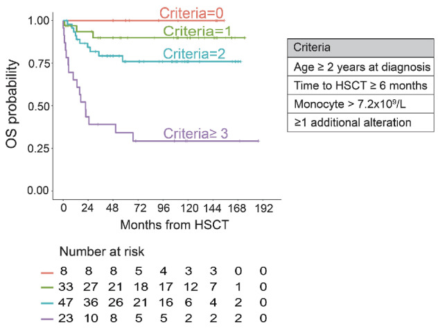
Prognostic classifier for overall survival. This classifier defined 4 prognostic groups of patients according to the 4 predictor factors from the multivariate analysis. OS: overall survival; HSCT: hematopoietic stem cell transplant.
Table 4.
Multivariate analyses for overall survival, event-free survival and relapse.
Five patients with autologous reconstitution survived. In CBL patients, as in NRAS patients, this outcome is expected as many of them show a spontaneously favorable evolution without HSCT. On the contrary, patients with KRAS mutations typically have an aggressive disease course and require HSCT to be cured. Nevertheless, case reports have described prolonged remissions in some KRAS patients treated with azacitidine or without any treatment at all.36-38 It has also been reported that patients with Ras-associated autoimmune leukoproliferative disorder (RALD), carrying the same somatic KRAS mutations as in JMML, can present an indolent condition difficult to distinguish from JMML.39 In our cohort, the 3 KRAS patients who survived with autologous reconstitution had no clinical autoimmune manifestations or immunophenotypic abnormalities. While the conditioning regimen and the transient presence of allogeneic stem cells could have contributed to disease control, our findings are consistent with observations in RALD and indolent KRAS JMML cases, suggesting that KRAS-mutated patients might encompass a spectrum of conditions ranging from mild to highly aggressive diseases. Exploring this matter further remains essential to refine treatment strategies and accurately differentiate patients who truly require transplantation from those who could potentially avoid it.40
Age at diagnosis over one, two or four years and time between diagnosis and HSCT over six months, which have been consistently described by independent study groups to influence the outcome of transplanted JMML patients, were also identified in our cohort to alter survival in multivariate analysis (Online Supplementary Table S7). Conversely, other classical prognostic factors such as platelet counts <33x109/L, elevated HbF for age, elevated BM blast percentage and abnormal karyotype were not found to influence outcome. Some of these prognostic markers have been demonstrated to be closely related to the genetic background of the disease, PTPN11 or NF1-JMML tending to harbor significantly lower platelet count, higher HbF or more frequent karyotypic alteration than CBL or NRAS-JMML.41 As previously discussed, when considering the comparability of studies conducted over time, the prognostic impact of some markers may have been lost in this selected aggressive group of JMML patients. Additionally, it is plausible that the relatively limited number of patients included in our study may have resulted in insufficient statistical power to detect their influence on survival. The role of pre-transplant chemotherapy in survival for patients with JMML remains controversial. In line with previous studies, we did not find any difference in EFS, or relapse incidence based on the chemotherapy regimen received before transplant.6,8 This finding reaffirms that patients with a clinical condition compatible with low-intensity treatment do not require intensive chemotherapy. Due to the infrequent use of azacitidine in our cohort, we were unable to compare the outcomes of this subgroup with those of patients who received 6-mercaptopurine.
Among our 8 patients who received azacitidine, 3 relapsed after transplant and 4 died consequently to relapse or TRM (N=1). The European Working Group (EWOG) recently published the prospective AZA-JMML-001 trial, investigating the impact of pre-transplant azacitidine in 18 patients. After 3 cycles, 61% of patients showed a partial response, and 14 achieved complete remission (CR) after HSCT during a 2-year follow-up.42 The recent wider use of azacitidine will allow us to determine on a larger scale whether this approach indeed yields a positive impact on post-transplant outcomes. In JMML, secondary genetic alterations including karyotype anomalies and additional mutations of the RAS pathway or SETBP1, ASXL1 and JAK3 have been demonstrated to be associated with an unfavorable prognosis. This has been highlighted by studies conducted by our group and others in unbiased cohorts of JMML patients, including transplanted and non-transplanted ones.13,14 In this study, we confirm the relevance of these secondary alterations in a selected population of transplanted patients and found that the number of genetic alterations, rather than the type of the alteration, was the main determinant factor for OS. However, secondary alterations did not impact the incidence of relapse. Unlike secondary alterations, the initiating RAS-pathway alterations were not associated with the outcome in our study, possibly due to insufficient statistical power. Indeed, CBL patients exhibited an excellent prognosis while NF1 patients had the worst survival rates due to a high incidence of relapse, in line with results from previous studies.13,14 In recent years, DNA methylation profiling has emerged as a novel prognostic marker in JMML, as demonstrated in 3 distinct patient cohorts.41,43,44 This methylation profile has shown associations with disease biology and clinical outcomes. Methylation has not been investigated in the current cohort, but it would be intriguing to explore its relevance as a prognostic marker in a cohort of transplanted JMML patients, predominantly comprising patients with high-risk features.
In addition, our study shows that monocyte count >7.2x10⁹/L was an independent factor of adverse outcomes and relapse. To our knowledge, this is the first time that monocyte count has been identified as a prognostic factor for JMML, although this variable has not often been evaluated in past series. Monocytosis in PB is the hallmark of JMML and is a mandatory criterion to ascertain the diagnosis. Elevated monocyte count has been linked to disease aggressiveness and worse survival in adult myelodysplastic syndromes, myeloproliferative neoplasms, and chronic myelomonocytic leukemia (CMML).45-48 In CMML, the closest adult counterpart of JMML, different subsets of monocytes, as well as their levels, may play a role in the outcome of patients, with a specific inflammatory fraction being associated with a poor prognosis.49-51 The distribution of the different monocyte fractions in JMML has not yet been thoroughly explored, and, therefore, we could not correlate our findings with a comprehensive immunophenotypic and functional analysis. It would be valuable to investigate this in the future to determine if the adverse outcomes are linked to a specific subset.
Overall, this study underscores promising survival outcome for transplanted JMML patients that can primarily be attributed to the remarkably low TRM. However, it is important to note the persistent high relapse rate, which reflects the limited availability of novel and effective anti-tumoral agents. We confirmed, on a genetically tested cohort, the significant impact of additional alterations on prognosis and found that elevated monocyte count is independently correlated with poor outcome. The prognostic classifier we developed identifies transplanted patients who are most susceptible to relapse and who could benefit from post-transplant interventions. This personalized approach holds promise for improving the outcomes and long-term survival of high-risk JMML patients after transplantation.
Supplementary Material
Data-sharing statement
The data that support the findings of this study are available on request from the corresponding author.
References
- 1.Arfeuille C, Vial Y, Cadenet M, et al. Germline bi-allelic SH2B3/ LNK alteration predisposes to a neonatal juvenile myelomonocytic leukemia-like disorder. Haematologica. 2023. Nov 16. doi: 10.3324/haematol.2023.283917. [Epub ahead of print]. [DOI] [PMC free article] [PubMed] [Google Scholar]
- 2.Khoury JD, Solary E, Abla O, et al. The 5th edition of the World Health Organization Classification of Haematolymphoid Tumours: Myeloid and Histiocytic/Dendritic Neoplasms. Leukemia. 2022;36(7):1703-1719. [DOI] [PMC free article] [PubMed] [Google Scholar]
- 3.Niemeyer CM, Flotho C. Juvenile myelomonocytic leukemia: who’s the driver at the wheel? Blood. 2019;133(10):1060-1070. [DOI] [PubMed] [Google Scholar]
- 4.Loh ML. Recent advances in the pathogenesis and treatment of juvenile myelomonocytic leukaemia. Br J Haematol. 2011;152(6):677-687. [DOI] [PubMed] [Google Scholar]
- 5.De Vos N, Hofmans M, Lammens T, De Wilde B, Van Roy N, De Moerloose B. Targeted therapy in juvenile myelomonocytic leukemia: where are we now? Pediatr Blood Cancer. 2022;69(11):e29930. [DOI] [PubMed] [Google Scholar]
- 6.Locatelli F, Nöllke P, Zecca M, et al. Hematopoietic stem cell transplantation (HSCT) in children with juvenile myelomonocytic leukemia (JMML): results of the EWOG-MDS/ EBMT trial. Blood. 2005;105(1):410-419. [DOI] [PubMed] [Google Scholar]
- 7.Locatelli F, Crotta A, Ruggeri A, et al. Analysis of risk factors influencing outcomes after cord blood transplantation in children with juvenile myelomonocytic leukemia: a EUROCORD, EBMT, EWOG-MDS, CIBMTR study. Blood. 2013;122(12):2135-2141. [DOI] [PMC free article] [PubMed] [Google Scholar]
- 8.Yoshida N, Sakaguchi H, Yabe M, et al. Clinical outcomes after allogeneic hematopoietic stem cell transplantation in children with juvenile myelomonocytic leukemia: a report from the Japan Society for Hematopoietic Cell Transplantation. Biol Blood Marrow Transplant. 2020;26(5):902-910. [DOI] [PubMed] [Google Scholar]
- 9.Yi ES, Kim SK, Ju HY, et al. Allogeneic hematopoietic cell transplantation in patients with juvenile myelomonocytic leukemia in Korea: a report of the Korean Pediatric Hematology-Oncology Group. Bone Marrow Transplant. 2023;58(1):20-29. [DOI] [PubMed] [Google Scholar]
- 10.Yabe M, Ohtsuka Y, Watanabe K, et al. Transplantation for juvenile myelomonocytic leukemia: a retrospective study of 30 children treated with a regimen of busulfan, fludarabine, and melphalan. Int J Hematol. 2015;101(2):184-190. [DOI] [PubMed] [Google Scholar]
- 11.Niemeyer CM, Arico M, Basso G, et al. Chronic myelomonocytic leukemia in childhood: a retrospective analysis of 110 cases. European Working Group on Myelodysplastic Syndromes in Childhood (EWOG-MDS). Blood. 1997;89(10):3534-3543. [PubMed] [Google Scholar]
- 12.Hasle H, Baumann I, Bergsträsser E, et al. The International Prognostic Scoring System (IPSS) for childhood myelodysplastic syndrome (MDS) and juvenile myelomonocytic leukemia (JMML). Leukemia. 2004;18(12):2008-2014. [DOI] [PubMed] [Google Scholar]
- 13.Caye A, Strullu M, Guidez F, et al. Juvenile myelomonocytic leukemia displays mutations in components of the RAS pathway and the PRC2 network. Nat Genet. 2015;47(11):1334-1340. [DOI] [PubMed] [Google Scholar]
- 14.Stieglitz E, Taylor-Weiner AN, Chang TY, et al. The genomic landscape of juvenile myelomonocytic leukemia. Nat Genet. 2015;47(11):1326-1333. [DOI] [PMC free article] [PubMed] [Google Scholar]
- 15.Sakaguchi H, Okuno Y, Muramatsu H, et al. Exome sequencing identifies secondary mutations of SETBP1 and JAK3 in juvenile myelomonocytic leukemia. Nat Genet. 2013;45(8):937-941. [DOI] [PubMed] [Google Scholar]
- 16.Sugimoto Y, Muramatsu H, Makishima H, et al. Spectrum of molecular defects in juvenile myelomonocytic leukaemia includes ASXL1 mutations. Br J Haematol. 2010;150(1):83-87. [DOI] [PubMed] [Google Scholar]
- 17.Pérez B, Kosmider O, Cassinat B, et al. Genetic typing of CBL, ASXL1, RUNX1, TET2 and JAK2 in juvenile myelomonocytic leukaemia reveals a genetic profile distinct from chronic myelomonocytic leukaemia. Br J Haematol. 2010;151(5):460-468. [DOI] [PubMed] [Google Scholar]
- 18.Przepiorka D, Weisdorf D, Martin P, et al. 1994 Consensus Conference on Acute GVHD Grading. Bone Marrow Transplant. 1995;15(6):825-828. [PubMed] [Google Scholar]
- 19.Jagasia MH, Greinix HT, Arora M, et al. National Institutes of Health Consensus Development Project on Criteria for Clinical Trials in Chronic Graft-versus-Host Disease: I. The 2014 Diagnosis and Staging Working Group report. Biol Blood Marrow Transplant. 2015;21(3):389-401. [DOI] [PMC free article] [PubMed] [Google Scholar]
- 20.Prentice RL. The statistical analysis of failure time data. 2nd ed. Hoboken, NJ: J. Wiley; 2010. [Google Scholar]
- 21.Prentice RL, Kalbfleisch JD, Peterson AV, Flournoy N, Farewell VT, Breslow NE. The analysis of failure times in the presence of competing risks. Biometrics. 1978;34(4):541-554. [PubMed] [Google Scholar]
- 22.White IR, Royston P. Imputing missing covariate values for the Cox model. Stat Med. 2009;28(15):1982-1998. [DOI] [PMC free article] [PubMed] [Google Scholar]
- 23.Grambsch PM, Therneau TM. Proportional hazards tests and diagnostics based on weighted residuals. Biometrika. 1994;81(3):515-526. [Google Scholar]
- 24.Niemeyer CM. JMML genomics and decisions. Hematology Am Soc Hematol Educ Program. 2018;2018(1):307-312. [DOI] [PMC free article] [PubMed] [Google Scholar]
- 25.Yoshimi A, Kamachi Y, Imai K, et al. Wiskott-Aldrich syndrome presenting with a clinical picture mimicking juvenile myelomonocytic leukaemia. Pediatr Blood Cancer. 2013;60(5):836-841. [DOI] [PubMed] [Google Scholar]
- 26.Manabe A, Okamura J, Yumura-Yagi K, et al. Allogeneic hematopoietic stem cell transplantation for 27 children with juvenile myelomonocytic leukemia diagnosed based on the criteria of the International JMML Working Group. Leukemia. 2002;16(4):645-649. [DOI] [PubMed] [Google Scholar]
- 27.Stieglitz E, Ward AF, Gerbing RB, et al. Phase II/III trial of a pre-transplant farnesyl transferase inhibitor in juvenile myelomonocytic leukemia: a report from the Children’s Oncology Group. Pediatr Blood Cancer. 2015;62(4):629-636. [DOI] [PMC free article] [PubMed] [Google Scholar]
- 28.Dvorak CC, Satwani P, Stieglitz E, et al. Disease burden and conditioning regimens in ASCT1221, a randomized phase II trial in children with juvenile myelomonocytic leukemia: a Children’s Oncology Group study. Pediatr Blood Cancer. 2018;65(7):e27034. [DOI] [PMC free article] [PubMed] [Google Scholar]
- 29.Lin Y-C, Luo C-J, Miao Y, et al. Human leukocyte antigen disparities reduce relapse after hematopoietic stem cell transplantation in children with juvenile myelomonocytic leukemia: a single-center retrospective study from China. Pediatr Transplant. 2021;25(2):e13825. [DOI] [PubMed] [Google Scholar]
- 30.Smith FO, King R, Nelson G, et al. Unrelated donor bone marrow transplantation for children with juvenile myelomonocytic leukaemia. Br J Haematol. 2002;116(3):716-724. [DOI] [PubMed] [Google Scholar]
- 31.Locatelli F, Niemeyer C, Angelucci E, et al. Allogeneic bone marrow transplantation for chronic myelomonocytic leukemia in childhood: a report from the European Working Group on Myelodysplastic Syndrome in Childhood. J Clin Oncol. 1997;15(2):566-573. [DOI] [PubMed] [Google Scholar]
- 32.Niemeyer CM, Kang MW, Shin DH, et al. Germline CBL mutations cause developmental abnormalities and predispose to juvenile myelomonocytic leukemia. Nat Genet. 2010;42(9):794-800. [DOI] [PMC free article] [PubMed] [Google Scholar]
- 33.Hecht A, Meyer J, Chehab FF, et al. Molecular assessment of pretransplant chemotherapy in the treatment of juvenile myelomonocytic leukemia. Pediatr Blood Cancer. 2019;66(11):e27948. [DOI] [PMC free article] [PubMed] [Google Scholar]
- 34.Vinci L, Flotho C, Noellke P, et al. Second allogeneic stem cell transplantation can rescue a significant proportion of patients with JMML relapsing after first allograft. Bone Marrow Transplant. 2023;58(5):607-609. [DOI] [PMC free article] [PubMed] [Google Scholar]
- 35.Yabe M, Sako M, Yabe H, et al. A conditioning regimen of busulfan, fludarabine, and melphalan for allogeneic stem cell transplantation in children with juvenile myelomonocytic leukemia. Pediatr Transplant. 2008;12(8):862-867. [DOI] [PubMed] [Google Scholar]
- 36.Hashmi SK, Punia JN, Marcogliese AN, et al. Sustained remission with azacitidine monotherapy and an aberrant precursor B-lymphoblast population in juvenile myelomonocytic leukemia. Pediatr Blood Cancer. 2019;66(10):e27905. [DOI] [PMC free article] [PubMed] [Google Scholar]
- 37.Furlan I, Batz C, Flotho C, et al. Intriguing response to azacitidine in a patient with juvenile myelomonocytic leukemia and monosomy 7. Blood. 2009;113(12):2867-2868. [DOI] [PubMed] [Google Scholar]
- 38.Matsuda K, Shimada A, Yoshida N, et al. Spontaneous improvement of hematologic abnormalities in patients having juvenile myelomonocytic leukemia with specific RAS mutations. Blood. 2007;109(12):5477-5480. [DOI] [PubMed] [Google Scholar]
- 39.Calvo KR, Price S, Braylan RC, et al. JMML and RALD (Rasassociated autoimmune leukoproliferative disorder): common genetic etiology yet clinically distinct entities. Blood. 2015;125(18):2753-2758. [DOI] [PMC free article] [PubMed] [Google Scholar]
- 40.Mayerhofer C, Niemeyer CM, Flotho C. Current treatment of juvenile myelomonocytic leukemia. J Clin Med. 2021;10(14):3084. [DOI] [PMC free article] [PubMed] [Google Scholar]
- 41.Murakami N, Okuno Y, Yoshida K, et al. Integrated molecular profiling of juvenile myelomonocytic leukemia. Blood. 2018;131(14):1576-1586. [DOI] [PubMed] [Google Scholar]
- 42.Niemeyer CM, Flotho C, Lipka DB, et al. Response to upfront azacitidine in juvenile myelomonocytic leukemia in the AZAJMML-001 trial. Blood Adv. 2021;5(14):2901-2908. [DOI] [PMC free article] [PubMed] [Google Scholar]
- 43.Stieglitz E, Mazor T, Olshen AB, et al. Genome-wide DNA methylation is predictive of outcome in juvenile myelomonocytic leukemia. Nat Commun. 2017;8(1):2127. [DOI] [PMC free article] [PubMed] [Google Scholar]
- 44.Lipka DB, Witte T, Toth R, et al. RAS-pathway mutation patterns define epigenetic subclasses in juvenile myelomonocytic leukemia. Nat Commun. 2017;8(1):2126. [DOI] [PMC free article] [PubMed] [Google Scholar]
- 45.Tefferi A, Shah S, Mudireddy M, et al. Monocytosis is a powerful and independent predictor of inferior survival in primary myelofibrosis. Br J Haematol. 2018;183(5):835-838. [DOI] [PubMed] [Google Scholar]
- 46.Patnaik MM, Tefferi A. Chronic myelomonocytic leukemia: 2022 update on diagnosis, risk stratification, and management. Am J Hematol. 2022;97(3):352-372. [DOI] [PubMed] [Google Scholar]
- 47.Patnaik MM, Padron E, LaBorde RR, et al. Mayo prognostic model for WHO-defined chronic myelomonocytic leukemia: ASXL1 and spliceosome component mutations and outcomes. Leukemia. 2013;27(7):1504-1510. [DOI] [PubMed] [Google Scholar]
- 48.Barraco D, Cerquozzi S, Gangat N, et al. Monocytosis in polycythemia vera: clinical and molecular correlates. Am J Hematol. 2017;92(7):640-645. [DOI] [PubMed] [Google Scholar]
- 49.Jestin M, Tarfi S, Duchmann M, et al. Prognostic value of monocyte subset distribution in chronic myelomonocytic leukemia: results of a multicenter study. Leukemia. 2021;35(3):893-896. [DOI] [PubMed] [Google Scholar]
- 50.Selimoglu-Buet D, Wagner-Ballon O, Saada V, et al. Characteristic repartition of monocyte subsets as a diagnostic signature of chronic myelomonocytic leukemia. Blood. 2015;125(23):3618-3626. [DOI] [PMC free article] [PubMed] [Google Scholar]
- 51.Selimoglu-Buet D, Rivière J, Ghamlouch H, et al. A miR-150/ TET3 pathway regulates the generation of mouse and human non-classical monocyte subset. Nat Commun. 2018;9(1):5455. [DOI] [PMC free article] [PubMed] [Google Scholar]
Associated Data
This section collects any data citations, data availability statements, or supplementary materials included in this article.
Supplementary Materials
Data Availability Statement
The data that support the findings of this study are available on request from the corresponding author.


