Abstract
A newly recognized histiocytosis entity, encompassing clinical and histopathologic features of Rosai-Dorfman disease (RDD) and Erdheim-Chester disease (ECD), is driven by MAP2K1 mutations [1, 2]. [18F]fluorodeoxyglucose ([18F]FDG) positron emission tomography (PET) features have not yet been reported.
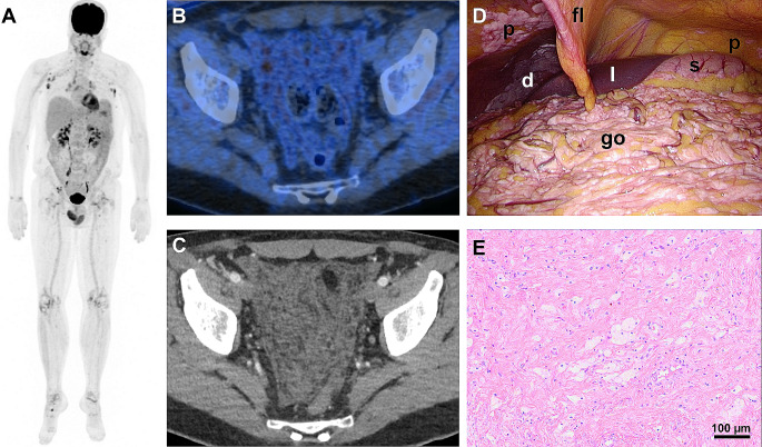
This 46 year-old man presented with a two-year history of clinical hallmarks resembling RDD rather than ECD, including lymphadenopathy and painless testicle enlargement [3], being also visible on [18F]FDG-PET (A). Testicular RDD-ECD involvement was also reported in 6/13 patients by Razanamahery et al. [2]. Diffuse omental proliferations, manifesting as faintly [18F]FDG-avid omental thickening resembling a fishing net (SUVmax 5.5; A, B, C), and symmetric large-joint synovitis were reported as specific features of RDD-ECD [1, 2], Notably, none of these features are characteristic of RDD or hitherto known ECD subtypes. Other RDD and/or ECD features were absent [4–7].
Open biopsy targeted peritoneal lesions (D) localized on the diaphragm (d), peritoneum (p) and greater omentum (go). Histopathology revealed nodular fibrosis, foamy cell infiltrates, pigment deposits and chronic perivascular inflammatory infiltrates (E). Molecular genetic analyses confirmed presence of a characteristic MAP2K1 mutation (p.Q56P).
Diamond et al. effectively treated a patient harboring the identical mutation with MEK inhibitors [8]. FAPI-PET focusing on fibrosis aspects of histiocytosis might help determining disease extent and assessing treatment response [9, 10].
In summary, the newly recognized RDD-ECD overlap histiocytosis demonstrates distinct [18F]FDG-PET features setting it apart from RDD and ECD. The concurrent presence of omental proliferations, symmetric large-joint synovitis, and high testicular uptake should raise suspicion for this yet uncharacterized disease.
Author contributions
All authors contributed to the conception and design. Image analysis / image compilation and design of the final image were performed by M.H. The first draft of the manuscript was written by M.H: and all authors commented on previous versions of the manuscript. All authors read and approved the final manuscript.
Funding
Open access funding provided by University of Zurich. The authors declare that no funds, grants, or other support was received during the preparation of this manuscript.
Open access funding provided by University of Zurich
Declarations
Competing Interests
The authors have no relevant financial or non-financial interests to disclose.
Footnotes
Publisher’s Note
Springer Nature remains neutral with regard to jurisdictional claims in published maps and institutional affiliations.
References
- 1.Portegys J, Heidemeier A, Rosenwald A, Gernert M, Frohlich M, Hueper S, et al. Erdheim-Chester disease with Rosai-Dorfman-Like lesions: treatment with methotrexate, anakinra and upadacitinib. RMD Open. 2023;9. 10.1136/rmdopen-2022-002852. [DOI] [PMC free article] [PubMed]
- 2.Razanamahery J, Diamond EL, Cohen-Aubart F, Plate KH, Lourida G, Charlotte F, et al. Erdheim-Chester disease with concomitant Rosai-Dorfman like lesions: a distinct entity mainly driven by MAP2K1. Haematologica. 2020;105:e5–8. 10.3324/haematol.2019.216937. 10.3324/haematol.2019.216937 [DOI] [PMC free article] [PubMed] [Google Scholar]
- 3.Abla O, Jacobsen E, Picarsic J, Krenova Z, Jaffe R, Emile JF, et al. Consensus recommendations for the diagnosis and clinical management of Rosai-Dorfman-Destombes disease. Blood. 2018;131:2877–90. 10.1182/blood-2018-03-839753. 10.1182/blood-2018-03-839753 [DOI] [PMC free article] [PubMed] [Google Scholar]
- 4.Mazor RD, Manevich-Mazor M, Shoenfeld Y. Erdheim-Chester Disease: a comprehensive review of the literature. Orphanet J Rare Dis. 2013;8:137. 10.1186/1750-1172-8-137. 10.1186/1750-1172-8-137 [DOI] [PMC free article] [PubMed] [Google Scholar]
- 5.Shamburek RD, Brewer HB Jr., Gochuico BR. Erdheim-Chester disease: a rare multisystem histiocytic disorder associated with interstitial lung disease. Am J Med Sci. 2001;321:66–75. 10.1097/00000441-200101000-00010. 10.1097/00000441-200101000-00010 [DOI] [PubMed] [Google Scholar]
- 6.Rosai J, Dorfman RF. Sinus histiocytosis with massive lymphadenopathy. A newly recognized benign clinicopathological entity. Arch Pathol. 1969;87:63–70. [PubMed] [Google Scholar]
- 7.Ahuja J, Kanne JP, Meyer CA, Pipavath SN, Schmidt RA, Swanson JO, Godwin JD. Histiocytic disorders of the chest: imaging findings. Radiographics. 2015;35:357–70. 10.1148/rg.352140197. 10.1148/rg.352140197 [DOI] [PubMed] [Google Scholar]
- 8.Diamond EL, Durham BH, Ulaner GA, Drill E, Buthorn J, Ki M, et al. Efficacy of MEK inhibition in patients with histiocytic neoplasms. Nature. 2019;567:521–4. 10.1038/s41586-019-1012-y. 10.1038/s41586-019-1012-y [DOI] [PMC free article] [PubMed] [Google Scholar]
- 9.Guo L, Shen G. [(68)Ga]Ga-FAPI versus [(18)F]FDG PET/CT in the evaluation of Langerhans cell histiocytosis. Eur J Nucl Med Mol Imaging. 2024. 10.1007/s00259-024-06671-4. 10.1007/s00259-024-06671-4 [DOI] [PubMed] [Google Scholar]
- 10.Pan Q, Zhang H, Cao X, Li J, Luo Y. Langerhans Cell Histiocytosis showed intense uptake of 68 Ga-FAPI. Clin Nucl Med. 2023;48:894–5. 10.1097/RLU.0000000000004786. 10.1097/RLU.0000000000004786 [DOI] [PubMed] [Google Scholar]


