Abstract
The tannery industry produces one of the worst contaminants, and unsafe disposal in nearby waterbodies and landfills has become an imminent threat to public health, especially when the resulting multidrug-resistant bacteria and heavy metals enter community settings and animal food chains. In this study, we have collected 10 tannery wastewater (TWW) samples and 10 additional non-tannery wastewater (NTW) samples to compare the chemical oxygen demand (COD), pH, biological oxygen demand (BOD), dissolved oxygen (DO), total dissolved solids (TDS), chromium concentration, bacterial load, and antibiotic resistance profiles. While COD, pH, and chromium concentration data were previously published from our lab, this part of the study uncovers that TWW samples had a significantly higher bacterial load, compared to the non-tannery wastewater samples (5.89 × 10⁴ and 9.38 × 10³ cfu/mL, respectively), higher BOD and TDS values, and significantly lower DO values. The results showed that 53.4, 46.7, 40.0, and 40.0% of the TWW isolates were resistant to ceftriaxone, erythromycin, nalidixic acid, and azithromycin, respectively. On the other hand, 20.0, 30.0, 50.0, and 40.0% of the NTW isolates were resistant to the same antibiotics, respectively. These findings suggest that the TWW isolates were more resistant to antibiotics than the NTW isolates. Moreover, the TWW isolates exhibited higher multidrug resistance than the NTW isolates, 33.33, and 20.00%, respectively. Furthermore, spearman correlation analysis depicts that there is a negative correlation between BOD and bacterial load up to a certain level (r = − 0.7749, p = 0.0085). In addition, there is also a consistent negative correlation between COD and bacterial load (r = − 0.7112, p = 0.0252) and TDS and bacterial load (r = − 0.7621, p = 0.0104). These findings suggest that TWW could pose a significant risk to public health and the environment and highlight the importance of proper wastewater treatment in tannery industries.
Keywords: Bacterial load, Multidrug-resistance, Tannery wastewater, Chromium, Chemical oxygen demand, Biological oxygen demand, Total dissolved solids, Dissolved oxygen
Subject terms: Biochemistry, Chemical biology, Microbiology, Environmental sciences
Introduction
The environmental impact of tannery wastes on water, terrestrial, and atmospheric systems is severe due to their high oxygen demand, discoloration, toxic chemical constituents, and bad odor. Tannery industry waste is a complex mixture of organic and inorganic pollutants, resulting in a pungent odor, high COD, biological oxygen demand, low dissolved oxygen, high dissolved salts, and high pH levels1–3. Over 200 chemicals are used during the tanning process, leading to a complex mixture of toxic substances such as chromium, polychlorinated biphenyl, sulfides, nitrates, and other harmful compounds discharged as waste4. Among these, the most dominant heavy metal in tannery effluent is chromium5, primarily as a result of its extensive use in the chrome tanning process, which is considered the most toxic to both human health and the environment1,3,5. Additionally, tannery wastes contaminate nearby water with zinc, lead, cadmium, copper, mercury, copper, nickel, and several other metals1,6. The discharge of untreated tannery wastewater containing these bio-toxic heavy metals into the environment, such as rivers and other waterbodies, therefore leads to toxicity in both terrestrial and aquatic biosystems, including those of agriculture, cattle, and poultry farming, which consequently poses risks to human health by threatening skin diseases, respiratory illnesses, and even cancer7–9. Furthermore, these heavy metals pose a survival threat to bacteria and have been reported to induce antibiotic resistance. For example, Gupta et al. demonstrated significant associations between heavy metals and antibiotic resistance, indicating that the resistance mechanisms are similar for both10. Moreover, pollution with heavy metals such as chromium, lead, cadmium, and mercury is well-documented to induce antibiotic resistance through co-selection10–12.
According to the Asian Development Bank (ADB), there are roughly 220 tanneries in Bangladesh, with a majority situated in Hazaribagh, Dhaka, which collectively hire around 850,000 laborers. Recently, these tanneries have been relocated to Savar, a location near the city of Dhaka. Most of these tanneries do not have proper effluent treatment plants (ETP) and release untreated wastewater into the river Buriganga and nearby landfills, causing serious threats to the environment and ecosystem, and are the most prominent sources of environmental chromium in Bangladesh, as indicated by a prior study by the researchers of this study13. The team found chromium content in TWW of different tannery wastewater disposal sites as high as 1.85 × 10 mg/L and COD as high as 6.5 × 10³ mg/L and the direct correlation between chromium content and chemical oxygen demand (COD) was observed statistically significant13.
Since tannery wastes contain a wide variety of contaminants adverse to sustained life14, these possess significant stresses to the microbial community and potentially give rise to heavy metal tolerance as well as antimicrobial resistance to the surviving microbes15. Multidrug resistance (MDR) is a rapidly growing public health concern in Bangladesh16. It is defined as a microbe being resistant to at least three different antimicrobial classes17. There has been research on MDR in Bangladeshi hospital wastes, urinary tract infection (UTI) patients, and other settings18–21, but no systematic investigation has been made to identify MDR linked to tannery wastes in the context of the Hazaribagh tannery industrial area of Bangladesh.
The origin of antibiotic resistance is the process of natural selection and the innate capacity of bacteria to adapt and endure antimicrobial threats22. Bacteria undergo natural genetic changes over time, and certain alterations can provide immunity to antibiotics. When antibiotics are used, vulnerable bacteria are eliminated, whereas resistant ones survive and proliferate. This process is worsened by the excessive use of antibiotics in humans and animals. Improper prescribing practices, such as administering antibiotics for viral infections, significantly contribute to resistance. The Centers for Disease Control and Prevention (CDC) underline that as many as 30% of antibiotic prescriptions in outpatient environments are not needed23. Furthermore, antibiotics used in farming to enhance livestock growth are a significant factor contributing to antibiotic resistance24. Additionally, environmental stress can cause resistance development in bacteria25. Resistant bacteria have the capability to transfer from animals to humans through the food chain26.
To determine the resistance of bacterial strains isolated from TWW and NTW, we tested the sensitivity of the isolates with 10 antibiotics of 6 classes. These antibiotics were selected focusing on their various mode of action. Macrolides, for example, work by attaching themselves to the 50S subunit of bacterial ribosomes27,28, which stops protein production. This prevents peptidyl tRNA from moving, an essential step in making new proteins, ultimately killing the bacteria27,29. β-lactams, including penicillin and cephalosporins, disrupt bacterial cell wall construction by binding to penicillin-binding proteins (PBPs)20,30,31. These proteins help link peptidoglycan chains that give the cell wall its strength. When this process is blocked, the cell wall weakens, and the bacteria burst due to osmotic pressure30.
Protein synthesis inhibitors include different antibiotic classes like macrolides, tetracyclines, and aminoglycosides. They interfere with various steps of protein production32. Tetracyclines, for example, bind to the 30S ribosomal subunit, stopping aminoacyl-tRNA from attaching, while aminoglycosides cause errors in reading mRNA33. Quinolones, such as ciprofloxacin, target bacterial enzymes DNA gyrase and topoisomerase IV, which are vital for DNA replication and repair34,35. By inhibiting these enzymes, quinolones cause breaks in bacterial DNA, leading to cell death36. Cephalosporins, a type of β-lactam, also block cell wall synthesis by targeting PBPs30,31. They are similar to penicillin but usually have a wider range of activity and are more resistant to β-lactamases. Aminoglycosides, like gentamicin, bind to the 30S ribosomal subunit and cause errors in mRNA reading, leading to the incorporation of incorrect amino acids into proteins37,38. This disrupts protein production and kills the bacteria.
Antibiotic resistance mechanisms involve diverse strategies, including regulating the expression of efflux and influx proteins39, horizontal gene transfer from already resistant bacteria40,41, mutation of antibiotic resistance genes like gyrA, gyrB, parC, parE, etc.42. Many of these genes are plasmid-encoded or within mobile genetic elements43,44, which helps spread resistance genes swiftly.
Macrolides, such as erythromycin, work by binding to the 50S ribosomal subunit, thereby inhibiting bacterial protein synthesis. Bacterial resistance to macrolides can occur through several mechanisms45. One such mechanism is the methylation of 23S rRNA by Erm enzymes, which lowers the drug’s binding affinity45,46. Additionally, efflux pumps, such as Mef, can expel the drug from the bacterial cell47. β-lactam resistance mechanisms include the production of β-lactamases, which hydrolyze the β-lactam ring, rendering the antibiotic ineffective48. Alterations in PBPs can reduce drug binding, while changes in porin channels can decrease drug uptake31. For tetracyclines and chloramphenicol, resistance mechanisms involve efflux pumps that expel the antibiotic and ribosomal protection proteins that dislodge the antibiotic from its target49. Quinolones, including ciprofloxacin, target DNA gyrase and topoisomerase IV, which are essential for DNA replication50. Resistance can occur through mutations in the genes encoding these enzymes, reducing drug binding, and through efflux pumps that lower intracellular drug concentrations50,51. Cephalosporin resistance mechanisms include the production of β-lactamases specific to cephalosporins, such as AmpC β-lactamases, and modifications in PBPs52.
In this study, we isolated and cultured bacterial colonies from 10 tannery wastewater (TWW) samples collected from 10 different tanneries, as well as from 10 non-tannery wastewater (NTW) samples. We quantified the bacterial load in each sample and further characterized 15 bacterial isolates from both TWW and NTW samples. We conducted antimicrobial susceptibility tests on the isolated bacteria using 10 antibiotics from 6 different classes, and constructed antimicrobial resistance (AMR) and multidrug resistance (MDR) profiles. We then examined the correlation between microbial load, multidrug resistance in bacteria, and the physicochemical characteristics of TWW samples (such as COD, BOD, TDS, and DO), as well as chromium levels, in comparison to NTW samples. Finally, we identified a potential role of untreated tannery wastewater in the evolution of multidrug resistance in bacteria and the consequent microbial pollution of surface water in Bangladesh.
Methods and materials
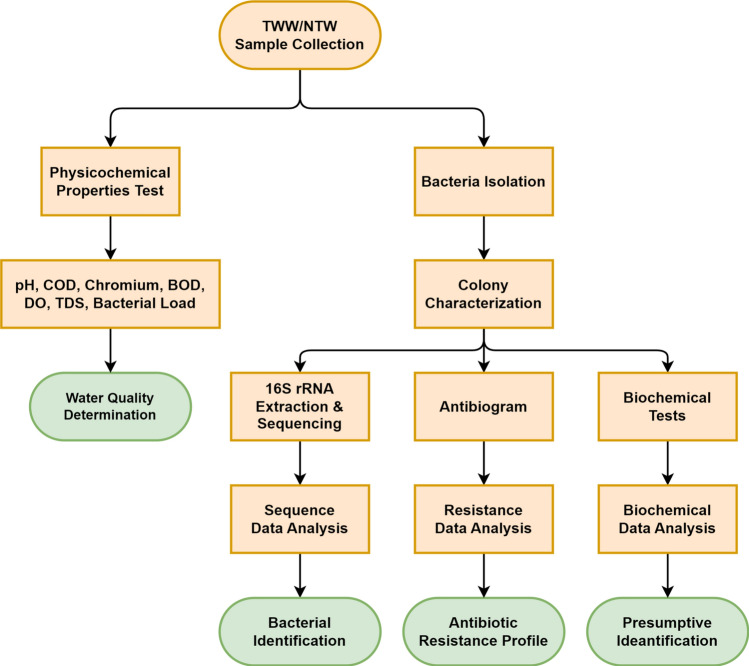
The study commenced with the collection of TWW and NTW samples from tannery industries and non-tannery water sources respectively, followed by physicochemical properties determination and bacterial load determination from diluted samples. Bacterial isolates were then characterized and identified via both biochemical tests and 16S rRNA sequence analysis. Additionally, isolated and identified bacterial strains underwent antibiotic susceptibility testing and a comprehensive resistance profile was established (Fig. 1).
Fig. 1.
Flowchart of the study.
Sample collection
Ten samples of tannery wastewater (TWW) were collected from the main drains of 10 tanneries of Hazaribagh, Dhaka, and 10 non-tannery wastewater (NTW) control samples were collected from the Buriganga River and the University of Dhaka area. The samples were collected during the monsoon season (June to September period) to capture peak tannery industrial activity. From each of the sampling points, 500 mL of wastewater and control water was collected.
Determination of physicochemical properties of TWW and NTW
The TWW and NTW samples were analyzed to measure pH, chromium content, COD, BOD, DO, TDS, and bacterial load. pH was measured with a bench-top Hanna pH meter and chromium content was determined using a Varian 240 Atomic Absorption Spectrometer (240 AAS). COD was determined by oxidizing organic matter with potassium dichromate and measuring excess potassium dichromate using ferrous ammonium sulfate following the back-titration method. COD value was equivalent to the consumed dichromate. To determine the bacterial load, each 1.0 mL sample of tannery wastewater was diluted 1000 times (1:1000), while each 1.0 mL sample of non-tannery water was diluted 100 times (1:100). From these diluted samples, 100 µL (0.1 mL) was plated and incubated at 37 °C for 24 h. Following incubation, bacterial colonies were manually counted using a colony counter.
BOD was measured using a BOD Trak II respirometric apparatus (HACH, USA). Samples were incubated at 20 °C for 5 days in sealed bottles. The BOD Trak II system automatically recorded the pressure drop caused by oxygen consumption, which was then converted to BOD5 values. Dissolved oxygen (DO) was determined using a portable YSI ProODO optical dissolved oxygen meter. The probe was calibrated before each use according to the manufacturer's instructions. Measurements were taken in situ to minimize atmospheric oxygen interference. Total Dissolved Solids (TDS) were quantified using a gravimetric method. A well-mixed sample was filtered through a standard glass fiber filter. A known volume of the filtrate was then evaporated to dryness in a weighed dish and dried to a constant weight at 180 °C. The increase in dish weight represented TDS. This process was carried out using a Mettler Toledo XS205 analytical balance for precise weight measurements and a Thermo Scientific Heratherm oven for the drying process.
All measurements were performed in triplicate to ensure reliability and reproducibility of results. Standard quality control procedures, including the use of blanks and standard solutions, were implemented throughout the analytical process to maintain accuracy and precision.
Isolation and identification of bacterial isolates
To isolate common bacterial isolates from TWW, 10 µL of 100 times diluted samples were inoculated at 37 °C in nutrient agar media for 24 h and a total of 15 colonies were selected considering varied colony morphologies (size, shape, pigmentation, elevation, and texture) on nutrient agar. These morphological characteristics included variations in pigmentation (e.g., white, yellow, orange, pink), elevation (flat, raised), and texture (smooth, oily). For instance, some colonies were small and light-colored, while others were large and bright white with a transparent appearance.
Additionally, 10 control bacterial isolates were randomly selected from ten NTW samples for further investigation. To ensure their viability over a longer period, bacterial isolates were cryopreserved at a temperature of − 80 °C to enable us to explore more about these isolates later.
Alongside observing colony morphology in culture media, fifteen biochemical tests were conducted to identify bacterial strains presumptively. Detailed results of the biochemical tests are presented in Table 2. For confirmatory identification, 16S rRNA was sequenced and analyzed via Geneous Prime software and NCBI BLAST tool.
Table 2.
Identification of TWW and NTW isolates based on biochemical tests and 16S rRNA analysis.
| Sample code | Gram Stain | Lactose | Glucose | Sucrose | H2S production | NO3 production | Indole production | MR reaction | VP reaction | Citrate use | Urease activity | Catalase activity | Oxidase activity | Starch hydrolysis | Casein hydrolysis | Presumptive identification |
|---|---|---|---|---|---|---|---|---|---|---|---|---|---|---|---|---|
| 16S rRNA based identification | ||||||||||||||||
| SD | + | – | A | A | – | – | – | + | – | – | – | + | + | + | + | – |
| – | ||||||||||||||||
| SC | – | + | – | – | – | – | – | – | – | – | – | + | – | – | – | Corynebacterium spp. |
| – | ||||||||||||||||
| LD (A) | – | – | – | – | + | + | – | – | – | – | – | + | – | – | – | Salmonella spp. |
| Salmonella enterica | ||||||||||||||||
| LE (A) | – | + | + | + | + | + | – | + | – | + | – | – | – | – | – | – |
| Escherichia coli | ||||||||||||||||
| LA (A) | – | A/G++ | A/G++ | A | – | + | + | + | – | – | – | + | + | – | – | Escherichia coli |
| Escherichia coli | ||||||||||||||||
| LB (A) | – | – | A | A | + | + | + | + | – | – | + | + | – | – | – | Salmonella spp. |
| Salmonella enterica | ||||||||||||||||
| LC (F) | + | A | A | A | – | – | – | + | – | – | – | – | – | – | + | Streptococcus spp. |
| – | ||||||||||||||||
| SA (A) | – | A/G++ | A/G++ | – | – | + | – | + | – | – | – | + | – | + | – | – |
| Escherichia coli | ||||||||||||||||
| SB (A) | + | – | + | + | – | – | + | – | – | – | + | + | – | – | – | |
| – |
A:acid production, G:gas production.
Genomic DNA extraction and 16S rRNA sequencing
The Qiagen Powerfecal Pro DNA kit was used to extract genomic DNA from bacterial isolates cultivated overnight, with subsequent amplification using universal primers 27F/1492R. Primer sequences were 16S rRNA-Forward: AGAGTTTGATCCTGGCTCAG and 16S rRNA-Reverse: GGTTACCTTGTTACGACTT. Validation of the presence and size of the PCR amplicons was verified via agarose gel electrophoresis subsequent to the rRNA being purified utilizing a PCR purification kit. Subsequently, the amplified PCR product of 16S rRNA of bacterial isolates underwent sequencing by BTSeq™ Contiguous Sequencing by Celemics, Inc., located in Ge-umcheon-gu, Seoul, Korea.
Antibiotic susceptibility testing
A single colony was picked from each of the freshly cultured bacterial isolates and was incubated in MHB for two hours at 37 °C, and microbial growth was adjusted to the 0.5 McFarland standard before preparing the lawn on Mueller Hinton agar (MHA) plates, placing antibiotic discs, and incubating at 37 °C for 18 h. Furthermore, since a 0.5 McFarland turbidity standard approximates the optical density of a bacterial suspension containing approximately 1.5 × 10⁸ colony-forming units (CFU) per milliliter, we measured the absorbance of bacterial cultures at 600 nm. An absorbance value within the range of 0.08–0.135 at 600 nm corresponds to a bacterial density of around 1.5 × 10⁸ cfu/mL. The assessment of antibiotic susceptibility among bacterial isolates from both the TWW and NTW was conducted via exposure to ten commercially available antibiotics belonging to six distinct classes (Supplementary Table 1). This was accomplished through the use of the standard Kirby-Bauer disc diffusion method as described previously53, with interpretation being carried out in accordance with the Clinical and Laboratory Standards Institute (CLSI-2022) and European Committee on Antimicrobial Susceptibility Testing (EUCAST-2022) guidelines. Isolates that showed resistance to three or more classes of antibiotics were classified as multidrug resistant (Supplementary Table 2). In cases where an antibiotic class included multiple antibiotics, resistance was defined as non-sensitivity to at least one antibiotic within that class.
Statistical analysis
Spearman's rank correlation analysis was employed to investigate the associations among chromium concentrations, chemical oxygen demand (COD), and bacterial load. This methodology yielded correlation coefficients and p values, which provided valuable insights. The dataset, in conjunction with antimicrobial resistance data, underwent Spearman’s rank correlation analysis using the statistical software R, while GraphPad Prism (version 8) was utilized for graphical depiction.
Results
Physicochemical characterization of TWW and NTW
Our investigation concentrated on the analysis of both tannery wastewater (TWW) and non-tannery wastewater (NTW) to ascertain essential physicochemical parameters, such as COD, BOD, DO, TDS, and Cr concentration, alongside bacterial load. This comparative assessment was undertaken to elucidate the distinct attributes of TWW in relation to NTW. The outcomes, delineated in Table 1, unveil notable distinctions between the two water categories. TWW manifested significantly heightened levels of BOD, TDS, and bacterial load when juxtaposed with NTW. Conversely, the concentration of dissolved oxygen (DO) was notably greater in NTW compared to TWW.
Table 1.
Physicochemical parameters determined from TWW and NTW samples.
| TWW ID | BOD (mg/L) | DO (mg/L) | TDS (mg/L) | Bacterial load (× 103 cfu/mL) | NTW ID | BOD (mg/L) | DO (mg/L) | TDS (mg/L) | Bacterial load (× 103 cfu/mL) |
|---|---|---|---|---|---|---|---|---|---|
| TWW1 | 470 | 0.25 | 11,500 | 41 | NTW1 | 8 | 5 | 220 | 0.925 |
| TWW2 | 160 | 0.6 | 3500 | 29.3 | NTW2 | 2.5 | 14 | 55 | 5.075 |
| TWW3 | 70 | 3.5 | 3200 | 50 | NTW3 | 3.5 | 8 | 135 | 0.675 |
| TWW4 | 270 | 1.0 | 6250 | 47 | NTW4 | 5 | 7 | 150 | 5.75 |
| TWW5 | 520 | 0.5 | 13,000 | 24 | NTW5 | 3 | 11 | 70 | 10 |
| TWW6 | 610 | 0.25 | 20,250 | 25.5 | NTW6 | 12.5 | 6 | 300 | 23 |
| TWW7 | 335 | 0.6 | 5300 | 38 | NTW7 | 7 | 9 | 110 | 19.5 |
| TWW8 | 1900 | 0.5 | 35,000 | 0.925 | NTW8 | 9 | 8 | 175 | 0.3 |
| TWW9 | 220 | 0.5 | 5000 | 58 | NTW9 | 9 | 7 | 250 | 15.5 |
| TWW10 | 390 | 0.5 | 7600 | 18.5 | NTW10 | 4 | 12 | 60 | 13 |
| Mean | 494.5 | 0.8 | 11,060 | 33.2 | Mean | 6.4 | 8.7 | 152.5 | 9.4 |
The increased BOD and TDS concentrations in TWW additionally signify a substantial organic load and dissolved solids content, correspondingly. These elements have the potential to foster eutrophication and impact the overall water quality of recipient water systems. Moreover, the heightened bacterial load in TWW gives rise to apprehensions regarding plausible pathogenic contamination.
Assessment of physicochemical properties and bacterial load in TWW and NTW
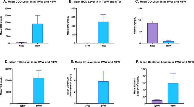
The assessment of pH, COD, and chromium was previously reported by Islam et al. The pH of the TWW samples ranged from 3.93 to 8.36, two samples being highly acidic in nature (pH below 7.0), while the pH of the control water sample was mostly close to basic (ranging from 6.82 to 8.25)13. The results of this study demonstrate that the mean values of BOD, TDS, chromium level, and bacterial load were considerably higher in TWW as compared to NTW control samples, as illustrated in Fig. 2.
Fig. 2.
Comparison of physicochemical properties in TWW and NTW. The mean COD level (A), mean BOD (B), mean TDS (D), mean Cr level (E), and mean bacterial load (F) were substantially higher in TWW compared to non-tannery control wastewater with the exception to Mean DO values (C). NTW: non-tannery wastewater; TWW: tannery wastewater.
The Chemical Oxygen Demand (COD) in TWW (1.4 × 10³ mg/L) was found to be substantially higher than in NTW (5.0 × 10 mg/L), indicating a significant organic pollutant load in tannery effluents. Biological Oxygen Demand (BOD) showed a similar trend, with TWW exhibiting a much higher value (4.9 × 10² mg/L) compared to NTW (6.35 mg/L), further confirming the high organic content in tannery wastewater. Dissolved Oxygen (DO) levels are inversely related to the previous parameters. NTW shows higher DO levels (8.7 mg/L) compared to TWW (0.82 mg/L), suggesting potential hypoxic conditions in tannery effluents. Total Dissolved Solids (TDS) in TWW (1.1 × 10⁴ mg/L) far exceed those in NTW (1.5 × 10² mg/L), indicating a high concentration of dissolved substances in tannery wastewater. Chromium concentration is notably higher in TWW (3.83 mg/L) compared to NTW (0.0026 mg/L), likely due to the use of chromium in tanning processes. TWW shows a substantially higher bacterial load (5.9 × 10⁴ cfu/mL) compared to NTW (9.3 × 10³ cfu/mL), suggesting increased microbial activity in tannery effluents (Fig. 2A–F).
Association of bacterial load with COD, BOD, TDS, DO, and chromium level
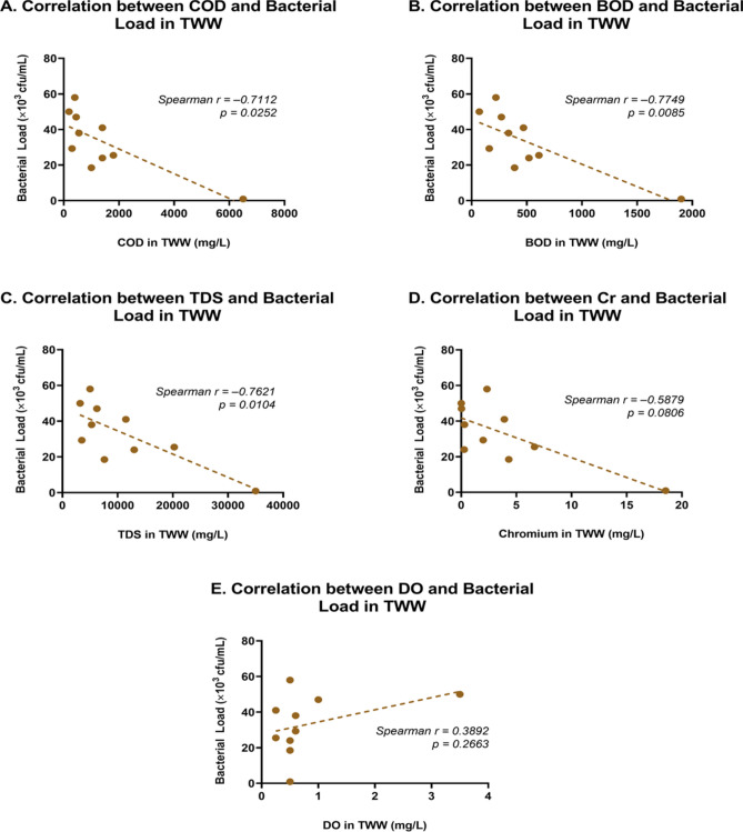
The bacterial load had a positive but insignificant correlation with the COD level in NTW samples (r = 0.2092, p = 0.5611) (Fig. 3A). However, the elevated COD level significantly inhibited the growth of bacteria in TWW, showing a strong negative correlation (Spearman r = − 0.7112, p = 0.0252) between COD and bacterial load. As COD levels increase, there is a significant decrease in bacterial load. The relationship appeared non-linear, with bacterial load dropping more rapidly at lower COD levels (Fig. 3A). This inverse relationship suggests that higher levels of organic pollutants (indicated by COD) may inhibit bacterial growth in tannery effluents. A strong negative correlation (Spearman r = − 0.7749, p = 0.0085) was observed between BOD and bacterial load (Fig. 3B). This relationship was slightly stronger than that of COD, indicating that the biodegradable organic matter (measured by BOD) may have a more direct impact on bacterial populations than the total organic content (measured by COD).
Fig. 3.
Association of COD, BOD, TDS, DO, and chromium with bacterial load in TWW. (A) Bacterial load decreases as the COD level increases beyond a certain level. (B) BOD showed a slightly stronger negative correlation than that of COD. (C) TDS shows a strong negative correlation with bacterial load, suggesting that as the concentration of dissolved solids increases, the bacterial population tends to decrease. (D) Bacterial load is inversely proportional to the chromium concentration in TWW. (E) DO level shows a weak positive correlation with bacterial load.
TDS showed a strong negative correlation (Spearman r = − 0.7621, p = 0.0104) with bacterial load (Fig. 3C). This suggests that as the concentration of dissolved solids increases, the bacterial population tends to decrease, possibly due to osmotic stress or the presence of inhibitory substances in the dissolved solids fraction. Chromium concentration exhibited a moderate negative correlation (Spearman r = − 0.5879, p = 0.0806) with bacterial load (Fig. 3D). While the relationship was not as strong as the other parameters, it still indicates a trend where higher chromium levels are associated with lower bacterial populations, likely due to the toxic effects of chromium on microorganisms.
In addition, dissolved oxygen (DO) level shows a weak positive correlation (Spearman r = 0.3892, p = 0.2663) with bacterial load (Fig. 3E). This relationship is not statistically significant, indicating that DO levels may not be a strong predictor of bacterial load in this TWW system.
Bacterial identification
The amplified products of 16S rRNA genes from the bacterial isolates were subjected to Sanger sequencing. Following this, sequence analysis was executed through the use of Genious Prime software, aligning these sequences with those from the 16S rRNA gene available in the National Center for Biotechnology Information (NCBI) database using the basic local alignment search tool (BLAST). The classification of species was accomplished by contrasting the acquired sequences with those existing in the GenBank repository and choosing the sequence that exhibited the greatest maximum identity score. The primary bacterial strains identified through 16S rRNA sequencing were Escherichia coli and Salmonella enterica (Table 2).
Resistance profile of TWW and NTW isolates
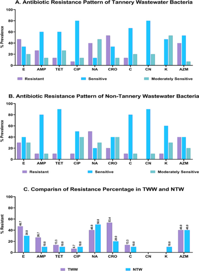
The TWW isolates were resistant to ceftriaxone, erythromycin, azithromycin, and nalidixic acid with 53.4, 46.7, 40.0, and 40.0% resistant isolates respectively. All the 15 TWW isolates exhibited absolute sensitivities to Gentamycin and Kanamycin (Supplementary Table 2). This efficacy was followed by ciprofloxacin, the protein synthesis inhibitors (namely tetracycline and chloramphenicol), ampicillin, nalidixic acid, and kanamycin at 6.7, 13.3, 26.7, 46.7 and 53.4% of isolates showing resistance respectively (Fig. 4A).
Fig. 4.
Antimicrobial resistance profile of TWW and NTW isolates. Amongst the TWW isolates, CN exhibited the highest antimicrobial activity, while CRO was found the least effective (A). CN showed the greatest effectiveness against NTW isolates while NA was the least (B). A comparison of resistance prevalence in TWW and NTW isolates reveals that the overall resistance to most antibiotics was higher in TWW isolates than of NTW isolates (C). Antibiotic susceptibility was determined against erythromycin (E), ampicillin (AMP), tetracycline (TET), ciprofloxacin (CIP), nalidixic acid (NA), ceftriaxone (CRO), chloramphenicol (C), gentamycin (CN), kanamycin (K), and azithromycin (AZM). TWW: Tannery wastewater, NTW: Non-tannery wastewater.
The resistance pattern in NTW isolates for erythromycin (30% resistant), azithromycin (40% resistant), and nalidixic acid (50% resistant) resemble that of TWW isolates. On the other hand, the NTW isolates showed great sensitivity towards four antibiotics—tetracycline, chloramphenicol, gentamycin, and kanamycin with 90, 80, 90, and 60% being sensitive, respectively. However, ceftriaxone was found to be significantly more effective against NTW isolates (20% resistant) compared to TWW isolates (53.4% resistant) (Fig. 4B).
Except for nalidixic acid, kanamycin, and ciprofloxacin, the other 7 antibiotics were found to be less effective against TWW isolates compared to their efficiency against NTW isolates (Fig. 4C). Interestingly, none of the isolates from either source showed any resistance against gentamycin (CN). The difference in resistance is most notable for ceftriaxone (CRO) and erythromycin (E), where TWW isolates showed alarming higher resistance than NTW isolates.
Resistance against different antimicrobial classes
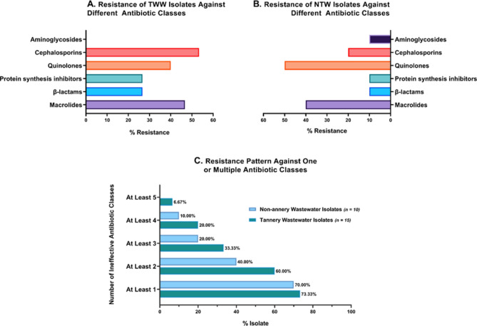
The aminoglycosides, namely gentamycin and kanamycin were extremely effective against both TWW and NTW isolates, with no resistant isolates (0%) and 10% resistant isolates respectively. Resistance to cephalosporins was considerably higher among TWW isolates (53.33%) than in NTW isolates (20%). Bacterial isolates both from the control and experimental wastewaters showed similar resistance toward quinolones, 40% among TWW and 50% among NTW isolates (Fig. 5A, B). About 47% of the TWW isolates were resistant to macrolides, whereas a slightly lower, 40% of the NTW isolates had conferred resistance against the macrolide group of antibiotics. Resistance against both protein synthesis inhibitor and β-lactam among the TWW isolates was more than double (26.67%) the resistance among the NTW isolates (10%).
Fig. 5.
Resistance pattern of TWW and NTW isolates against different antimicrobial classes. (A) Antibiotic resistance pattern of bacterial strains collected from TWW samples. Aminoglycosides and cephalosporins were the most and least sensitive antimicrobial classes against TWW isolates. (B) Antibiotic resistance pattern of bacterial strains collected from NTW samples. The NTW isolates were most resistant to quinolones. (C) Resistance pattern against one or multiple classes of antibiotics. The comparative analysis reveals that TWW isolates were constantly multiple drug-resistant compared to NTW.
MDR profile of TWW and NTW bacteria
Among the TWW isolates, 33.33% were resistant to at least three different classes of antimicrobial, making them multidrug-resistant (MDR), much higher than the NTW isolates, 20% of which were MDR. Figure 5C compares bacterial resistance to multiple classes of antibiotics, where resistance in TWW isolates was always worse than in NTW isolates. 20% of the isolates in TWW had developed resistance against at least 4 classes of antibiotics, and the corresponding value in the NTW isolates was 10%. While no isolates were resistant to more than 4 classes of antibiotics in control wastewater, 6.67% of the TWW isolates were extensively drug-resistant (XDR), since they showed resistance against 5 out of 6 antibiotic classes.
Discussion
Despite being a significant contributor to export earnings, employment facilities, and the overall country’s economy, tanneries have been associated with severe environmental pollution and health hazards in Bangladesh. Tanneries included in this study are spread in the area of Hazaribagh and the team carefully selected 10 sites for sample collection. All of these sites had a thousand times higher concentration of environmental chromium than the safe concentration, which was reported to be 0.1 mg/L in the environment5. The chromium concentration in the wastewater disposed of by one particular Leather industry which was very close to the river Buriganga was found to be 1.85 × 10 mg/L. The absence of bioaccumulation of chromium in aquatic flora, fauna, and microorganisms implies that elevated levels of chromium in the environment can have a severe toxic impact on the affected plants and animals54. Toxicity in humans can manifest as cutaneous lesions or rashes, as well as renal and hepatic impairment54.
However, the concentration of chromium in tannery effluent determined in our study was significantly lower (ranging from 0.002 mg/L to 1.85 × 10 mg/L) than that of another study (ranging from 5.25 × 10 mg/L to 3.74 × 10² mg/L) conducted in the same tannery industrial area but comprised of different tanneries55. This implied that not all tanneries contribute to chromium contamination equally, which was also observed in our collection sites. These researchers also noted that the concentration of chromium in wastewater was recorded highest during January–February, when the rivers were dried, and the concentration was the lowest during May–June, the monsoon period of the year55.
Recent studies have highlighted the effect of elevated levels of heavy metals, especially chromium, on the co-selection with antibiotic resilience56–58. Chromium was identified to impose pressure on the bacterial population, impacting the electrochemical system and leading to elevated horizontal transfer of antibiotic resistance genes, consequently elevating the likelihood of augmented horizontal transmission of resistance genes56. This may potentially account for the heightened antibiotics resistance in TWW in comparison to NTW.
One study reported that significant quantities of this heavy metal can be eliminated through conventional physicochemical and biological procedures59. In contemporary times, there has been a surge in the utilization of cutting-edge technologies like nanomaterials, membrane technology, electrocoagulation, ion exchange, and electrodialysis for the treatment of TWW60,61. This trend can be attributed to the comparative benefits that these technologies offer over other conventional methods, particularly in the area of sustainability61.
Higher COD values in the TWW make aquatic life difficult to sustain3. It has been suggested that chromium in TWW along with other toxic compounds and higher COD has created an environmental pressure causing naturally present bacteria in the water to develop resistance to various commonly used antibiotics62. These bacteria may transfer their resistance genes through horizontal transmission and allow the development of superbugs and pathogenic bacteria that are resistant to the most widely used antibiotics63,64.As to our findings, the TWW contains an alarmingly high level of multidrug-resistant (MDR) isolates, which is likely to spread into other waterbodies and nearby landfills. Most bacterial isolates from the NTW showed moderate sensitivity to commonly used antibiotics whereas bacterial isolates from the TWW showed resistance to an increased number of antibiotics. A few isolates have been found to be resistant to 5 classes of antibiotics out of 6 classes, making them extensively drug-resistant (XDR), which is even more concerning than those of MDR (Fig. 5).
It is noteworthy to highlight that with the exception of a few samples of TWW and a solitary NTW sample, all instances of TWW and NTW wastewater exhibited a pH range between 7.4 and 8.34, which is optimal for bacterial proliferation. This observation implies two critical aspects. The COD and chromium concentration are the two key factors that determine bacterial load. Furthermore, it should be noted that these factors do not impact the pH level of the water.
The mean chromium concentration in the NTW samples was 0.002 mg/L. Despite the evidence that the pH, COD values, and chromium concentration are favorable for microbial growth, the bacterial load observed in the NTW samples was considerably lower than that of the TWW samples. This can be attributed to the NTW sources being less contaminated by other wastes that provide bacterial nutrients.
The findings of this study illustrate a marked increase in average BOD levels in TWW compared to NTW. Elevated BOD signifies a higher concentration of biodegradable organic substances65, which can result in heightened microbial activity. This increased microbial presence may contribute to an elevated probability of antibiotic resistance emergence22, as a larger number of bacteria are exposed to potential antibiotic remnants and conditions that induce resistance in the wastewater. It is noteworthy that the levels of dissolved oxygen (DO) are conspicuously lower in TWW than in NTW. Reduced DO levels can create conducive environments for anaerobic bacteria66, potentially causing alteration to the microbial community67. This oxygen-deficient setting could exert selective pressure on bacteria to develop survival mechanisms22,66, indirectly influencing the development of antibiotic resistance. The data also indicates significantly higher Total Dissolved Solids (TDS) levels in TWW. Elevated TDS values may suggest the existence of various dissolved substances, including organic and inorganic matters and potential antibiotic remnants or their byproducts68. Increased TDS concentrations might offer more nutrients for bacterial proliferation69, potentially leading to an augmented bacterial population as depicted in Fig. 2F.
The combined impact of elevated BOD and COD, decreased DO, and increased TDS in TWW could have substantial ecological implications if discharged into natural aquatic systems70. This discharge may trigger eutrophication, disrupt aquatic ecosystems, and potentially create environments conducive to the proliferation of antibiotic-resistant bacteria.
Antibiotic resistance emerges when bacteria develop mechanisms to endure the medications formulated to eradicate them, resulting in treatments being less efficacious and escalating the likelihood of severe infections and mortality. Apart from a sheer number of multidrug-resistant isolates, many isolates in our study were found to be intermediately sensitive (designated as “I” in Supplementary Table 2), which has a high probability of being resistant to further stress caused by chromium and other contaminants in the TWW. This has already occurred in TWW as was shown in Fig. 4A and B, where it was obvious that the TWW isolates showed significantly higher resistance against β-lactam, protein synthesis inhibitors, and cephalosporins compared to those of the NTW isolates. This is particularly alarming since members of β-lactam, protein synthesis inhibitors, and cephalosporin classes of antibiotics, such as ampicillin, tetracycline, chloramphenicol, and ceftriaxone are among the most commonly prescribed antibiotics in our country.
Multidrug-resistant bacteria have previously been associated with severe human illnesses like cholera, diarrhea, and other water-borne diseases in the Bangladeshi population18,71–74. These water-borne diseases have a high mortality rate, especially for infants and children under 5 years of age (37%)75–78. Since, as of now, most tannery industries are located very close to a running river, the carriage risks the health of nearby community people.
The investigation underscores the pressing requirement for implementing actions to efficiently diminish the employment of chemicals throughout the process of leather production, while simultaneously purifying the previously contaminated water sources to forestall the buildup of heavy metals in the food chain. Additionally, decontamination of the previously polluted water sources is crucial to guarantee that no dissemination of MDR bacteria arises, which could potentially endanger public health.
Conclusions and recommendations
The water quality parameters revealed in this investigation indicate that TWW presents conditions that could potentially support the emergence and dissemination of antibiotic-resistant bacteria. The alarmingly elevated levels of COD, BOD, and TDS and the decreased levels of DO in TWW compared to NTW pose a significant survival threat to bacteria (marked by a strong negative correlation), and this threat is one causative agent for induced antibiotic resistance. This is evident in the resistance profile of the bacteria isolated from the tannery industry area, with markedly high multidrug resistance and antibiotic resistance in general. TWW isolates were substantially more resistant to cephalosporins, β-lactams, macrolides, and protein synthesis inhibitors. Additionally, 33.33% of the TWW isolates were MDR, compared to 20.00% of the NTW isolates. However, the bacterial load in TWW was over 500% greater than that of NTW, indicating bacteria are able to evade harsh conditions and carry antibiotic resistance mechanisms, and pose a significant risk to public health once they contaminate the community setting.
Overall, the findings of this study underscore the pressing necessity for a cohesive strategy to supervise and regulate antimicrobial utilization across all domains. Enforcing stringent effluent management procedures and promoting exploration into substitute therapies and preventive practices are pivotal measures in tackling this predicament. The government of Bangladesh has meanwhile taken the initiative to dedicate a facility for tannery industries with facilities like efficient water treatment modules and separate tannery waste from community sewage lines to prevent contamination. However, the massive pile of tannery waste in our study area has remained untreated, and the effectiveness of the government’s initiative needs to be studied. Appropriate treatment and handling of tannery wastewater are imperative to mitigate these risks prior to environmental discharge.
Supplementary Information
Author contributions
Z.M. and L.N.I. conceived the idea and supervised the research work. Z.M., R.K.M., A.R., and M.M.K. performed laboratory-based experiments. Z.M. and R.K.M. performed the data analysis. Z.M. and A.R. collected the samples. Z.M., L.N.I., and M.M.K. provided consumables to perform laboratory experiments. Z.M. and R.K.M. prepared the first draft. Z.M., L.N.I., and M.M.K. reviewed the first draft of the manuscript. Z.M., R.K.M., L.N.I. and M.M.K. addressed all the revisions and modifications required. All authors have read and approved the manuscript.
Funding
Authors did not receive any funding for this study.
Data availability
All data generated or analysed during this study are included in the submitted manuscript [and its supplementary information files].
Competing interests
The authors declare no competing interests.
Footnotes
Publisher's note
Springer Nature remains neutral with regard to jurisdictional claims in published maps and institutional affiliations.
Contributor Information
Zimam Mahmud, Email: zimam@du.ac.bd.
Laila N. Islam, Email: laila@du.ac.bd
Supplementary Information
The online version contains supplementary material available at 10.1038/s41598-024-71472-6.
References
- 1.Igiri, B. E. et al. Toxicity and bioremediation of heavy metals contaminated ecosystem from tannery wastewater: A review. J. Toxicol.2018, 2568038 (2018). 10.1155/2018/2568038 [DOI] [PMC free article] [PubMed] [Google Scholar]
- 2.Masood, F. & Malik, A. Environmental concerns of the tanning industry. In Environmental Deterioration and Human Health: Natural and Anthropogenic Determinants (eds Malik, A. et al.) 39–53 (Springer, 2014). [Google Scholar]
- 3.Patel, N. et al. Environmental impact and treatment of tannery waste. In Water Pollution and Remediation: Organic Pollutants (eds Inamuddin et al.) 577–595 (Springer International Publishing, 2021). [Google Scholar]
- 4.Morera, J. et al. Avoiding the production of polluting and toxic chemicals in the tanning process. J. Am. Leather Chem. Assoc.10.34314/jalca.v116i10.4617 (2021). 10.34314/jalca.v116i10.4617 [DOI] [Google Scholar]
- 5.Prasad, S. et al. Chromium contamination and effect on environmental health and its remediation: A sustainable approaches. J. Environ. Manag.285, 112174 (2021). 10.1016/j.jenvman.2021.112174 [DOI] [PubMed] [Google Scholar]
- 6.Silambarasan, T. S. et al. Bioremediation of tannery effluent contaminated soil: A green approach. In Advances in Bioremediation and Phytoremediation for Sustainable Soil Management: Principles, Monitoring and Remediation (ed. Malik, J. A.) 283–300 (Springer International Publishing, 2022). [Google Scholar]
- 7.Bari, M. L. et al. Potential human health risks of tannery waste-contaminated poultry feed. J. Health Pollut.5(9), 68–77 (2015). 10.5696/2156-9614-5-9.68 [DOI] [PMC free article] [PubMed] [Google Scholar]
- 8.Rastogi, S. K. et al. Occupational cancers in leather tanning industries: A short review. Indian J. Occup. Environ. Med.11(1), 3–5 (2007). 10.4103/0019-5278.32456 [DOI] [PMC free article] [PubMed] [Google Scholar]
- 9.Dalju, I. et al. Occupational risk factors associated with respiratory symptoms among tannery workers in Mojo town, Southeast Ethiopia, 2018: A comparative cross-sectional study. Multidiscip. Respir. Med.14(1), 27 (2019). 10.1186/s40248-019-0188-1 [DOI] [PMC free article] [PubMed] [Google Scholar]
- 10.Gupta, S. et al. Heavy metal and antibiotic resistance in four Indian and UK rivers with different levels and types of water pollution. Sci. Total Environ.857(Pt 1), 159059 (2023). 10.1016/j.scitotenv.2022.159059 [DOI] [PubMed] [Google Scholar]
- 11.Chen, S. et al. Heavy metal induced antibiotic resistance in bacterium LSJC7. Int. J. Mol. Sci.16(10), 23390–23404 (2015). 10.3390/ijms161023390 [DOI] [PMC free article] [PubMed] [Google Scholar]
- 12.Seiler, C. & Berendonk, T. U. Heavy metal driven co-selection of antibiotic resistance in soil and water bodies impacted by agriculture and aquaculture. Front. Microbiol.3, 399 (2012). 10.3389/fmicb.2012.00399 [DOI] [PMC free article] [PubMed] [Google Scholar]
- 13.Islam, L. et al. Assessment of physicochemical and biochemical qualities of tannery effluents of Hazaribagh, Dhaka, and comparison with non-tannery water samples. Int. J. Environ.4, 68–81 (2015). 10.3126/ije.v4i1.12179 [DOI] [Google Scholar]
- 14.Kumar, R. et al. Management of tannery waste effluents towards the reclamation of clean water using an integrated membrane system: A state-of-the-art review. Environ. Res.229, 115881 (2023). 10.1016/j.envres.2023.115881 [DOI] [PubMed] [Google Scholar]
- 15.McMahon, M. A. et al. Environmental stress and antibiotic resistance in food-related pathogens. Appl. Environ. Microbiol.73(1), 211–217 (2007). 10.1128/AEM.00578-06 [DOI] [PMC free article] [PubMed] [Google Scholar]
- 16.Hoque, R. et al. Tackling antimicrobial resistance in Bangladesh: A scoping review of policy and practice in human, animal and environment sectors. PLoS One15(1), e0227947 (2020). 10.1371/journal.pone.0227947 [DOI] [PMC free article] [PubMed] [Google Scholar]
- 17.Tanwar, J. et al. Multidrug resistance: An emerging crisis. Interdiscip. Perspect. Infect. Dis.2014, 541340 (2014). 10.1155/2014/541340 [DOI] [PMC free article] [PubMed] [Google Scholar]
- 18.Mahmud, Z. et al. Virotyping, genotyping, and molecular characterization of multidrug resistant Escherichia coli isolated from diarrheal patients of Bangladesh. Gene Rep.23, 101182 (2021). 10.1016/j.genrep.2021.101182 [DOI] [Google Scholar]
- 19.Akter, F. et al. Ciprofloxacin-resistant Escherichia coli in hospital wastewater of Bangladesh and prediction of its mechanism of resistance. World J. Microbiol. Biotechnol.28(3), 827–834 (2012). 10.1007/s11274-011-0875-3 [DOI] [PubMed] [Google Scholar]
- 20.Manik, R. K. et al. Multidrug resistance profiles and resistance mechanisms to β-lactams and fluoroquinolones in bacterial isolates from hospital wastewater in Bangladesh. Curr. Issues Mol. Biol.45(8), 6485–6502 (2023). 10.3390/cimb45080409 [DOI] [PMC free article] [PubMed] [Google Scholar]
- 21.Roy, A. et al. Prevalence of bacteriuria, and characterization of their antimicrobial sensitivity pattern and efflux pumps activity in diabetic and non-diabetic uti patients. Biores. Commun. BRC10(01), 1413–1419 (2023). [Google Scholar]
- 22.Munita, J. M. & Arias, C. A. Mechanisms of antibiotic resistance. Microbiol. Spectr.10.1128/microbiolspec.VMBF-0016-2015 (2016). 10.1128/microbiolspec.VMBF-0016-2015 [DOI] [PMC free article] [PubMed] [Google Scholar]
- 23.Hersh, A. L. et al. Unnecessary antibiotic prescribing in US ambulatory care settings, 2010–2015. Clin. Infect. Dis.72(1), 133–137 (2021). [DOI] [PMC free article] [PubMed] [Google Scholar]
- 24.Reardon, S. Antibiotic use in farming set to soar despite drug-resistance fears. Nature614(7948), 397 (2023). [DOI] [PubMed] [Google Scholar]
- 25.Wu, S. et al. Effects of acid, alkaline, cold, and heat environmental stresses on the antibiotic resistance of the Salmonella enterica serovar Typhimurium. Food Res. Int.144, 110359 (2021). 10.1016/j.foodres.2021.110359 [DOI] [PubMed] [Google Scholar]
- 26.Hickman, R. A. et al. Exploring the antibiotic resistance burden in livestock, livestock handlers and their non-livestock handling contacts: A one health perspective. Front. Microbiol.12, 651461 (2021). 10.3389/fmicb.2021.651461 [DOI] [PMC free article] [PubMed] [Google Scholar]
- 27.Dinos, G. P. The macrolide antibiotic renaissance. Br. J. Pharmacol.174(18), 2967–2983 (2017). 10.1111/bph.13936 [DOI] [PMC free article] [PubMed] [Google Scholar]
- 28.Mo, J. et al. Metabolomic profiles in a green alga (Raphidocelissubcapitata) following erythromycin treatment: ABC transporters and energy metabolism. J. Environ. Sci.124, 591–601 (2023). 10.1016/j.jes.2021.12.001 [DOI] [PubMed] [Google Scholar]
- 29.Kannan, K. et al. The general mode of translation inhibition by macrolide antibiotics. Proc. Natl. Acad. Sci. U. S. A.111(45), 15958–15963 (2014). 10.1073/pnas.1417334111 [DOI] [PMC free article] [PubMed] [Google Scholar]
- 30.Aurilio, C. et al. Mechanisms of action of carbapenem resistance. Antibiotics11(3), 421 (2022). 10.3390/antibiotics11030421 [DOI] [PMC free article] [PubMed] [Google Scholar]
- 31.Glen, K. A. & Lamont, I. L. β-lactam resistance in Pseudomonas aeruginosa: Current status, future prospects. Pathogens10(12), 1638 (2021). 10.3390/pathogens10121638 [DOI] [PMC free article] [PubMed] [Google Scholar]
- 32.Arenz, S. & Wilson, D. N. Bacterial protein synthesis as a target for antibiotic inhibition. Cold Spring Harb. Perspect. Med.6(9), a025361 (2016). 10.1101/cshperspect.a025361 [DOI] [PMC free article] [PubMed] [Google Scholar]
- 33.Rusu, A. & Buta, E. L. The development of third-generation tetracycline antibiotics and new perspectives. Pharmaceutics13(12), 2085 (2021). 10.3390/pharmaceutics13122085 [DOI] [PMC free article] [PubMed] [Google Scholar]
- 34.Hooper, D. C. Quinolone mode of action. Drugs49(Suppl 2), 10–15 (1995). 10.2165/00003495-199500492-00004 [DOI] [PubMed] [Google Scholar]
- 35.Bhatt, S. & Chatterjee, S. Fluoroquinolone antibiotics: Occurrence, mode of action, resistance, environmental detection, and remediation—A comprehensive review. Environ. Pollut.315, 120440 (2022). 10.1016/j.envpol.2022.120440 [DOI] [PubMed] [Google Scholar]
- 36.Pham, T. D. M., Ziora, Z. M. & Blaskovich, M. A. T. Quinolone antibiotics. Medchemcomm10(10), 1719–1739 (2019). 10.1039/C9MD00120D [DOI] [PMC free article] [PubMed] [Google Scholar]
- 37.Prokhorova, I. et al. Aminoglycoside interactions and impacts on the eukaryotic ribosome. Proc. Natl. Acad. Sci. U. S. A.114(51), E10899-e10908 (2017). 10.1073/pnas.1715501114 [DOI] [PMC free article] [PubMed] [Google Scholar]
- 38.Srinivas, P. et al. 30S subunit recognition and G1405 modification by the aminoglycoside-resistance 16S ribosomal RNA methyltransferase RmtC. Proc. Natl. Acad. Sci. U. S. A.120(25), e2304128120 (2023). 10.1073/pnas.2304128120 [DOI] [PMC free article] [PubMed] [Google Scholar]
- 39.Darby, E. M. et al. Molecular mechanisms of antibiotic resistance revisited. Nat. Rev. Microbiol.21(5), 280–295 (2023). 10.1038/s41579-022-00820-y [DOI] [PubMed] [Google Scholar]
- 40.Ellabaan, M. M. H. et al. Forecasting the dissemination of antibiotic resistance genes across bacterial genomes. Nat. Commun.12(1), 2435 (2021). 10.1038/s41467-021-22757-1 [DOI] [PMC free article] [PubMed] [Google Scholar]
- 41.Kent, A. G. et al. Widespread transfer of mobile antibiotic resistance genes within individual gut microbiomes revealed through bacterial Hi-C. Nat. Commun.11(1), 4379 (2020). 10.1038/s41467-020-18164-7 [DOI] [PMC free article] [PubMed] [Google Scholar]
- 42.Marciano, D. C. et al. Evolutionary action of mutations reveals antimicrobial resistance genes in Escherichia coli. Nat. Commun.13(1), 3189 (2022). 10.1038/s41467-022-30889-1 [DOI] [PMC free article] [PubMed] [Google Scholar]
- 43.Carattoli, A. Plasmids and the spread of resistance. Int. J. Med. Microbiol.303(6–7), 298–304 (2013). 10.1016/j.ijmm.2013.02.001 [DOI] [PubMed] [Google Scholar]
- 44.Yao, Y. et al. Intra- and interpopulation transposition of mobile genetic elements driven by antibiotic selection. Nat. Ecol. Evol.6(5), 555–564 (2022). 10.1038/s41559-022-01705-2 [DOI] [PubMed] [Google Scholar]
- 45.Wang, N. et al. Novel mechanisms of macrolide resistance revealed by in vitro selection and genome analysis in Mycoplasma pneumoniae. Front. Cell Infect. Microbiol.13, 1186017 (2023). 10.3389/fcimb.2023.1186017 [DOI] [PMC free article] [PubMed] [Google Scholar]
- 46.Fyfe, C. et al. Resistance to macrolide antibiotics in public health pathogens. Cold Spring Harb. Perspect. Med.6(10), a025395 (2016). 10.1101/cshperspect.a025395 [DOI] [PMC free article] [PubMed] [Google Scholar]
- 47.Schroeder, M. R. et al. High-level macrolide resistance due to the mega element [mef(E)/mel] in Streptococcus pneumoniae. Front. Microbiol.10, 868 (2019). 10.3389/fmicb.2019.00868 [DOI] [PMC free article] [PubMed] [Google Scholar]
- 48.De Angelis, G. et al. Molecular mechanisms, epidemiology, and clinical importance of β-lactam resistance in Enterobacteriaceae. Int. J. Mol. Sci.21(14), 5090 (2020). 10.3390/ijms21145090 [DOI] [PMC free article] [PubMed] [Google Scholar]
- 49.Roberts, M. C. & Schwarz, S. Tetracycline and chloramphenicol resistance mechanisms. In Antimicrobial Drug Resistance: Mechanisms of Drug Resistance Vol. 1 (eds Mayers, D. L. et al.) 231–243 (Springer International Publishing, 2017). [Google Scholar]
- 50.Johnning, A. et al. Resistance mutations in gyrA and parC are common in Escherichia communities of both fluoroquinolone-polluted and uncontaminated aquatic environments. Front. Microbiol.6, 1355 (2015). 10.3389/fmicb.2015.01355 [DOI] [PMC free article] [PubMed] [Google Scholar]
- 51.Redgrave, L. S. et al. Fluoroquinolone resistance: Mechanisms, impact on bacteria, and role in evolutionary success. Trends Microbiol.22(8), 438–445 (2014). 10.1016/j.tim.2014.04.007 [DOI] [PubMed] [Google Scholar]
- 52.Owusu, F. A. et al. Occurrence of carbapenemases, extended-spectrum beta-lactamases and AmpCs among Beta-lactamase-producing Gram-negative bacteria from clinical sources in Accra, Ghana. Antibiotics12(6), 1016 (2023). 10.3390/antibiotics12061016 [DOI] [PMC free article] [PubMed] [Google Scholar]
- 53.Bauer, A. W. et al. Antibiotic susceptibility testing by a standardized single disk method. Am. J. Clin. Pathol.45(4), 493–496 (1966). 10.1093/ajcp/45.4_ts.493 [DOI] [PubMed] [Google Scholar]
- 54.Sharma, P. et al. Health hazards of hexavalent chromium (Cr (VI)) and its microbial reduction. Bioengineered13(3), 4923–4938 (2022). 10.1080/21655979.2022.2037273 [DOI] [PMC free article] [PubMed] [Google Scholar]
- 55.Asaduzzaman, M. et al. Impact of tannery effluents on the aquatic environment of the Buriganga River in Dhaka, Bangladesh. Toxicol. Ind. Health32(6), 1106–1113 (2014). 10.1177/0748233714548206 [DOI] [PubMed] [Google Scholar]
- 56.Zhang, K. et al. Effect of chromium and copper stress on antibiotic resistance genes transfer and associated metabolic mechanism in electrochemical system treating sulfanilamide wastewater. J. Environ. Chem. Eng.11(1), 109122 (2023). 10.1016/j.jece.2022.109122 [DOI] [Google Scholar]
- 57.Edet, U. O., Bassey, I. U. & Joseph, A. P. Heavy metal co-resistance with antibiotics amongst bacteria isolates from an open dumpsite soil. Heliyon9(2), e13457 (2023). 10.1016/j.heliyon.2023.e13457 [DOI] [PMC free article] [PubMed] [Google Scholar]
- 58.Mahmud, J. et al. Co-resistance to chromium and antibiotics in bacteria isolated from tannery wastes. Dhaka Univ. J. Pharm. Sci.14, 193–197 (2015). 10.3329/dujps.v14i2.28510 [DOI] [Google Scholar]
- 59.Genawi, N. M. et al. Chromium removal from tannery wastewater by electrocoagulation: Optimization and sludge characterization. Water12, 1374. 10.3390/w12051374 (2020). 10.3390/w12051374 [DOI] [Google Scholar]
- 60.Almeida, J. C. et al. Chromium removal from contaminated waters using nanomaterials—A review. TrAC Trends Anal. Chem.118, 277–291 (2019). 10.1016/j.trac.2019.05.005 [DOI] [Google Scholar]
- 61.Nur-E-Alam, M. et al. An overview of chromium removal techniques from tannery effluent. Appl. Water Sci.10(9), 205 (2020). 10.1007/s13201-020-01286-0 [DOI] [Google Scholar]
- 62.Larsson, D. G. J. & Flach, C.-F. Antibiotic resistance in the environment. Nat. Rev. Microbiol.20(5), 257–269 (2022). 10.1038/s41579-021-00649-x [DOI] [PMC free article] [PubMed] [Google Scholar]
- 63.Tao, S. et al. The spread of antibiotic resistance genes in vivo model. Can. J. Infect. Dis. Med. Microbiol.2022, 3348695 (2022). 10.1155/2022/3348695 [DOI] [PMC free article] [PubMed] [Google Scholar]
- 64.Dimitriu, T. Evolution of horizontal transmission in antimicrobial resistance plasmids. Microbiology10.1099/mic.0.001214 (2022). 10.1099/mic.0.001214 [DOI] [PubMed] [Google Scholar]
- 65.Hussain, F. et al. Real-time biomonitoring of oxygen uptake rate and biochemical oxygen demand using a novel optical biogas respirometric system. J. Environ. Manag.277, 111467 (2021). 10.1016/j.jenvman.2020.111467 [DOI] [PubMed] [Google Scholar]
- 66.Lei, Z. et al. Partial-nitritation of low-strength anaerobic effluent: A moderate-high dissolved oxygen concentration facilitates ammonia-oxidizing bacteria disinhibition and nitrite-oxidizing bacteria suppression. Sci. Total Environ.770, 145337 (2021). 10.1016/j.scitotenv.2021.145337 [DOI] [PubMed] [Google Scholar]
- 67.Rago, L. et al. Influences of dissolved oxygen concentration on biocathodic microbial communities in microbial fuel cells. Bioelectrochemistry116, 39–51 (2017). 10.1016/j.bioelechem.2017.04.001 [DOI] [PubMed] [Google Scholar]
- 68.Adjovu, G. E. et al. Measurement of total dissolved solids and total suspended solids in water systems: A review of the issues, conventional, and remote sensing techniques. Remote Sens.15, 3534 (2023). 10.3390/rs15143534 [DOI] [Google Scholar]
- 69.Foladori, P., Petrini, S. & Andreottola, G. How suspended solids concentration affects nitrification rate in microalgal-bacterial photobioreactors without external aeration. Heliyon6(1), e03088 (2020). 10.1016/j.heliyon.2019.e03088 [DOI] [PMC free article] [PubMed] [Google Scholar]
- 70.Dey, S. & Islam, A. A review on textile wastewater characterization in Bangladesh. Resour. Environ.5, 15–44 (2015). [Google Scholar]
- 71.Garbern, S. C. et al. Multidrug-resistant enteric pathogens in older children and adults with diarrhea in Bangladesh: Epidemiology and risk factors. Trop. Med. Health49(1), 34 (2021). 10.1186/s41182-021-00327-x [DOI] [PMC free article] [PubMed] [Google Scholar]
- 72.Garbern, S. C. et al. Clinical and socio-environmental determinants of multidrug-resistant vibrio cholerae 01 in older children and adults in Bangladesh. Int. J. Infect. Dis.105, 436–441 (2021). 10.1016/j.ijid.2021.02.102 [DOI] [PMC free article] [PubMed] [Google Scholar]
- 73.Jain, P. et al. High prevalence of multiple antibiotic resistance in clinical E. coli isolates from Bangladesh and prediction of molecular resistance determinants using WGS of an XDR isolate. Sci. Rep.11(1), 22859 (2021). 10.1038/s41598-021-02251-w [DOI] [PMC free article] [PubMed] [Google Scholar]
- 74.Mahmud, Z. et al. Unveiling the spectrum of antibiotic resistance and virulence genes in Escherichia coli: A comparative study of clinical and environmental isolates. Gene Rep.36, 101987 (2024). 10.1016/j.genrep.2024.101987 [DOI] [Google Scholar]
- 75.Khan, A. I. et al. Epidemiology of cholera in Bangladesh: Findings from nationwide hospital-based surveillance, 2014–2018. Clin. Infect. Dis.71(7), 1635–1642 (2020). 10.1093/cid/ciz1075 [DOI] [PubMed] [Google Scholar]
- 76.Khan, A. I. et al. Outbreak of diarrheal diseases causes mortality in different geographical locations of Bangladesh during the 2021 COVID-19 era. Front. Public Health11, 1103518 (2023). 10.3389/fpubh.2023.1103518 [DOI] [PMC free article] [PubMed] [Google Scholar]
- 77.Sack, D. A. et al. Contrasting epidemiology of cholera in Bangladesh and Africa. J. Infect. Dis.224(12 Suppl 2), S701-s709 (2021). 10.1093/infdis/jiab440 [DOI] [PMC free article] [PubMed] [Google Scholar]
- 78.Sarmin, M. et al. Predictor of death in diarrheal children under 5 years of age having severe sepsis in an urban critical care ward in Bangladesh. Glob. Pediatr. Health10.1177/2333794X19862716 (2019). 10.1177/2333794X19862716 [DOI] [PMC free article] [PubMed] [Google Scholar]
Associated Data
This section collects any data citations, data availability statements, or supplementary materials included in this article.
Supplementary Materials
Data Availability Statement
All data generated or analysed during this study are included in the submitted manuscript [and its supplementary information files].


