Abstract
Purpose:
In this single-institution phase II investigator-initiated study, we assessed the ability of MAPK and VEGF pathway blockade to overcome resistance to immunotherapy in microsatellite-stable metastatic colorectal cancer (MSS mCRC).
Patients and Methods:
Patients with MSS, BRAF wild-type mCRC who progressed on ≥2 prior lines of therapy received pembrolizumab, binimetinib, and bevacizumab until disease progression or unacceptable toxicity. After a safety run-in, patients were randomized to a 7-day run-in of binimetinib or simultaneous initiation of all study drugs, to explore whether MEK inhibition may increase tumor immunogenicity. The primary endpoint was objective response rate (ORR) in all patients combined (by Response Evaluation Criteria in Solid Tumors v1.1).
Results:
Fifty patients received study drug treatment; 54% were male with a median age of 55 years (range, 31–79). The primary endpoint, ORR, was 12.0% [95% confidence interval (CI) 4.5%–24.3%], which was not statistically different than the historical control data of 5% (P = 0.038, exceeding prespecified threshold of 0.025). The disease control rate was 70.0% (95% CI, 55.4%–82.1%), the median progression-free survival 5.9 months (95% CI, 4.2–8.7 months), and the median overall survival 9.3 months (95% CI, 6.7–12.2 months). No difference in efficacy was observed between the randomized cohorts. Grade 3 and 4 adverse events were observed in 56% and 8% of patients, respectively; the most common were rash (12%) and increased aspartate aminotransferase (12%).
Conclusions:
Pembrolizumab, binimetinib, and bevacizumab failed to meet its primary endpoint of higher ORR compared with historical control data, demonstrated a high disease control rate, and demonstrated acceptable tolerability in refractory MSS mCRC.
Translational Relevance.
The combination of pembrolizumab, binimetinib, and bevacizumab failed to meet its primary endpoint of higher objective response rate compared with historical control data of current treatment options for refractory microsatellite-stable colorectal cancer. However, a signal of improved efficacy was observed in patients without baseline liver metastases, supporting further development of pembrolizumab, binimetinib, and bevacizumab in this population. This study identified baseline peripheral blood PD1+ CD8+ T cells and effector memory CD8+ T cells as potential biomarkers predictive of response, and an increase in TIGIT+ CD8+ T cells and monocyte abundance in the peripheral blood during treatment as potential mechanisms of resistance. These exploratory pharmacodynamic findings should be further evaluated in future immunotherapy clinical trials in microsatellite-stable colorectal cancer.
Introduction
Colorectal cancer has remained one of the most common and fatal cancers despite widespread population-based screening. It is estimated that in the Unites States in 2023, colorectal cancer will again rank among the top four in both new cases, with 153,020, and in deaths, with 52,550 (1).
Proficient mismatch-repair/microsatellite-stable (pMMR/MSS) metastatic colorectal cancer (mCRC) is treated with combinations of conventional chemotherapy, including fluoropyrimidine, oxaliplatin, and irinotecan, and biologic agents targeting VEGF receptor or, if appropriate based on tumor sidedness and molecular profile, EGFR (2–4). In the majority of patients without other actionable molecular findings including BRAF V600E mutation, HER-2 amplification, or mismatch-repair deficiency/microsatellite-instability high (dMMR/MSI-H), additional treatment options in the refractory setting are limited to regorafenib, fruquintinib, and trifluridine/tipiracil with or without bevacizumab; these are minimally effective with survival benefit of just a few months (5–9). In MSS mCRC, there is an enormous unmet need for additional safe and effective treatment options.
The emergence of immune checkpoint inhibitors has been one of the most promising advances in oncology in decades. Its efficacy in the fields of melanoma, renal cell carcinoma, non–small cell lung cancer, and other tumor types has been revolutionary. In colorectal cancer, immunotherapy has led to very impressive response rates in both metastatic (10) and neoadjuvant (11, 12) settings in patients with dMMR/MSI-H tumors, though unfortunately this population represents only approximately 5% of patients with mCRC (13). Ongoing research aims to identify effective immunotherapeutic approaches for pMMR/MSS tumors, which are immunologically cold with abundant inhibitory immune cells and insufficient cytotoxic T-cell activation and tumor infiltration. Combinations of targeted therapy, particularly multikinase and VEGF inhibitors, with immunotherapy have proven to be efficacious in many other tumor types and are the topic of much investigation in mCRC (14–18).
The MAPK pathway is activated by stimulation of membrane tyrosine kinase receptors including VEGF receptor and EGFR and leads to cell proliferation, angiogenesis, and metastasis in colorectal cancer (19, 20). Inhibition of MEK is already approved alone or in combination for BRAF-mutated melanoma and has been studied in mCRC (21, 22). Aside from disrupting a pivotal signaling pathway, inhibition of the MAPK pathway can have immunomodulatory properties on the tumor microenvironment (TME). In the TME of patients with melanoma, treatment with combined BRAF and MEK inhibition increases CD4+ and CD8+ lymphocytes and increases tumor PDL1 expression in some patients (23). In triple-negative breast cancer as well as other in vitro/in vivo studies in several tumor models, increased MAPK activation correlates with fewer tumor-infiltrating lymphocytes (24, 25). However, combined MEK and PD(L)1 inhibition seems insufficient to overcome the cold TME in pMMR/MSS mCRC; a phase III trial with cobimetinib and atezolizumab failed to meet its primary endpoint of improved overall survival (OS) compared with regorafenib, and objective response rate (ORR) was 0% in a small cohort of patients treated with binimetinib and pembrolizumab in a separate trial (26, 27).
Angiogenesis inhibition may further sensitize tumors to checkpoint blockade. Anti-VEGF therapy increases dendritic cell maturation, increases trafficking of CD8+ T cells into the TME, and decreases expression of inhibitory molecules mediating CD8+ T-cell exhaustion (28–32). In patients with melanoma, anti-CTLA4 therapy induces immune-mediated vasculopathy with associated tumor necrosis, which may synergize with VEGF blockade, and combination VEGF and CTLA4 blockade increases CD8+ T-cell infiltration into the tumor compared with anti-CTLA4 alone (33, 34). It is based on these data and other similar findings that there is a rationale for studying a three-drug regimen targeting three pillars of cancer biology: proliferative signaling, immune evasion, and angiogenesis. We hypothesized that dual blockade of MAPK and VEGF pathways would potentiate immune checkpoint blockade in typically recalcitrant refractory MSS colorectal cancer.
Patients and Methods
Patients
Patients were eligible for this study if they were 18 years of age or older, had an Eastern Cooperative Oncology Group (ECOG) performance status of 0 to 1, and had histologically confirmed pMMR/MSS mCRC with a measurable tumor burden as defined by Response Evaluation Criteria in Solid Tumors v1.1 (RECIST v1.1; Supplementary Table S1; ref. 35). Additionally, patients must have had disease progression or intolerance on at least two prior lines of chemotherapy given in the metastatic setting. Prior treatment with bevacizumab and/or EGFR inhibitors were allowed. Key exclusion criteria included known BRAF V600E mutation; personal history of autoimmune disease or autoimmune disease requiring systemic treatment in the prior 2 years; prior treatment with CD137 agonists, immune checkpoint blockade agents (anti-PD1, anti-PDL1), or inhibitors of the MAPK pathway (BRAF, MEK, or ERK inhibitors); current or recent use of aspirin (>325 mg/day), clopidogrel, or therapeutic anticoagulation unless coagulation studies were within parameters and dose was stable for over 2 weeks; diagnosis of immunodeficiency or chronic need for steroids or immunosuppressive agents; and known retinal pathology.
Study design
This was a single-institution, open-label, and investigator-initiated phase II clinical trial conducted at the University of Colorado Comprehensive Cancer Center (NCT03475004). The trial was performed in two stages. In stage 1 (safety run-in), 10 patients were planned to enroll to ensure safety of this novel combination of drugs; patients in the safety run-in started all three agents concurrently. Following this, stage 2 (expansion) consisted of the planned enrollment and randomization of 40 additional patients into one of two cohorts: cohort A received a 7-day run-in of binimetinib prior to receiving pembrolizumab and bevacizumab, and cohort B started all three agents simultaneously. A binimetinib run-in was included to explore the hypothesis that MEK inhibition may increase tumor immunogenicity, as was previously observed in a mCRC clinical trial with a run-in of MEK inhibition combined with Wnt pathway modulation (36).
Patients received pembrolizumab at a dose of 200 mg and bevacizumab at a dose of 7.5 mg/kg on day 1 of every 21-day cycle. In addition, they received binimetinib continuously at a starting dose of 45 mg twice daily (BID), with patients in cohort A beginning binimetinib 7 days prior to the start of cycle 1 day 1. In stage 2, peripheral blood was collected for biomarker analysis prior to infusion on cycle 1 day 1 (after the 7-day binimetinib run-in in cohort A and prior to starting infusions in all patients) and either prior to infusion on cycle 4 day 1 (if response or stable disease) or end of study. Required paired tumor biopsies were performed prior to binimetinib run-in and prior to cycle 1 day 1 (cohort A), and prior to cycle 1 day 1 and on cycle 1 day 21 (±5 days, cohort B). To be eligible, patients must have had disease amenable to biopsy and stated willingness to undergo study-related biopsies. The on-study biopsy was performed as long as the tumor was accessible and it would not expose the patient to substantially increased risk of complications. Treatment was continued until disease progression by RECIST v1.1, death, unacceptable toxicity, or a decision to withdraw from the study by the patient or the treating physician. The maximum number of cycles of pembrolizumab was 35, whereas there was no maximum duration of binimetinib and bevacizumab. Under certain circumstances in which there seemed to be a clinical benefit despite progressive disease by imaging, patients could consent to continuing the study treatments beyond progression.
Assessments for the efficacy outcomes were made by CT which was performed within 4 weeks prior to initiating treatment and then every 9 (±1) weeks thereafter. Progression, stable disease, and complete or partial response were determined by RECIST v1.1 (37). Assessments for adverse events (AE) as classified by Common Terminology Criteria for Adverse Events (CTCAE) version 4.03 were carried out by the treating clinician at the start of every 21-day cycle based on patient-reported symptoms and laboratory analysis (38). Echocardiograms and ophthalmologic examinations were also mandated at regular intervals. This study of human investigations was conducted in accordance with the Belmont Report after approval by an institutional review board and in accordance with an assurance filed with and approved by the U.S. Department of Health and Human Services. The investigators obtained written informed consent from each participant or each participant’s guardian and data were anonymized to protect the identities of patients involved in the research. The study followed the CONSORT statement guidelines.
Study objectives and statistical methods
The primary objective of the study was to assess the ORR (partial response or complete response) by RECIST v1.1 in patients treated with this triple combination of drugs. Additional efficacy endpoints included progression-free survival (PFS), OS, and disease control rate (DCR; stable disease, partial response, or complete response). In addition to these efficacy endpoints, safety, tolerability, and pharmacodynamics were evaluated.
The trial was designed as a superiority trial with ORR as the primary endpoint and tested using an upper one-sided exact binomial test to test the hypotheses H0: P ≤ 0.05 versus H1: P > 0.05, in which P represents the population ORR. The null value of 0.05 was chosen based on historical results in trials of regorafenib or trifluridine/tipiracil in the third line or beyond setting. The triple combination of drugs would be considered worthy of further study if the ORR (including all patients combined) is significantly larger than 0.05. A sample size of 40 subjects was chosen, which would provide the exact binomial test with 83.9% power to detect an alternative ORR of 0.20, controlling the type 1 error rate at 0.025. Additionally, the ORR would be summarized using the sample proportion as the point estimate along with a Clopper–Pearson exact binomial 95% confidence interval (CI). Estimates and 95% CIs would also be calculated for each of the two randomized treatment cohorts to elucidate the effect of the binimetinib safety 7-day run-in. The secondary endpoint of DCR would be summarized in the same manner (excluding the hypothesis test). The time-to-event secondary endpoints of PFS and OS would be summarized using the nonparametric Kaplan–Meier methods to estimate the median time-to-event along with the associated 95% CI; results would again be generated for each randomized treatment cohort. Carcinoembryonic antigen (CEA) kinetics were evaluated by performing a paired t test on the CEA change from cycle 1 to cycle 4. The safety outcomes would be presented using descriptive statistics of counts and percentages. All analyses were conducted in the safety-evaluable population, defined as patients who received any amount of study drugs.
Exploratory correlative analyses
Immune cells in the peripheral blood at both pre- and posttreatment timepoints were analyzed by mass cytometry (Helios, Standard BioTools). Samples were individually stained with DNA-intercalating barcodes using Standard BioTools Barcoding kit for simultaneous sample processing, then mixed and stained with cell surface antibodies for 30 minutes at room temperature as previously described (39). Cells were stained with intracellular antibodies following permeabilization in Transcription Factor Phospho Buffer Set (BD Biosciences) and resuspended in DNA intercalator. Samples were normalized with internal calibration beads (Standard BioTools) and de-barcoded using ParkerICI/premessa (version 0.3.4) R packages (github.com/ParkerICI/premessa). Cell populations were analyzed with traditional Boolean gating using FlowJo 10.9 software (BD Biosciences, RRID: SCR_008520) and frequencies were exported for further statistical analyses.
Data availability
The data generated in this study are available upon request from the corresponding author and on ClinicalTrials.gov (NCT03475004), as a community-recognized, structured repository does not exist.
Results
Patient characteristics
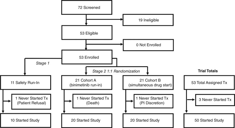
Between October 2018 and June 2021, 72 patients were screened, 53 patients were enrolled in the study, and 50 patients received at least one dose of the study drug (Fig. 1).
Figure 1.
CONSORT diagram illustrating the trial flow. The diagram indicates patients who were screened, eligible, and enrolled into stages 1 and 2 of the trial. PI, principal investigator; Tx, treatment.
The data cutoff was October 16, 2023. Twenty-seven (54%) patients were male, and the median age was 55 years (range, 31–79). Most patients had an ECOG performance status of 0 (66%), with the remainder of patients (34%) having an ECOG performance status of 1. A mutation in KRAS was noted in 24 (48%) patients, with 25 (50%) found to be KRAS wild type and one patient with unknown KRAS status. All patients with known BRAF (n = 42) and NRAS (n = 40) status were wild type. The population was heavily pretreated, with a median of six prior lines of therapy. Baseline characteristics of all patients who received at least one dose of study drug are summarized in Table 1. Forty-five patients completed the first response evaluation imaging (after 9 weeks); 5 patients exited the study prior to this and were considered to have disease progression for response evaluation.
Table 1.
Baseline characteristics of all patients who received at least one dose of study drug.
| Characteristic | Number of patients (N = 50), n (%) |
|---|---|
| Age (years; median, min–max) | 55 (31–79) |
| Sex | |
| Male | 27 (54) |
| Female | 23 (46) |
| ECOG performance status | |
| 0 | 33 (66) |
| 1 | 17 (34) |
| Tumor sidednessa | |
| Right | 11 (22) |
| Left | 39 (78) |
| Gene mutation | |
| KRAS (n = 49 known) | 24 (49) |
| BRAF (n = 43 known) | 0 (0) |
| NRAS (n = 41 known) | 0 (0) |
| Prior bevacizumabb | 46 (92) |
| Baseline liver metastases | |
| Yes | 38 (76) |
| No | 12 (24) |
| Prior lines of therapy (median, min‐max) | 6 (2–10) |
Tumor sidedness is defined as right (ascending colon, hepatic flexure, and transverse colon) or left (splenic flexure, descending colon, and rectum).
Among the n = 4 patients who did not previously receive bevacizumab, bevacizumab was avoided in 1 patient due to an intracranial aneurysm (later evaluated and deemed appropriate to proceed on this trial) and the rationale for avoiding bevacizumab in the other 3 patients was unknown (treated at outside facilities).
Efficacy
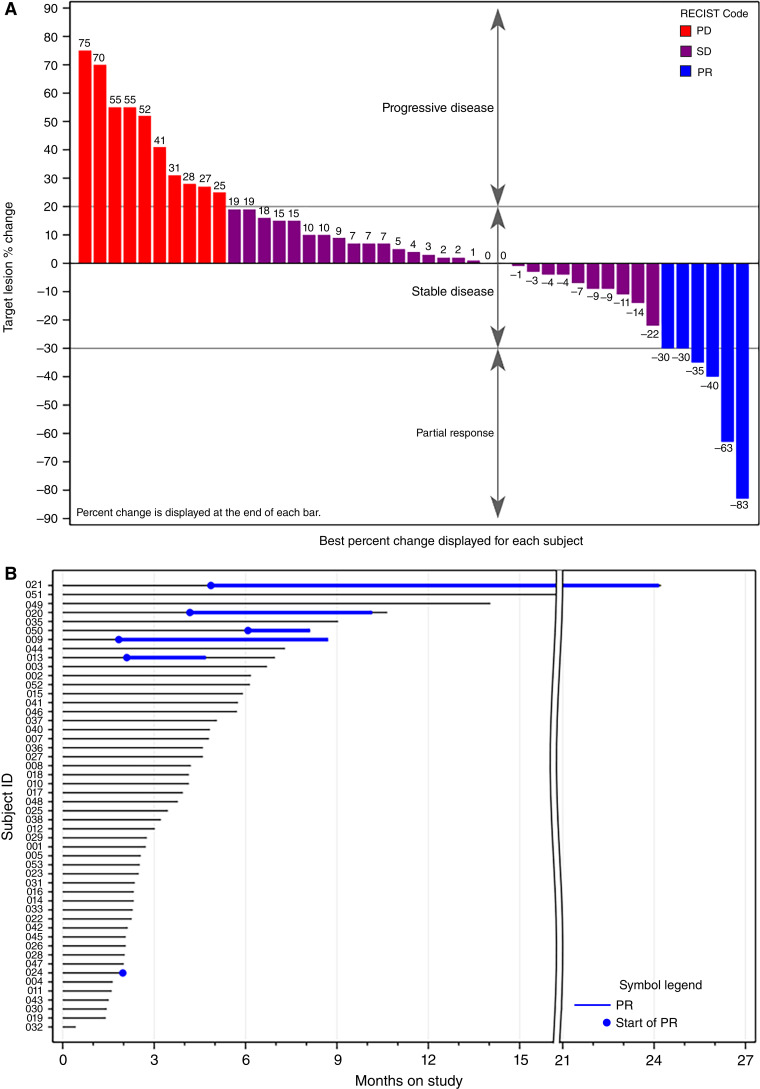
The primary endpoint of this study was ORR per RECIST v1.1. No patients had a complete response. A partial response was seen in 6 patients (12.0%; 95% CI, 4.5%–24.3%), which was not statistically significantly different than the historical control data with regorafenib and trifluridine/tipiracil of 5% (P = 0.038, exceeding the prespecified threshold of 0.025). Stable disease was seen in an additional 29 patients (58.0%). The disease control rate was 70.0% (95% CI, 55.4%–82.1%). Fifteen patients (30.0%) had disease progression as the best response; among these, 2 patients elected to continue study treatments beyond progression and neither had a subsequent response. Using Fisher exact test, there was no statistically significant difference between the two randomized cohorts in ORR (15.0% in cohort A, 10.0% in cohort B, P = 1.00) or DCR (65.0% in cohort A, 70.0% in cohort B, P = 1.00). Among the 6 patients with response, the mean duration of response was 7.3 months, with a range of 2.0 to 20.1 months. Depth and duration of response are demonstrated in Fig. 2A and B, respectively. Stable disease by RECIST v1.1 was further supported by CEA kinetics. Among the 25 patients with stable disease by RECIST at the first response assessment (around cycle 4 day 1) who also had CEA values available at cycle 1 day 1 and cycle 4 day 1, CEA decreased by a mean 130 ng/mL from cycle 1 day 1 to cycle 4 day 1 (95% CI, −263 to +3 ng/mL; P = 0.055; Supplementary Fig. S1A and S1B).
Figure 2.
Depth and duration of response by subject. A, Waterfall plot of best percent change in aggregate size of target lesions. B, Swimmer plot of duration of response. Note: No patients remain on study treatment. PD, progressive disease; PR, partial response; SD, stable disease.
Of the 6 patients with a response, 4 (66.7%) had a KRAS mutation. Disease control (stable disease or better) at the first restaging was seen in 14/23 (60.9%) patients with KRAS mutation and 20/26 (76.9%) patients with KRAS wild type. The ORR in patients with baseline liver metastases was 7.9% (3/38 responses), whereas the ORR in patients with no baseline liver metastases was 25.0% (3/12 responses; P = 0.141). Objective response was observed regardless of tumor sidedness, with ORR 20.0% (2/10), 100.0% (1/1; distal transverse colon with 20.1-month duration of response), and 6.8% (3/44) for patients with right, transverse, and left-sided tumors. The patient with 20.1-month duration of response was enrolled in cohort A and the tumor was KRAS G12V, NRAS wild type, and BRAF wild type. This patient had previously undergone resection of the primary tumor and resection of metastatic disease to the liver (approximately 2.5 years prior to enrollment) and peritoneum. At the time of enrollment, the patient had only lung metastases present. Two other patients had lung-only metastases at the study entry, both of whom experienced stable disease as best response. Of the 4 patients who had not received prior bevacizumab, 3 of them had a response, whereas the other had stable disease.
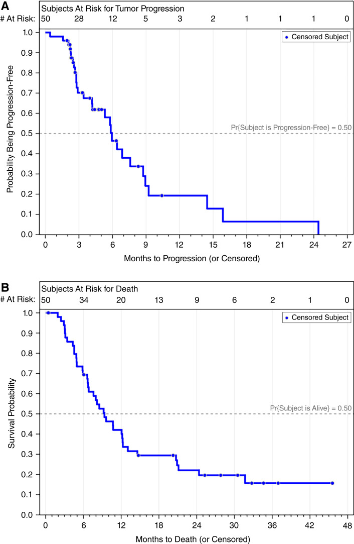
The observed median PFS was 5.9 months (95% CI, 4.2–8.7 months; Fig. 3A) and median OS was 9.3 months (95% CI, 6.7–12.2 months; Fig. 3B). There was no statistically significant difference between the randomized cohorts in median PFS (5.8 months in both cohort A and cohort B, P = 0.18) or median OS (9.3 months in cohort A vs. 8.5 months in cohort B, P = 0.49; Supplementary Figs. S2 and S3). Although median PFS was comparable between patients without and with liver metastases (5.3 months vs. 5.8 months, respectively, P = 0.63), patients without liver metastases had significantly longer median OS (20.9 months vs. 8.0 months, P = 0.03; Supplementary Figs. S4 and S5).
Figure 3.
Survival outcomes of the trial. Kaplan–Meier curves for PFS (A) and OS (B).
Safety and tolerability
Safety and tolerability were as expected for this three-drug regimen. Thirteen patients (27%) had no dose reduction in binimetinib, 22 (45%) required one dose reduction to 30 mg BID, 13 (37%) had a further dose reduction to 15 mg BID, and 1 (2%) patient had to discontinue binimetinib completely (N = 49, as one patient did not receive binimetinib). Among patients who required dose reduction, the most common reasons were rash (51%), vision changes/retinopathy (18%), diarrhea (18%), and fatigue (5%).
Treatment-emergent AEs were common but also as expected. All patients experienced at least one AE. AEs were limited to grade 1 or 2 in 26% of patients; grade 3 and 4 AEs were seen in 56% and 8% of patients, respectively. There were five grade 5 AEs (10% of patients), all of which were assessed as unrelated to study treatments. Acneiform rash was the most common AE, occurring in 39 (78%) patients though with the vast majority (85%) limited to grade 1 or 2. Following rash, the most common AEs included diarrhea (60%), nausea (40%), proteinuria (32%), increased serum creatinine phosphokinase (CPK, 28%), hypertension (26%), fatigue (24%), increased aspartate aminotransferase (22%), increased alkaline phosphatase (20%), and vomiting (20%). Of all grade 3 and 4 AEs, the most common per patient were acneiform rash (12%), increased aspartate aminotransferase (12%), diarrhea (8%), hypertension (8%), increased alanine aminotransferase (8%), anemia (8%), increased serum CPK (6%), and increased alkaline phosphatase (6%). Table 2 summarizes the AE profile per patient.
Table 2.
Summary of treatment-emergent AEs occurring in at least 10% of patients by CTCAE v4.03, by patient (N=50).
| AE term | Grade 1/2 (%) | Grade 3/4 (%) | Total (%) |
|---|---|---|---|
| Rash acneiform | 33 (66) | 6 (12) | 39 (78) |
| Diarrhea | 26 (52) | 4 (8) | 30 (60) |
| Nausea | 18 (36) | 2 (4) | 20 (40) |
| Proteinuria | 16 (32) | 0 | 16 (32) |
| CPK increased | 11 (22) | 3 (6) | 14 (28) |
| Hypertension | 9 (18) | 4 (8) | 13 (26) |
| Fatigue | 11 (22) | 1 (2) | 12 (24) |
| Aspartate aminotransferase increased | 5 (10) | 6 (12) | 11 (22) |
| Alkaline phosphatase increased | 7 (14) | 3 (6) | 10 (20) |
| Vomiting | 9 (18) | 1 (2) | 10 (20) |
| Alanine aminotransferase increased | 5 (10) | 4 (8) | 9 (18) |
| Urinary tract infection | 7 (14) | 1 (2) | 8 (16) |
| Retinopathy | 7 (14) | 1 (2) | 8 (16) |
| Constipation | 6 (12) | 1 (2) | 7 (14) |
| Hypothyroidism | 7 (14) | 0 | 7 (14) |
| Blurred vision | 7 (14) | 0 | 7 (14) |
| Fever | 7 (14) | 0 | 7 (14) |
| Anemia | 1 (2) | 4 (8) | 5 (10) |
| Dehydration | 4 (8) | 1 (2) | 5 (10) |
| Abdominal pain | 4 (8) | 1 (2) | 5 (10) |
| Edema limbs | 5 (10) | 0 | 5 (10) |
| Mucositis oral | 5 (10) | 0 | 5 (10) |
Correlative analyses
Immune cells in the peripheral blood from all study participants with paired samples (N = 27 patients, including N = 14 in cohort A and N = 13 in cohort B) were analyzed at baseline (in cohort A, after the 7-day binimetinib run-in and prior to infusion on cycle 1 day 1, and in cohort B, prior to starting all treatments on cycle 1 day 1) and posttreatment (either prior to infusion on cycle 4 day 1, if partial response or stable disease, or end of study) by mass cytometry. At baseline, there were no differences in any measured cell population between patients in cohorts A and B and between patients with and without liver metastases (data not shown, due to numerous cell populations analyzed and no differences observed), except phospho-ERK+ monocytes were significantly lower in cohort A (after the binimetinib run-in) compared with cohort B (prior to initiation of all treatments; Supplementary Fig. S6). Although this indicates that the binimetinib run-in induced pharmacodynamic effect, markers of immune activation related to this run-in were not identified in the peripheral blood.
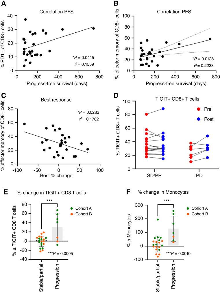
In all patients at baseline, a higher percentage of PD1+ CD8+ T cells and effector memory CD8+ T cells correlated with longer PFS; the latter also correlated with increased tumor shrinkage from baseline (Fig. 4A–C). The coefficients of determination (R2) were low. Similar correlation was observed between baseline percentage of PD1+ CD8+ T cells and effector memory CD8+ T cells and PFS in cohort A but not cohort B (Supplementary Fig. S7A–F); this seems to be driven by several patients with long PFS in cohort A. No correlation was observed between baseline percentage of PD1+ CD8+ T cells and effector memory CD8+ T cells and PFS or best tumor response in patients with or without liver metastases (Supplementary Fig. S8A–F). Patients with disease progression as best response, compared with disease control (stable disease or partial response), demonstrated an increase in T-cell immunoreceptor with immunoglobulin and immunoreceptor tyrosine-based inhibitory motif domain (TIGIT) expression on CD8+ T cells and an increase in monocytes posttreatment versus baseline (Fig. 4D–F). Changes in TIGIT+ CD8+ T cells and monocytes seem to be driven by patients in cohort A (Fig. 4E and F); however, the low number of patients in cohort B with disease progression as best response limits conclusions. This suggests that increased TIGIT expression on T cells and increased monocyte abundance may mediate resistance to this treatment regimen. Correlative analyses from paired tumor biopsies are ongoing and will be separately reported.
Figure 4.
Correlative peripheral blood immune cell characterization pre- and posttreatment. Correlation of (A and B) baseline % PD1+ CD8+ T cells and % effector memory CD8+ T cells (CD8+CD45RO+/−CD27lowCD127lowPD1lowCD11b+CD25+TbetlowCCR4HighCCR6+CCR7−) with PFS; (C) baseline % effector memory CD8+ T cells with best tumor response; (D) % TIGIT+ CD8+ T cells at baseline and posttreatment in patients with disease control (SD or PR and PD); and (E and F) correlation of change in % TIGIT+ CD8+ T cells and monocytes (CD3−CD19−CD56−CD11c+) posttreatment vs. baseline (% in the posttreatment sample − % in the baseline sample)/% in the baseline sample, with a positive number indicating an increase from baseline to posttreatment.
Discussion
pMMR/MSS mCRC does not respond to single-agent anti-PD1 therapy, with response rates of 0% (40). Our study is one of the several recent trials to investigate novel immunotherapy combinations in MSS mCRC; although efficacy has varied, patients without liver metastases seem to achieve more favorable outcomes.
The phase Ib REGONIVO trial evaluated regorafenib and nivolumab in Japanese patients with refractory gastric and colorectal cancer (all but one patient was MSS). Among 25 patients with colorectal cancer, the ORR was 36% with median PFS 7.9 months; ORR was higher in patients without (58%) versus with (15%) liver metastases (17). A subsequent study in MSS colorectal cancer with the same treatments in the United States was disappointing, with an ORR of 7% in all patients; ORR was higher (22%) in patients without liver metastases (41). In patients without liver metastases, the combination of regorafenib, ipilimumab, and nivolumab led to 36% ORR and median OS was more than 22 months (42). Trials evaluating regorafenib + pembrolizumab or regorafenib + avelumab (REGOMUNE) were unsuccessful, with 0% ORR (43, 44). Some responses were observed in patients with MSS colorectal cancer in the phase II LEAP-005 trial with lenvatinib + pembrolizumab (ORR 22% and median PFS 2.3 months) and phase II CAMILLA trial with cabozantinib + durvalumab (ORR 28% and median PFS 4.4 months; refs. 16, 45). Among patients without liver metastases treated with botensilimab and balstilimab in the phase I trial, the ORR was 23%, DCR was 80%, and median OS was 20.9 months (46). To date, there are two randomized phase III trials in this patient population. The IMblaze370 trial randomized patients with refractory colorectal cancer (95% MSS) to atezolizumab and cobimetinib, atezolizumab alone, or standard-of-care regorafenib. ORR, PFS, and OS were similar among all three treatment groups; partial responses were observed in 3% of patients treated with atezolizumab and cobimetinib, and 2% of patients treated with atezolizumab alone or regorafenib (26). The second randomized phase III trial was LEAP-017, which randomized patients with refractory pMMR/MSS colorectal cancer (70% with liver metastases) to either lenvatinib and pembrolizumab or standard-of-care therapy (investigator’s choice of regorafenib or trifluridine/tipiracil). In abstract format at final analysis, lenvatinib and pembrolizumab trended toward longer OS, PFS, and ORR but did not meet statistical significance thresholds (47). The multikinase inhibitor XL092 with atezolizumab is being evaluated in the ongoing phase III STELLAR-303 trial (48), as are fruquintinib with tislelizumab in another phase II trial (NCT04716634).
The ORR of 12% in our trial was numerically higher than prior studies with anti-PD1 checkpoint blockade alone (0%) and a prior study with binimetinib and pembrolizumab (0%). Although the primary endpoint, ORR, was not statistically significant better than the prespecified 5% historical control threshold in refractory MSS colorectal cancer (regorafenib, fruquintinib, and trifluridine/tipiracil ± bevacizumab, 1%–6%), a trend was observed (5–9, 27). The patient population was heavily pretreated (median 6 prior lines of therapy), which may contribute to the relatively low ORR observed. Acknowledging limitations of cross-trial comparisons and the single-institution nature of this study, ORR observed here is similar to that observed in multiple recent studies investigating combination immunotherapy in MSS colorectal cancer. We also observed a potential signal of increased effectiveness of pembrolizumab, binimetinib, and bevacizumab in patients without liver metastases, with a trend toward better ORR, statistically longer median OS than patients with liver metastases, and a 20.1-month duration of response in one patient; this is similar to results of other studies in this patient population. There was no difference in efficacy between the randomized cohorts (binimetinib run-in vs. concurrent treatment initiation), which were included to explore the hypothesis that MEK inhibitor “run-in” may increase tumor immunogenicity. Although the primary endpoint was not met, the DCR of 70% (supported by both imaging and CEA kinetics), median PFS of 5.9 months, and longest partial response of 20.1 months are encouraging that pembrolizumab, binimetinib, and bevacizumab may be immunomodulatory and synergistically reprogram the TME in some heavily pretreated patients and confer improved efficacy compared with prior results with cobimetinib and atezolizumab.
Toxicity was consistent with the known profiles of pembrolizumab, binimetinib, and bevacizumab. The most common AE was acneiform rash, occurring in 78% of patients, and was grade 3 to 4 in 12%. Other common AEs included diarrhea, nausea, vomiting, proteinuria, increased CPK, hypertension, fatigue, and increased transaminases. Binimetinib dose reduction was frequently required (73% of patients), whereas only one patient required permanent discontinuation.
Investigation of mechanisms of response, mechanisms of resistance, and predictive biomarkers are critically important to better identify patients most likely to benefit from this or similar regimens. Correlative analysis suggests that higher baseline levels of peripheral blood PD1+ CD8+ T cells and effector memory CD8+ T cells correlated with response to therapy, however the coefficients of determination are low and may be due to small sample size. Higher baseline PD1+ CD8+ T cells and PD1+ CD8+ T-cell receptor diversity in the peripheral blood has been correlated with improved outcomes in patients with non–small cell lung cancer treated with immunotherapy (49, 50). In melanoma, baseline peripheral blood effector memory CD8+ T cells were higher in responders to anti-CTLA4 therapy (51). These findings are intriguing and could serve as predictive biomarkers if validated in future immunotherapy clinical trials in MSS colorectal cancer. Although the binimetinib run-in demonstrated a pharmacodynamic effect in the peripheral blood (lower pERK+ monocytes compared with cohort B), there was no evidence of an immunomodulatory effect in the peripheral blood; this was consistent with similar efficacy in the randomized cohorts. Although patients in this trial without liver metastases experienced numerically higher ORR and statistically longer survival, there were no baseline peripheral blood predictive immune biomarkers identified. Additionally, increased TIGIT expression on CD8+ T cells and increased monocyte abundance in the peripheral blood on-treatment versus baseline was associated with disease progression. This highlights TIGIT expression and monocytes as possible mechanisms of resistance to pembrolizumab, binimetinib, and bevacizumab, and potential treatment targets in the future.
There are several limitations with peripheral blood correlative analyses. First, a true “pretreatment” sample in cohort A was not obtained (the first sample was obtained after the binimetinib run-in), precluding intrapatient comparison of the pharmacodynamic effect of the binimetinib run-in. Second, the small sample size of patients with primary disease progression in cohort B (N = 2) limited additional analyses between cohorts A and B. Finally, the peripheral blood immune profile may not reflect the TME. Correlative analyses from paired tumor biopsies and ctDNA are ongoing, will be separately reported, and may further elucidate the impact of the binimetinib run-in and impact of liver metastases on treatment efficacy. Additional study limitations include the modest sample size and single-arm design, which makes efficacy comparison with historical and contemporary controls and subgroup analyses difficult. Detailed information on sites of metastatic disease outside the liver and lungs was not available, further limiting subgroup analyses in these populations. Tumor molecular profiling was not repeated prior to study entry, raising the possibility of undetected treatment-emergent BRAF, MAPK, or other alterations (52). Most patients had an ECOG performance status of 0, limiting the generalizability of results in a heavily pretreated population. Although responses were seen regardless of KRAS mutational status, the small number of responses similarly limits further interpretation. Data on reason for trial discontinuation, performance status at trial discontinuation, and subsequent therapy are not available, making it difficult to comment on explanations for the observation that the median OS was only 3.4 months longer than median PFS.
Conclusions
The combination of pembrolizumab, binimetinib, and bevacizumab failed to meet its primary endpoint of higher ORR compared with historical control data of current treatment options for refractory pMMR/MSS BRAF wild type mCRC while demonstrating a high disease control rate with an expected safety profile in this single-institution study. There was no difference in efficacy between MEK inhibitor run-in and concurrent treatment initiation in the randomized cohorts. A signal of improved efficacy was observed in patients without liver metastases at study entry, supporting further investigation of pembrolizumab, binimetinib, and bevacizumab in this population. At baseline, a higher percentage of peripheral blood PD1+ CD8+ T cells and effector memory CD8+ T cells correlated with longer PFS, which may serve as an accessible biomarker for selecting patients more likely to benefit from immunotherapy regimens if appropriately validated. An increase in TIGIT expression on CD8+ T cells and increase in monocyte abundance in the peripheral blood during treatment may mediate resistance to this treatment regimen, providing rationale to investigate the inclusion of an anti-TIGIT agent in immunotherapy combinations in this patient population. Correlative analyses from paired tumor biopsies are ongoing and may further elucidate mechanisms of response and resistance.
Supplementary Material
Supplementary Table S1. Representativeness of Study Participants.
Supplementary Figure S1. CEA kinetics in patients with stable disease at first response assessment.
Supplementary Figure S2. Progression-free survival in the randomized cohorts.
Supplementary Figure S3. Overall survival in the randomized cohorts.
Supplementary Figure S4. Progression-free survival in patients with or without baseline liver metastases.
Supplementary Figure S5. Overall survival in patients with or without baseline liver metastases.
Supplementary Figure S6. Correlative peripheral blood immune cell characterization at baseline.
Supplementary Figure S7. Correlation of baseline immune cell populations with response to treatment in Cohorts A and B.
Supplementary Figure S8. Correlation of baseline immune cell populations with response to treatment in patients with and without liver metastases.
Acknowledgments
This work was supported by NIH R01CA229259-01 (to C.H. Lieu), NIH 5K12CA086913-21 (to R.W. Lentz), the University of Colorado Cancer Center Support Grant (P30CA046934), Merck, and Pfizer. The authors thank additional laboratory technicians including Brian Gittleman, Betelehem Yacob, Jessica Pafford, Natalie Navarro, and Stephen Smoots.
Footnotes
Note: Supplementary data for this article are available at Clinical Cancer Research Online (http://clincancerres.aacrjournals.org/).
Authors’ Disclosures
R.W. Lentz reports grants from NIH during the conduct of the study; R.W. Lentz also reports grants from ALX Oncology, Guardant, and EDDC; nonfinancial support from Merck and Eli Lilly and Company; other support from Myeloid Therapeutics and Agenus; and grants and other support from Boehringer Ingelheim outside the submitted work. T.M. Pitts reports grants from NIH during the conduct of the study. S.L. Davis reports other support from Merck during the conduct of the study, as well as other support from Bristol Myers Squibb, Symphogen, I-Mab, TriSalus Life Sciences, Tvardi Therapeutics, EMD Serono, and ORIC Pharmaceuticals outside the submitted work. S.S. Kim reports grants from Merck and other support from Eisai, Bristol Myers Squibb, Daiichi Sankyo, and Astellas outside the submitted work. A.D. Leal reports nonfinancial support and other support from Bristol Myers Squibb and Exelixis; nonfinancial support from Elicio Therapeutics, Inc., AbbVie, FameWave Ltd., Conjupro Biotherapeutics, Inc., and Corcept Therapeutics; and other support from Elsevier outside the submitted work. S.G. Eckhardt reports other support from Exelixis, Amgen, and OnKure outside the submitted work. C.H. Lieu reports grants from NCI R01CA229259-01, Merck, and Pfizer during the conduct of the study. No disclosures were reported by the other authors.
Authors’ Contributions
R.W. Lentz: Resources, data curation, formal analysis, funding acquisition, validation, investigation, visualization, methodology, writing–original draft, writing–review and editing. T.J. Friedrich: Data curation, formal analysis, validation, investigation, visualization, methodology, writing–original draft, writing–review and editing. P.J. Blatchford: Data curation, software, formal analysis, visualization, writing–original draft, writing–review and editing. K.R. Jordan: Data curation, software, formal analysis, investigation, visualization, writing–original draft, writing–review and editing. T.M. Pitts: Conceptualization, writing–review and editing. H.R. Robinson: Investigation, writing–review and editing. S.L. Davis: Investigation, writing–review and editing. S.S. Kim: Investigation, writing–review and editing. A.D. Leal: Investigation, writing–review and editing. M.R. Lee: Investigation, project administration, writing–review and editing. M.R.N. Waring: Investigation, project administration, writing–review and editing. A.C. Martin: Investigation, project administration, writing–review and editing. A.T.A. Dominguez: Investigation, writing–review and editing. S.M. Bagby: Investigation, writing–review and editing. S.J. Hartman: Investigation, writing–review and editing. S.G. Eckhardt: Conceptualization, supervision, writing–review and editing. W.A. Messersmith: Investigation, writing–review and editing. C.H. Lieu: Conceptualization, resources, data curation, formal analysis, supervision, funding acquisition, validation, investigation, visualization, methodology, writing–original draft, writing–review and editing.
References
- 1. Siegel RL, Miller KD, Wagle NS, Jemal A. Cancer statistics, 2023. CA Cancer J Clin 2023;73:17–48. [DOI] [PubMed] [Google Scholar]
- 2. Douillard JY, Siena S, Cassidy J, Tabernero J, Burkes R, Barugel M, et al. Randomized, phase III trial of panitumumab with infusional fluorouracil, leucovorin, and oxaliplatin (FOLFOX4) versus FOLFOX4 alone as first-line treatment in patients with previously untreated metastatic colorectal cancer: the PRIME study. J Clin Oncol 2010;28:4697–705. [DOI] [PubMed] [Google Scholar]
- 3. Douillard JY, Oliner KS, Siena S, Tabernero J, Burkes R, Barugel M, et al. Panitumumab-FOLFOX4 treatment and RAS mutations in colorectal cancer. N Engl J Med 2013;369:1023–34. [DOI] [PubMed] [Google Scholar]
- 4. Di Nicolantonio F, Martini M, Molinari F, Sartore-Bianchi A, Arena S, Saletti P, et al. Wild-type BRAF is required for response to panitumumab or cetuximab in metastatic colorectal cancer. J Clin Oncol 2008;26:5705–12. [DOI] [PubMed] [Google Scholar]
- 5. Mayer RJ, Van Cutsem E, Falcone A, Yoshino T, Garcia-Carbonero R, Mizunuma N, et al. Randomized trial of TAS-102 for refractory metastatic colorectal cancer. N Engl J Med 2015;372:1909–19. [DOI] [PubMed] [Google Scholar]
- 6. Grothey A, Van Cutsem E, Sobrero A, Siena S, Falcone A, Ychou M, et al. Regorafenib monotherapy for previously treated metastatic colorectal cancer (CORRECT): an international, multicentre, randomised, placebo-controlled, phase 3 trial. Lancet 2013;381:303–12. [DOI] [PubMed] [Google Scholar]
- 7. Tabernero J, Prager GW, Fakih M, Ciardiello F, Van Cutsem E, Elez E, et al. Trifluridine/tipiracil plus bevacizumab for third-line treatment of refractory metastatic colorectal cancer: the phase 3 randomized SUNLIGHT study. J Clin Oncol 2023;41:4. [Google Scholar]
- 8. Dasari A, Lonardi S, Garcia-Carbonero R, Elez E, Yoshino T, Sobrero A, et al. Fruquintinib versus placebo in patients with refractory metastatic colorectal cancer (FRESCO-2): an international, multicentre, randomised, double-blind, phase 3 study. Lancet 2023;402:41–53. [DOI] [PubMed] [Google Scholar]
- 9. Li J, Qin S, Xu RH, Shen L, Xu J, Bai Y, et al. Effect of fruquintinib vs placebo on overall survival in patients with previously treated metastatic colorectal cancer: the FRESCO randomized clinical trial. JAMA 2018;319:2486–96. [DOI] [PMC free article] [PubMed] [Google Scholar]
- 10. André T, Shiu K-K, Kim TW, Jensen BV, Jensen LH, Punt C, et al. Pembrolizumab in microsatellite-instability–high advanced colorectal cancer. N Engl J Med 2020;383:2207–18. [DOI] [PubMed] [Google Scholar]
- 11. Cercek A, Lumish M, Sinopoli J, Weiss J, Shia J, Lamendola-Essel M, et al. PD-1 blockade in mismatch repair-deficient, locally advanced rectal cancer. N Engl J Med 2022;386:2363–76. [DOI] [PMC free article] [PubMed] [Google Scholar]
- 12. Verschoor YL, Van Den Berg J, Beets G, Sikorska K, Aalbers A, Van Lent A, et al. Neoadjuvant nivolumab, ipilimumab, and celecoxib in MMR-proficient and MMR-deficient colon cancers: final clinical analysis of the NICHE study. J Clin Oncol 2022;40:3511–11. [Google Scholar]
- 13. Buchler T. Microsatellite instability and metastatic colorectal cancer—a clinical perspective. Front Oncol 2022;12:888181. [DOI] [PMC free article] [PubMed] [Google Scholar]
- 14. Finn RS, Qin S, Ikeda M, Galle PR, Ducreux M, Kim TY, et al. Atezolizumab plus bevacizumab in unresectable hepatocellular carcinoma. N Engl J Med 2020;382:1894–905. [DOI] [PubMed] [Google Scholar]
- 15. Choueiri TK, Powles T, Burotto M, Escudier B, Bourlon MT, Zurawski B, et al. Nivolumab plus cabozantinib versus sunitinib for advanced renal-cell carcinoma. N Engl J Med 2021;384:829–41. [DOI] [PMC free article] [PubMed] [Google Scholar]
- 16. Gomez-Roca C, Yanez E, Im S-A, Castanon Alvarez E, Senellart H, Doherty M, et al. LEAP-005: a phase II multicohort study of lenvatinib plus pembrolizumab in patients with previously treated selected solid tumors—results from the colorectal cancer cohort. J Clin Oncol 2021;39:94. [Google Scholar]
- 17. Fukuoka S, Hara H, Takahashi N, Kojima T, Kawazoe A, Asayama M, et al. Regorafenib plus nivolumab in patients with advanced gastric or colorectal cancer: an open-label, dose-escalation, and dose-expansion phase Ib trial (REGONIVO, EPOC1603). J Clin Oncol 2020;38:2053–61. [DOI] [PubMed] [Google Scholar]
- 18. Ganesh K, Stadler ZK, Cercek A, Mendelsohn RB, Shia J, Segal NH, et al. Immunotherapy in colorectal cancer: rationale, challenges and potential. Nat Rev Gastroenterol Hepatol 2019;16:361–75. [DOI] [PMC free article] [PubMed] [Google Scholar]
- 19. Fang JY, Richardson BC. The MAPK signalling pathways and colorectal cancer. Lancet Oncol 2005;6:322–7. [DOI] [PubMed] [Google Scholar]
- 20. Zhang W, Liu HT. MAPK signal pathways in the regulation of cell proliferation in mammalian cells. Cell Res 2002;12:9–18. [DOI] [PubMed] [Google Scholar]
- 21. Flaherty KT, Robert C, Hersey P, Nathan P, Garbe C, Milhem M, et al. Improved survival with MEK inhibition in BRAF-mutated melanoma. N Engl J Med 2012;367:107–14. [DOI] [PubMed] [Google Scholar]
- 22. Kopetz S, Grothey A, Yaeger R, Van Cutsem E, Desai J, Yoshino T, et al. Encorafenib, binimetinib, and cetuximab in BRAF V600e–mutated colorectal cancer. N Engl J Med 2019;381:1632–43. [DOI] [PubMed] [Google Scholar]
- 23. Kakavand H, Wilmott JS, Menzies AM, Vilain R, Haydu LE, Yearley JH, et al. PD-L1 expression and tumor-infiltrating lymphocytes define different subsets of MAPK inhibitor-treated melanoma patients. Clin Cancer Res 2015;21:3140–8. [DOI] [PubMed] [Google Scholar]
- 24. Loi S, Dushyanthen S, Beavis PA, Salgado R, Denkert C, Savas P, et al. RAS/MAPK activation is associated with reduced tumor-infiltrating lymphocytes in triple-negative breast cancer: therapeutic cooperation between MEK and PD-1/PD-L1 immune checkpoint inhibitors. Clin Cancer Res 2016;22:1499–509. [DOI] [PMC free article] [PubMed] [Google Scholar]
- 25. Liu L, Mayes PA, Eastman S, Shi H, Yadavilli S, Zhang T, et al. The BRAF and MEK inhibitors dabrafenib and trametinib: effects on immune function and in combination with immunomodulatory antibodies targeting PD-1, PD-L1, and CTLA-4. Clin Cancer Res 2015;21:1639–51. [DOI] [PubMed] [Google Scholar]
- 26. Eng C, Kim TW, Bendell J, Argilés G, Tebbutt NC, Di Bartolomeo M, et al. Atezolizumab with or without cobimetinib versus regorafenib in previously treated metastatic colorectal cancer (IMblaze370): a multicentre, open-label, phase 3, randomised, controlled trial. Lancet Oncol 2019;20:849–61. [DOI] [PubMed] [Google Scholar]
- 27. Chen EX, Kavan P, Tehfe M, Kortmansky JS, Sawyer MB, Chiorean EG, et al. Pembrolizumab (pembro) plus binimetinib (bini) with or without chemotherapy (chemo) for metastatic colorectal cancer (mCRC): results from KEYNOTE-651 cohorts A, C, and E. J Clin Oncol 2022;40:3573.35724342 [Google Scholar]
- 28. Oyama T, Ran S, Ishida T, Nadaf S, Kerr L, Carbone DP, et al. Vascular endothelial growth factor affects dendritic cell maturation through the inhibition of nuclear factor-kappa B activation in hemopoietic progenitor cells. J Immunol 1998;160:1224–32. [PubMed] [Google Scholar]
- 29. Ohm JE, Carbone DP. VEGF as a mediator of tumor-associated immunodeficiency. Immunol Res 2001;23:263–72. [DOI] [PubMed] [Google Scholar]
- 30. Voron T, Colussi O, Marcheteau E, Pernot S, Nizard M, Pointet AL, et al. VEGF-A modulates expression of inhibitory checkpoints on CD8+ T cells in tumors. J Exp Med 2015;212:139–48. [DOI] [PMC free article] [PubMed] [Google Scholar]
- 31. Dikov MM, Ohm JE, Ray N, Tchekneva EE, Burlison J, Moghanaki D, et al. Differential roles of vascular endothelial growth factor receptors 1 and 2 in dendritic cell differentiation. J Immunol 2005;174:215–22. [DOI] [PubMed] [Google Scholar]
- 32. Dirkx AE, oude Egbrink MG, Castermans K, van der Schaft DW, Thijssen VL, Dings RP, et al. Anti-angiogenesis therapy can overcome endothelial cell anergy and promote leukocyte-endothelium interactions and infiltration in tumors. FASEB J 2006;20:621–30. [DOI] [PubMed] [Google Scholar]
- 33. Hodi FS, Lawrence D, Lezcano C, Wu X, Zhou J, Sasada T, et al. Bevacizumab plus ipilimumab in patients with metastatic melanoma. Cancer Immunol Res 2014;2:632–42. [DOI] [PMC free article] [PubMed] [Google Scholar]
- 34. Hodi FS, Mihm MC, Soiffer RJ, Haluska FG, Butler M, Seiden MV, et al. Biologic activity of cytotoxic T lymphocyte-associated antigen 4 antibody blockade in previously vaccinated metastatic melanoma and ovarian carcinoma patients. Proc Natl Acad Sci U S A 2003;100:4712–7. [DOI] [PMC free article] [PubMed] [Google Scholar]
- 35. Eisenhauer EA, Therasse P, Bogaerts J, Schwartz LH, Sargent D, Ford R, et al. New response evaluation criteria in solid tumours: revised RECIST guideline (version 1.1). Eur J Cancer 2009;45:228–47. [DOI] [PubMed] [Google Scholar]
- 36. Krishnamurthy A, Dasari A, Noonan AM, Mehnert JM, Lockhart AC, Leong S, et al. Phase Ib results of the rational combination of selumetinib and cyclosporin A in advanced solid tumors with an expansion cohort in metastatic colorectal cancer. Cancer Res 2018;78:5398–407. [DOI] [PMC free article] [PubMed] [Google Scholar]
- 37. Schwartz LH, Litière S, de Vries E, Ford R, Gwyther S, Mandrekar S, et al. RECIST 1.1-update and clarification: from the RECIST committee. Eur J Cancer 2016;62:132–7. [DOI] [PMC free article] [PubMed] [Google Scholar]
- 38. US Department of Health and Human Services . Common terminology criteria for adverse events (CTCAE) version 4.03. [cited 2024]. Available from:https://evs.nci.nih.gov/ftp1/CTCAE/CTCAE_4.03/CTCAE_4.03_2010-06-14_QuickReference_8.5x11.pdf
- 39. Borgers JSW, Tobin RP, Vorwald VM, Smith JM, Davis DM, Kimball AK, et al. High-dimensional analysis of postsplenectomy peripheral immune cell changes. Immunohorizons 2020;4:82–92. [DOI] [PMC free article] [PubMed] [Google Scholar]
- 40. Le DT, Uram JN, Wang H, Bartlett BR, Kemberling H, Eyring AD, et al. PD-1 blockade in tumors with mismatch-repair deficiency. N Engl J Med 2015;372:2509–20. [DOI] [PMC free article] [PubMed] [Google Scholar]
- 41. Fakih M, Raghav KPS, Chang DZ, Bendell JC, Larson T, Cohn AL, et al. Single-arm, phase 2 study of regorafenib plus nivolumab in patients with mismatch repair-proficient (pMMR)/microsatellite stable (MSS) colorectal cancer (CRC). J Clin Oncol 2021;39:3560. [Google Scholar]
- 42. Fakih M, Sandhu J, Lim D, Li X, Li S, Wang C. Regorafenib, ipilimumab, and nivolumab for patients with microsatellite stable colorectal cancer and disease progression with prior chemotherapy: a phase 1 nonrandomized clinical trial. JAMA Oncol 2023;9:627–34. [DOI] [PMC free article] [PubMed] [Google Scholar]
- 43. Barzi A, Azad NS, Yang Y, Tsao-Wei D, Rehman R, Fakih M, et al. Phase I/II study of regorafenib (rego) and pembrolizumab (pembro) in refractory microsatellite stable colorectal cancer (MSSCRC). J Clin Oncol 2022;40:15. [Google Scholar]
- 44. Cousin S, Bellera CA, Guégan JP, Gomez-Roca CA, Metges J-P, Adenis A, et al. REGOMUNE: a phase II study of regorafenib plus avelumab in solid tumors—results of the non-MSI-H metastatic colorectal cancer (mCRC) cohort. J Clin Oncol 2020;38:4019.32986529 [Google Scholar]
- 45. Saeed A, Park R, Dai J, Al-Rajabi RMT, Kasi A, Saeed A, et al. Phase II trial of cabozantinib (Cabo) plus durvalumab (Durva) in chemotherapy refractory patients with advanced mismatch repair proficient/microsatellite stable (pMMR/MSS) colorectal cancer (CRC): CAMILLA CRC cohort results. J Clin Oncol 2022;40:135. [Google Scholar]
- 46. Bullock A, Fakih M, Gordon M, Tsimberidou A, El-Khoueiry A, Wilky B, et al. LBA-4 results from an expanded phase 1 trial of botensilimab (BOT), a multifunctional anti-CTLA-4, plus balstilimab (BAL; anti-PD-1) for metastatic heavily pretreated microsatellite stable colorectal cancer (MSS CRC). Ann Oncol 2023;34(Suppl 1):S178–9. [Google Scholar]
- 47. Kawazoe A, Xu R, Passhak M, Teng H, Shergill A, Gumus M, et al. LBA-5 Lenvatinib plus pembrolizumab versus standard of care for previously treated metastatic colorectal cancer (mCRC): the phase 3 LEAP-017 study. Ann Oncol 2023;34:S179. [DOI] [PMC free article] [PubMed] [Google Scholar]
- 48. Hecht JR, Tabernero J, Parikh AR, Wang Y, Wang Z, Schwickart M, et al. STELLAR-303: a phase 3 study of XL092 in combination with atezolizumab versus regorafenib in patients with previously treated metastatic colorectal cancer (mCRC). J Clin Oncol 2023;41:TPS267. [Google Scholar]
- 49. Han J, Duan J, Bai H, Wang Y, Wan R, Wang X, et al. TCR repertoire diversity of peripheral PD-1+CD8+ T cells predicts clinical outcomes after immunotherapy in patients with non-small cell lung cancer. Cancer Immunol Res 2020;8:146–54. [DOI] [PubMed] [Google Scholar]
- 50. Mazzaschi G, Minari R, Zecca A, Cavazzoni A, Ferri V, Mori C, et al. Soluble PD-L1 and circulating CD8+PD-1+ and NK cells enclose a prognostic and predictive immune effector score in immunotherapy treated NSCLC patients. Lung Cancer 2020;148:1–11. [DOI] [PubMed] [Google Scholar]
- 51. Subrahmanyam PB, Dong Z, Gusenleitner D, Giobbie-Hurder A, Severgnini M, Zhou J, et al. Distinct predictive biomarker candidates for response to anti-CTLA-4 and anti-PD-1 immunotherapy in melanoma patients. J Immunother Cancer 2018;6:18. [DOI] [PMC free article] [PubMed] [Google Scholar]
- 52. Parseghian CM, Sun R, Woods M, Napolitano S, Lee HM, Alshenaifi J, et al. Resistance mechanisms to anti-epidermal growth factor receptor therapy in RAS/RAF wild-type colorectal cancer vary by regimen and line of therapy. J Clin Oncol 2023;41:460–71. [DOI] [PMC free article] [PubMed] [Google Scholar]
Associated Data
This section collects any data citations, data availability statements, or supplementary materials included in this article.
Supplementary Materials
Supplementary Table S1. Representativeness of Study Participants.
Supplementary Figure S1. CEA kinetics in patients with stable disease at first response assessment.
Supplementary Figure S2. Progression-free survival in the randomized cohorts.
Supplementary Figure S3. Overall survival in the randomized cohorts.
Supplementary Figure S4. Progression-free survival in patients with or without baseline liver metastases.
Supplementary Figure S5. Overall survival in patients with or without baseline liver metastases.
Supplementary Figure S6. Correlative peripheral blood immune cell characterization at baseline.
Supplementary Figure S7. Correlation of baseline immune cell populations with response to treatment in Cohorts A and B.
Supplementary Figure S8. Correlation of baseline immune cell populations with response to treatment in patients with and without liver metastases.
Data Availability Statement
The data generated in this study are available upon request from the corresponding author and on ClinicalTrials.gov (NCT03475004), as a community-recognized, structured repository does not exist.


