Abstract
Background
Keen Osteoarthritis (KOA) is a common chronic disabling disease characterized by joint pain and dysfunction, which seriously affects patients’ quality of life. Recent studies have shown that transcranial direct current stimulation (tDCS) was a promising treatment for KOA.
Purpose
Investigate the effects of tDCS on pain and physical function in patients with KOA.
Methods
Randomized controlled trials related to tDCS and KOA were systematically searched in the PubMed, Embase, Medline, Cochrane Library, CINHL, and Web of Science databases from inception to July 23, 2024. The pain intensity was evaluated using the visual analog scale or the numeric rating scale, and the pain sensitivity was assessed using conditioned pain modulation, pressure pain threshold, heat pain threshold, or heat pain tolerance. The physical function outcome was evaluated using the Western Ontario and McMaster Universities Osteoarthritis Index or the Knee injury and Osteoarthritis Outcome Score. Statistical analysis was performed using Review Manager 5.4.
Results
Seven studies with a total of 503 participants were included. Compared to sham tDCS, tDCS was effective in reducing the short-term pain intensity (SMD: -0.58; 95% CI: -1.02, -0.14; p = 0.01) and pain sensitivity (SMD: -0.43; 95% CI: -0.70, -0.16; p = 0.002) but failed to significantly improve the long-term pain intensity (SMD: -0.26; 95% CI: -0.59, 0.08; p = 0.13) in KOA patients. In addition, tDCS did not significantly improve the short-term (SMD: -0.13; 95% CI: -0.35, 0.08; p = 0.22) and long-term (SMD: 0.02; 95% CI: -0.22, 0.25; p = 0.90) physical function in patients with KOA.
Conclusions
The tDCS can reduce short-term pain intensity and sensitivity but fails to significantly relieve long-term pain intensity and improve the physical function in patients with KOA. Thus, tDCS may be a potential therapeutic tool to reduce short-term pain intensity and pain sensitivity in patients with KOA.
Supplementary Information
The online version contains supplementary material available at 10.1186/s12891-024-07805-3.
Keywords: Transcranial direct current stimulation, Knee osteoarthritis, Pain, Physical function
Introduction
Osteoarthritis is a chronic, disabling disease of multiple etiologies, occurring primarily in joints with high loads and activities; the knee is the most complex and loaded joint in the body and, therefore, most prone to OA [1]. Knee osteoarthritis (KOA) is a common chronic disabling disease in the middle-aged and elderly population and has become the fourth leading cause of disability worldwide [2, 3]. The pathogenesis of KOA is complex and involves multiple factors, such as mechanical stress, inflammation, metabolism, immunity, and genetics, with age, genetics, body weight, gender, and race as risk factors [4, 5]. KOA is characterized by joint pain, stiffness, swelling, and limited joint function due to structural and functional failure of the synovial joint [6]. In particular, the joint pain and dysfunction caused by KOA can significantly impact the quality of life in severe cases [7, 8]. Currently, a variety of treatments have been applied to the treatment of KOA, including oral non-steroidal anti-inflammatory drugs (NSAIDs), weight loss, exercise, modification of daily living abilities, orthotics, physical therapy, and intra-articular injections for the early stage of patients and surgical intervention for the advanced stage of patients [9, 10]. However, the therapeutic effect is limited, and there is a need for more effective treatments for KOA [11–14].
Recent studies have shown that pain-related brain mechanisms are altered in patients with KOA pain and that altered central pain processing is an essential driver of joint pain and dysfunction in patients with KOA [15–18]. Therefore, non-pharmacological interventions targeting central nervous system pain processing are increasingly attractive. Transcranial direct current stimulation (tDCS) is a non-invasive brain stimulation procedure that has demonstrated efficacy in treating chronic pain by altering the cortical excitability of brain tissue [19–25]. However, the therapeutic effect of tDCS for KOA remains unclear. For example, Chang et al. concluded that tDCS was effective in relieving pain and improving physical function in patients with KOA [27]. However, the study by Azizi et al. found that tDCS was not effective in improving pain and physical function in KOA patients compared to the control group [26].
Some clinical studies with small samples have investigated the effect of tDCS in patients with KOA, but their results were inconsistent [26–32]. Thus, this systematic review and meta-analysis aimed to investigate the effect of tDCS on pain and physical function in patients with KOA.
Methods
This study was conducted according to the Preferred Reporting Items for Systematic Reviews and Meta-Analyses (PRISMA) guidelines [33]. The review protocol was registered on PROSPERO under the registration number CRD42022355451.
Study selection
Two reviewers (JMY and YQW) independently assessed and selected the literature according to the predetermined inclusion criteria. In case of disagreement, a third reviewer (YBZ) would be consulted. The inclusion criteria for the study were based on the PICOS principle: (1) population (P): patients were diagnosed with KOA according to the American College of Rheumatology criteria; (2) intervention (I): the intervention of the experiment group was tDCS; (3) comparison (C): the intervention of the control group was sham tDCS; (4) outcome (O): the outcomes of the study included pain and physical function; (5) study design (S): the study type was restricted to randomized controlled trials (RCTs).
Search strategies
The PubMed, Embase, Medline, Cochrane Library, CINAHL, and Web of Science databases were searched from inception to July 23, 2024. Combined medical terms were searched as follows: (“Transcranial Direct Current Stimulation” OR “tDCS”) AND (“Osteoarthritis”) AND (“knee”) AND (“randomized controlled trial” OR “RCT”). The detailed search strategy is described in Appendix S1. In addition, we manually searched the references of the identified studies to ensure the inclusion of all relevant papers.
Quality assessment
The methodological quality of each included study was assessed by two reviewers (HH and JHZ) using the Cochrane Risk of Bias Tool for risk of bias in the included studies [34]. In case of disagreement, a third reviewer (YBZ) was involved to reach a consensus. The following domains were assessed: (1) random sequence generation; (2) allocation concealment; (3) blinding of participants and personnel; (4) blinding of outcome assessment; (5) incomplete outcome data; (6) selective reporting; (7) other sources of bias. Each item was categorized as low risk, high risk, or unclear risk.
In addition, we assessed the quality of each piece of evidence using the Grading of Recommendations Assessment Development and Evaluation (GRADE) [35]. This system incorporates eight domains of risk of bias, directness of evidence, consistency and precision of results, publication bias, effect size, dose-response, and effect of confounding factors, rated as “high,” “moderate,” “low,” or “very low.”
Data extraction and meta-analysis
Data were independently screened and extracted by two reviewers (QZ and YL) using a standardized form. Data extraction included: (1) the name of the first author and the country and region of the author; (2) the age and sex of the participants; (3) the sample size of the intervention and control groups; (4) the intensity and duration of the intervention and the mode of use in the control group; (5) the time point of outcome assessment; (6) the outcome indicators; (7) Mean and standard deviation (SD) of the differences in visual analog scale (VAS), numeric rating scale (NRS), conditioned pain modulation (CPM), pressure pain threshold (PPT), heat pain threshold (HPTh), heat pain tolerance (HPTo), Western Ontario and McMaster Universities Osteoarthritis Index (WOMAC), and Knee injury and Osteoarthritis Outcome Score (KOOS) between the control and intervention groups. If the mean and SD of the differences were not available, we instead extracted the mean and SD of the pre-intervention and post-intervention values for the control and intervention groups. If consensus could not be reached, a third reviewer (YBZ) acted as an arbiter.
The pain intensity of patients with KOA was assessed by the VAS or the NRS. The VAS score and the NRS score range from 0 to 10 or 100, with higher scores indicating more pain [36]. The pain sensitivity of patients with KOA was assessed by CPM, PPT, HPTh, or HPTo. A multimodal Quantitative Sensory Testing (QST) battery was administered for experimental pain sensitivity [37]. Thermal stimulation was performed using the limit rise method to measure the HPTh and HPTo in the knee. Starting from a baseline of 32 °C, the thermode temperature was increased at a rate of 0.5 °C per second until the participant pressed a button to stop the thermal stimulation. Participants were asked to press a button to assess HPTh when they felt “the first pain” and to press a button to assess HPTo when they “no longer felt able to tolerate the pain.” The average of three trials was calculated to determine HPTo and HPTh. Knee PPT was then measured using blunt mechanical pressure delivered by a digital manometer. The pressure was continuously increased (at a rate of 0.3 kgf/cm2/s) while asking participants to notify the experimenter when they felt “the first time they became in pain” to assess PPT. After immersing the contralateral hand in a cold water bath at 12 °C for 1 min, CPM was assessed by determining the change in trapezius PPT. The physical function of patients with KOA was evaluated by the WOMAC or the KOOS. The WOMAC consists of three subscales related to pain during activity (range 0–20), stiffness during the day (range 0–8), and impairment of physical function (range 0–68), with higher scores indicating more pain, stiffness, and impairment of physical function severity. These scales have been widely used in clinical pain studies, and psychometric properties have been demonstrated [38–40]. The KOOS score consists of five sections, with a minimum answer score of 0 and a maximum score of 4 for each question. The scores for each section are calculated individually and then converted to a percentage score using a conversion formula, where a score of 0 means that the part of the joint is functioning very poorly, and a score of 100 means that the part of the joint is functioning perfectly well [41].
We counted data from two assessment time points: the end of the intervention and the end of the follow-up. Review Manager (RevMan) version 5.4 software was used for statistical analysis. The mean and SD of the differences and the sample size were entered into the statistical software. If the difference could not be obtained directly, the mean change was calculated by subtracting the final mean from the baseline mean. According to Cochrane’s recommendations, the SD of the baseline change was calculated using a correlation coefficient (r) estimated at 0.7, and the SD of the baseline and final means for each group was calculated using Equation 1[42]. If 95% confidence intervals (95% CI) were provided in the article, SD was calculated according to the Equation 2, where N represents the sample size; if the sample size of each group is small (≤ 60), then 3.92 needs to be replaced with 2 x t-value, t-value can be obtained by consulting the table of t-boundary values.
 |
1 |
 |
2 |
In this meta-analysis, we used mean differences (MD) and 95% CI to report effect sizes for studies using the same measure and standardized mean differences (SMD) and 95% CI for those continuous outcomes that measured the same outcome using different units. Heterogeneity was tested using the I2 statistic. As they were heterogeneous in terms of duration of interventions, time points of assessment, and risk of bias between studies, a random-effects model was used for meta-analysis [43].
Results
Study selection
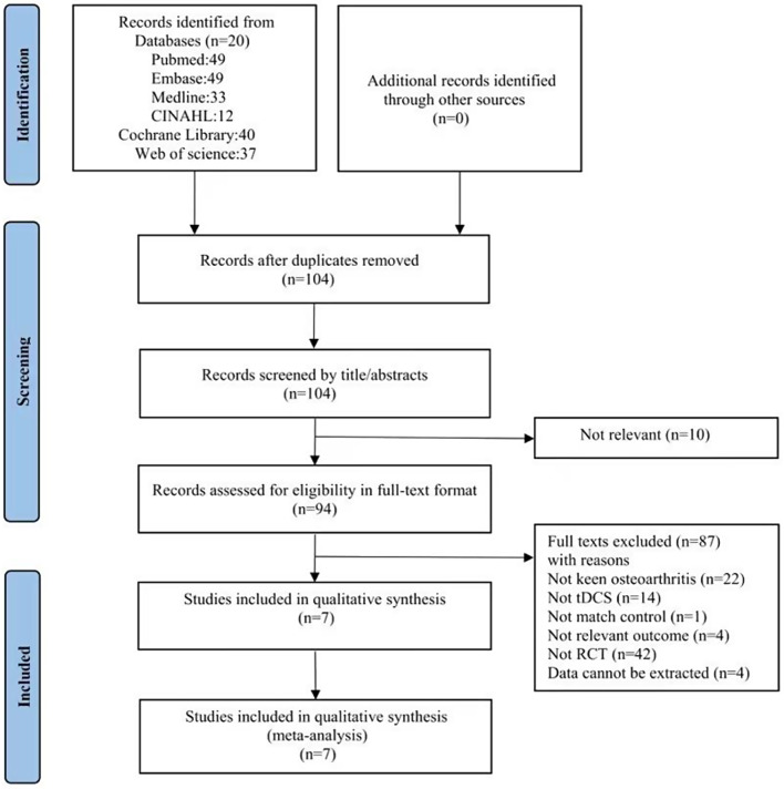
After a systematic search of the 6 databases, we found 220 articles, including 49 in PubMed, 49 in Embase, 37 in Web of Science, 33 in Medline, 12 in CINAHL, and 40 in the Cochrane Library database. Seven studies were finally included after a series of screenings. The detailed selection process of these trials is shown in Fig. 1. Detailed reasons for exclusion and references to excluded studies can be found in Appendix S2.
Fig. 1.
Flow chart of study selection
Study characteristics
The characteristics of the 7 included RCTs were shown in Table 1. A total of 503 participants were included, with sample sizes ranging from 25 to 120. Most of the included studies included patients older than 50, and only one study included those under 50 [26]. The main intervention parameters of tDCS were shown in Table 2. The active tDCS was performed using a pair of saline-saturated sponge electrodes placed on the skin. The anodal electrode was placed on C3 or C4 (10–20 systems of electroencephalography electrode placement) contralateral to the affected knee, and the cathodal electrode was placed on the supraorbital area (SO) contralateral to the anode. The active tDCS was used for active stimulation using a constant current intensity of 2 mA for 20 min per day. In contrast, for the sham tDCS, the electrode position was the same as for active tDCS, the stimulator provided only 2 mA of current, and the stimulation lasted 30 s in six studies and only 15 s in another study [27]. In all seven studies, the duration of the interventions was not identical, with three studies having a duration of 1 week and several interventions of 5 times a week [26, 30, 32], three studies having a duration of 3 weeks and several interventions of 5 times a week [28, 29, 31], and one study having a duration of 8 weeks and several interventions of 2 times a week [27]. Of all included studies, three studies used the VAS to assess pain intensity [26, 27, 29], and three studies used the NRS to assess pain intensity [28–30]. Two studies used CPM, PPT, HPTh, and HPTo to evaluate pain sensitivity [31, 32], and one study used CPM and PPT to assess pain sensitivity [29]. Four studies used WOMAC to evaluate physical function [27–30], and one study used KOOS to assess physical function [26].
Table 1.
Characteristics of the studies included in the review
| Study | Country | Age | Sex(M/F) | Intervention | Control group | Outcomes | Time points of assessment | |
|---|---|---|---|---|---|---|---|---|
|
Azizi et al. 2021 [26] |
Iran | 30–70 | 18/36 |
tDCS (n = 27) |
sham tDCS (n = 27) |
KOOS, VAS | Baseline, Week 1, Month 3 | |
|
Chang et al. 2017 [26] |
China | >50 | 8/17 |
tDCS + exercise (n = 13) |
sham tDCS + exercise(n = 12) | VAS, WOMAC | Baseline, Week 8 | |
|
Martorella et al. 2022 [28] |
American | 50–80 | 38/82 |
tDCS (n = 60) |
sham tDCS (n = 60) |
NRS, WOMAC | Baseline, Week 3, Month 3 | |
|
Regina et al. 2021 [29] |
Brazil | >60 | 16/88 |
tDCS (n = 51) |
sham tDCS (n = 53) |
VAS, NRS, WOMAC, PPT, CPM | Baseline, Week 3, Month 2 | |
|
Ahn et al. 2017 [30] |
American | 50–70 | 19/21 |
tDCS (n = 20) |
sham tDCS (n = 20) |
NRS, WOMAC |
Baseline, Days 1 ~ 5, Week 1 Week 2, Week 3 |
|
|
Martorella et al. 2022 [31] |
American | 50–85 | 38/82 |
tDCS (n = 60) |
sham tDCS (n = 60) |
HPTh, HPTo, PPT, CPM | Baseline, Week 3 | |
|
Ahn et al. 2018 [32] |
American | 50–70 | 19/21 |
tDCS (n = 20) |
sham tDCS (n = 20) |
HPTh, HPTo, PPT, CPM | Baseline, Week 1 |
Notes M: man; F: Female; tDCS: transcranial direct current stimulation; KOOS: Knee injury and Osteoarthritis Outcome Score; VAS: visual analog scale; NRS: numeric rating scale; WOMAC: Western Ontario and McMaster Universities Osteoarthritis Index; PPT: pressure pain threshold; CPM: conditioned pain modulation; HPTh: heat pain threshold; HPTo: heat pain tolerance
Table 2.
Main intervention parameters of tDCS
| Study | Stimulation site (anodal electrode) |
Stimulation site (cathodal electrode) |
Intensity of stimulation (m A) | Duration of stimulation (min) | Duration of intervention | Stimulation of sham tDCS |
|---|---|---|---|---|---|---|
|
Azizi et al. 2021 [26] |
C3 or C4 |
SO contralateral to the anode |
2 | 20 | 5 times per week for 1 week | 2 mA of current and the stimulation lasted 30 s |
|
Chang et al. 2017 [27] |
C3 or C4 |
SO contralateral to the anode |
2 | 20 | 2 times per week for 8 weeks | 2 mA of current and the stimulation lasted 15 s |
|
Martorella et al. 2022 [28] |
C3 or C4 |
SO contralateral to the anode |
2 | 20 | 5 times per week for 3 weeks | 2 mA of current and the stimulation lasted 30 s |
|
Regina et al. 2021 [29] |
C3 or C4 |
SO contralateral to the anode |
2 | 20 | 5 times per week for 3 weeks | 2 mA of current and the stimulation lasted 30 s |
|
Ahn et al. 2017 [30] |
C3 or C4 |
SO contralateral to the anode |
2 | 20 | 5 times per week for 1 week | 2 mA of current and the stimulation lasted 30 s |
|
Martorella et al. 2022 [31] |
C3 or C4 |
SO contralateral to the anode |
2 | 20 | 5 times per week for 3 weeks | 2 mA of current and the stimulation lasted 30 s |
|
Ahn et al. 2018 [32] |
C3 or C4 |
SO contralateral to the anode |
2 | 20 | 5 times per week for 1week | 2 mA of current and the stimulation lasted 30 s |
Note SO: supraorbital area
Quality of included studies
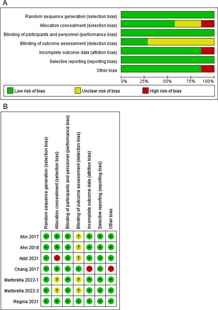
The risk of bias in the included studies was assessed according to the Cochrane tool for seven studies, as shown in Fig. 2. One study did not use allocation concealment (high risk of bias) [26]. Two studies did not mention allocation concealment (unclear risk of bias) [28, 31]. Five studies did not mention blinding of outcome assessment (unclear risk of bias) [26, 28, 30–32]. One study had incomplete outcome data because the follow-up rate was less than 85% (high risk of bias) [27]. One study used tDCS intervention and exercise therapy, which may have impacted the outcome and led to a risk of bias (high risk of bias) [27]. No studies had selection bias or reporting bias.
Fig. 2.
Risk of bias graph and summary of included studies. (A) The risk of bias graph shows the overall risk of bias in each domain. (B) The risk of bias summary indicates the risk of bias in each domain for each study
Quality of outcome indicators
We used the GRADE level of evidence to assess the critical outcome indicators of the included studies. We found a high risk of bias for the outcome indicator used to assess pain intensity and physical function, allowing a risk of bias rating of severe. The outcome indicator used to assess KOA physical function involved a small sample size (n < 400), enabling imprecise risk ratings of severe. No serious risk was identified for the remaining items, so the final rating for the indicator used to assess pain intensity was moderate, the final rating for the indicator used to evaluate pain sensitivity was high, and the final rating for the indicator used to assess physical function was low, as detailed in Table 3.
Table 3.
GRADE evidence profile for outcomes among trials included in the systematic review
| Outcomes | Number of studies | Design | Risk of bias | Inconsistency | Indirectness | Imprecision | Publication bias |
Absolute effect |
GRADE quality |
Symbolic expression |
|---|---|---|---|---|---|---|---|---|---|---|
|
Short-term pain intensity: (VAS or NRS) |
5 | RCT | -1# | 0 | 0 | 0 | 0 | SMD 0.58 lower (1.02 to 0.14 lower) | Moderate | ⨁⨁⨁⊖ |
|
Long-term pain intensity: (VAS or NRS) |
4 | RCT | -1# | 0 | 0 | 0 | 0 | SMD 0.26 lower (0.59 lower to 0.08 higher) | Moderate | ⨁⨁⨁⊖ |
| Pain sensitivity: (CPM, PPT, HPTh, or HPTo) | 3 | RCT | 0 | 0 | 0 | 0 | 0 | SMD 0.43 lower (0.7 to 0.16 lower) | High | ⨁⨁⨁⨁ |
| Short-term physical function: (WOMAC or KOOS) | 5 | RCT | -1# | 0 | 0 | -1* | 0 | SMD 0.13 lower (0.35 lower to 0.08 higher) | Low | ⨁⨁⊖⊖ |
| Long-term physical function: (WOMAC or KOOS) | 3 | RCT | -1# | 0 | 0 | -1* | 0 | SMD 0.02 higher (0.22 lower to 0.25 higher) | Low | ⨁⨁⊖⊖ |
Notes VAS: visual analog scale; NRS: numeric rating scale; CPM: conditioned pain modulation; PPT: pressure pain threshold; HPTh: heat pain threshold; HPTo: heat pain tolerance; WOMAC: Western Ontario and McMaster Universities Osteoarthritis Index; KOOS: Knee injury and Osteoarthritis Outcome Score; RCT: randomized controlled trial; SMD: standardized mean differences; #Downgraded by levels due to a high risk of bias; *Downgraded by levels due to small sample size (n<400)
Effect of tDCS on pain intensity
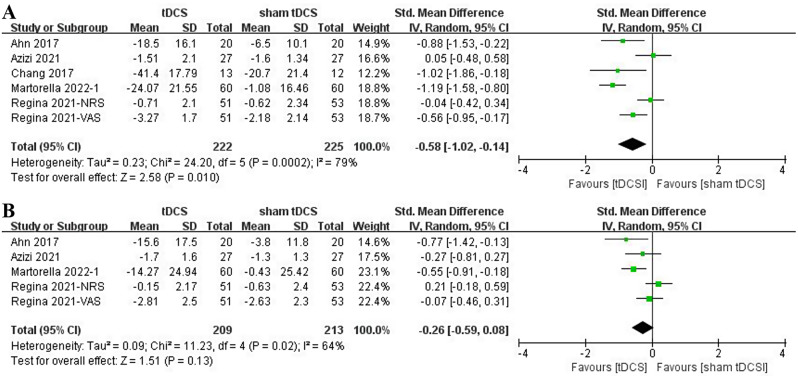
Five studies assessed the effect of tDCS on short-term pain intensity in patients with KOA using the VAS score or the NRS score [26–30]. Meta-analysis (Fig. 3A) showed that tDCS was effective in reducing short-term pain intensity in patients with KOA (SMD: -0.58; 95% CI: -1.02, -0.14; p = 0.01). Four studies assessed the effect of tDCS on long-term pain intensity in patients with KOA using the VAS scores or the NRS scores. Meta-analysis (Fig. 3B) showed that tDCS did not significantly improve long-term pain intensity in patients with KOA (SMD: -0.26; 95% CI: -0.59, 0.08; p = 0.13).
Fig. 3.
Forest plot of the effect of tDCS on pain intensity in patients with KOA. (A) The effect of tDCS on short-term pain intensity. (B) The effect of tDCS on long-term pain intensity. NRS: numeric rating scale; VAS: visual analog scale
Effect of tDCS on pain sensitivity
Three studies assessed the effect of tDCS on short-term pain sensitivity in patients with KOA by CPM, PPT, HPTh, or HPTo [29, 31, 32]. Meta-analysis (Fig. 4) showed that tDCS was effective in reducing short-term pain sensitivity in patients with KOA compared with the control group (SMD: -0.43; 95% CI: -0.70, -0.16; p = 0.002). Only one study evaluated the effect of tDCS on long-term pain sensitivity in patients with KOA, and the results of this study showed that tDCS failed to improve long-term pain sensitivity in patients with KOA [29].
Fig. 4.
Forest plot of the effect of tDCS on short-term pain sensitivity in patients with KOA. CPM: conditioned pain modulation; HPTh: heat pain threshold; HPTo: heat pain tolerance; PPT: pressure pain threshold
Effect of tDCS on physical function
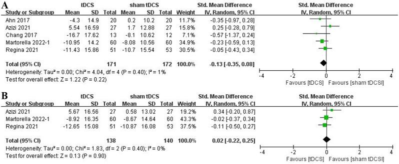
Five studies assessed the effects of tDCS on short-term physical function in patients with KOA through WOMAC or KOOS [26–30]. Meta-analysis (Fig. 5A) showed that tDCS did not significantly improve short-term physical function in patients with KOA (SMD: -0.13; 95% CI: -0.35, 0.08; p = 0.22). Three studies evaluated the effects of tDCS on long-term physical function in patients with KOA via WOMAC or KOOS [26. 28–29]. Meta-analysis (Fig. 5B) showed that tDCS did not significantly improve long-term physical function in patients with KOA (SMD: 0.02; 95% CI: -0.22, 0.25; p = 0.90).
Fig. 5.
Forest plot of the effect of tDCS on physical function in patients with KOA. (A) The effect of tDCS on short-term physical function. (B) The effect of tDCS on long-term physical function
Discussion
Pain is the predominant symptom of patients with KOA, and severe joint pain can affect the quality of life [44, 45]. Previous studies thought that the pain of KOA patients is caused by regional peripheral afferents injury [46]. However, recent studies have found that central nociceptive sensitization plays a crucial role in KOA, leading to local and widespread nociceptive hyperalgesia in these patients [47–49]. tDCS is a non-invasive neuromodulator acting on the central nervous system and can alter neuronal excitability [50–52]. Therefore, tDCS may improve endogenous central pain inhibition in elderly KOA patients by attenuating the effects of central sensitization and modulating brain activity that processes pain, resulting in pain relief [53]. Also, it interacts with various neurotransmitters associated with pain processing, such as dopamine, 5-hydroxytryptamine, acetylcholine, and g-aminobutyric acid [54–58]. In addition, several studies have suggested that inhibition of thalamic sensory neurons and de-inhibition of periaqueductal grey matter neurons may be responsible for pain relief [59]. The results of our meta-analysis showed that tDCS was effective in relieving short-term pain intensity and pain sensitivity in patients with KOA but failed to significantly improve long-term pain intensity in patients. This may be due to the following reasons: Firstly, it may be that tDCS was used alone rather than in combination with another treatment; most of the included studies used tDCS only as an intervention; only the study by Chang et al. combined exercise therapy, but it did not evaluate the long-term effects of tDCS [27]. Secondly, the number of tDCS interventions in most studies may be too low, resulting in tDCS being able to modulate pain control in the short term, but its therapeutic effects are not maintained. Therefore, future studies on tDCS in combination with other therapies are needed, as well as more studies to determine the optimal duration and number of tDCS treatments to achieve maintenance of the treatment effect.
KOA patients often suffer from recurrent disease, so their physical functions are usually affected [60, 61]. Our findings suggest that tDCS did not significantly improve physical function in patients with KOA. The lack of statistical significance for physical function may be due to three reasons: Firstly, most of the included studies had a low number of interventions, Chang and colleagues conducted a 16-session tDCS intervention and found that tDCS improved overall physical function (as assessed by WOMAC) in patients with KOA [27]. Secondly, tDCS intervention alone may not improve physical function in patients with KOA, but one study that combined tDCS intervention with exercise therapy showed that tDCS improved physical function in patients with KOA [27]. Finally, the WOMAC includes pain, stiffness, and joint function. However, we only analyzed the total scores of the WOMAC because only one study examined all three aspects [30]. Therefore, tDCS with an increased number of interventions or in combination with other interventions (e.g., exercise therapy) may be able to improve patients’ physical function.
In addition, there are some limitations to this study. Firstly, the number and sample sizes of studies included in this analysis were minimal, which may impact the accuracy of the results. Secondly, the high degree of heterogeneity between studies reduces the quality of that evidence, making comparability of studies difficult. Thirdly, because only one study evaluated the long-term effects of tDCS on pain sensitivity in patients with KOA [29], we could not assess the long-term effects of tDCS on pain sensitivity in patients with KOA. Finally, because the intervention sites, intervention parameters, and duration of tDCS were essentially the same in the included studies, we were unable to explore the effects of tDCS on patients with KOA with different intervention sites, intervention parameters, and duration of intervention.
Conclusions
Our findings indicate that tDCS can reduce short-term pain intensity and sensitivity but fails to significantly relieve long-term pain intensity and improve the physical function in patients with KOA. Thus, tDCS may be a potential therapeutic tool to reduce short-term pain intensity and pain sensitivity in patients with KOA. In addition, we found that combining tDCS with other therapies (e.g., exercise therapy) or increasing the number of interventions may improve physical function in patients with KOA. Future studies will require larger sample sizes, longer follow-up times, longer durations of tDCS treatment, and studies of different stimulation sites to determine the optimal tDCS dose and parameters for patients with KOA.
Electronic supplementary material
Below is the link to the electronic supplementary material.
Acknowledgements
Not applicable.
Abbreviations
- KOA
Knee osteoarthritis
- tDCS
Transcranial direct current stimulation
- RCT
Randomized controlled trial
- GARDE
Grading of Recommendations Assessment Development and Evaluation
- SD
Standard deviation
- VAS
Visual analog scale
- NRS
Numeric rating scale
- CPM
Conditioned pain modulation
- PPT
Pressure pain threshold
- HPTh
Heat pain threshold
- HPTo
Heat pain tolerance
- QST
Quantitative Sensory Testing
- WOMAC
Western Ontario and McMaster Universities Osteoarthritis Index
- KOOS
Knee injury and Osteoarthritis Outcome Score
- 95% CI
95% confidence intervals
- MD
Mean differences
- SMD
Standardized mean differences
- SO
Supraorbital area
Author contributions
Yanlin Wu collected the data and wrote the manuscript. Yun Luo supervised this study and revised the manuscript. Jiaming Yang and Yongqiang Wu selected the literature. Qiang Zhu and Yi Li extracted the data. Hao Hu and Jiahong Zhang assessed the quality of each included study. Yanbiao Zhong solved the difference. Maoyuan Wang provided the idea and designed this study.
Funding
Mao-yuan Wang is supported by the National Natural Science Foundation of China (82060420); the Natural Science Foundation of Jiangxi Province, China (20212BAB206004); Key Laboratory of the Ministry of Education for the Prevention and Treatment of Cardiovascular and Cerebrovascular Diseases, Gannan Medical University (NX202022).
Data availability
Data are available on reasonable request to the corresponding author at: wmy.gmu.kf@gmail.com.
Declarations
Ethics approval and consent to participate
Not applicable.
Consent for publication
Not applicable.
Competing interests
The authors declare no competing interests.
Footnotes
Publisher’s note
Springer Nature remains neutral with regard to jurisdictional claims in published maps and institutional affiliations.
Yanlin Wu and Yun Luo are the co-first authors of this work.
References
- 1.Prieto-Alhambra D, Judge A, Javaid MK, Cooper C, Diez-Perez A, Arden NK. Incidence and risk factors for clinically diagnosed knee, hip and hand osteoarthritis: influences of age, gender and osteoarthritis affecting other joints. Ann Rheum Dis. 2014;73(9):1659–64. 10.1136/annrheumdis-2013-203355 [DOI] [PMC free article] [PubMed] [Google Scholar]
- 2.Zhou M, Wang H, Zeng X, Yin P, Zhu J, Chen W, Li X, Wang L, Wang L, Liu Y, et al. Mortality, morbidity, and risk factors in China and its provinces, 1990–2017: a systematic analysis for the global burden of Disease Study 2017. Lancet. 2019;394(10204):1145–58. 10.1016/S0140-6736(19)30427-1 [DOI] [PMC free article] [PubMed] [Google Scholar]
- 3.Zhang L, Wen YL, He CY, Zeng Y, Wang JQ, Wang GY. Relationship between classification of Fabellae and the severity of knee osteoarthritis: a relevant Study in the Chinese Population. Orthop Surg. 2022;14(2):274–9. 10.1111/os.13006 [DOI] [PMC free article] [PubMed] [Google Scholar]
- 4.Busija L, Bridgett L, Williams SR, Osborne RH, Buchbinder R, March L, Fransen M. Osteoarthritis. Best Pract Res Clin Rheumatol. 2010;24(6):757–68. 10.1016/j.berh.2010.11.001 [DOI] [PubMed] [Google Scholar]
- 5.Felson DT, Lawrence RC, Dieppe PA, Hirsch R, Helmick CG, Jordan JM, Kington RS, Lane NE, Nevitt MC, Zhang Y, et al. Osteoarthritis: new insights. Part 1: the disease and its risk factors. Ann Intern Med. 2000;133(8):635–46. 10.7326/0003-4819-133-8-200010170-00016 [DOI] [PubMed] [Google Scholar]
- 6.Cherian JJ, Jauregui JJ, Leichliter AK, Elmallah RK, Bhave A, Mont MA. The effects of various physical non-operative modalities on the pain in osteoarthritis of the knee. Bone Joint J 2016, 98–b(1 Suppl A):89–94. [DOI] [PubMed]
- 7.Kolasinski SL, Neogi T, Hochberg MC, Oatis C, Guyatt G, Block J, Callahan L, Copenhaver C, Dodge C, Felson D, et al. 2019 American College of Rheumatology/Arthritis Foundation Guideline for the Management of Osteoarthritis of the Hand, hip, and Knee. Arthritis Care Res (Hoboken). 2020;72(2):149–62. 10.1002/acr.24131 [DOI] [PMC free article] [PubMed] [Google Scholar]
- 8.Liu Q, Niu J, Li H, Ke Y, Li R, Zhang Y, Lin J. Knee symptomatic osteoarthritis, walking disability, NSAIDs use and all-cause Mortality: Population-based Wuchuan Osteoarthritis Study. Sci Rep. 2017;7(1):3309. 10.1038/s41598-017-03110-3 [DOI] [PMC free article] [PubMed] [Google Scholar]
- 9.Roos EM, Arden NK. Strategies for the prevention of knee osteoarthritis. Nat Rev Rheumatol. 2016;12(2):92–101. 10.1038/nrrheum.2015.135 [DOI] [PubMed] [Google Scholar]
- 10.Van Manen MD, Nace J, Mont MA. Management of primary knee osteoarthritis and indications for total knee arthroplasty for general practitioners. J Am Osteopath Assoc. 2012;112(11):709–15. [PubMed] [Google Scholar]
- 11.Matharu GS, Garriga C, Rangan A, Judge A. Does Regional Anesthesia reduce complications following total hip and knee replacement compared with General Anesthesia? An analysis from the National Joint Registry for England, Wales, Northern Ireland and the Isle of Man. J Arthroplasty. 2020;35(6):1521–e15281525. 10.1016/j.arth.2020.02.003 [DOI] [PubMed] [Google Scholar]
- 12.Saltzman BM, Frank RM, Davey A, Cotter EJ, Redondo ML, Naveen N, Wang KC, Cole BJ. Lack of standardization among clinical trials of injection therapies for knee osteoarthritis: a systematic review. Phys Sportsmed. 2020;48(3):266–89. 10.1080/00913847.2020.1726716 [DOI] [PubMed] [Google Scholar]
- 13.Allen KD, Arbeeva L, Callahan LF, Golightly YM, Goode AP, Heiderscheit BC, Huffman KM, Severson HH, Schwartz TA. Physical therapy vs internet-based exercise training for patients with knee osteoarthritis: results of a randomized controlled trial. Osteoarthritis Cartilage. 2018;26(3):383–96. 10.1016/j.joca.2017.12.008 [DOI] [PMC free article] [PubMed] [Google Scholar]
- 14.Gregori D, Giacovelli G, Minto C, Barbetta B, Gualtieri F, Azzolina D, Vaghi P, Rovati LC. Association of pharmacological treatments with Long-Term Pain Control in patients with knee osteoarthritis: a systematic review and Meta-analysis. JAMA. 2018;320(24):2564–79. 10.1001/jama.2018.19319 [DOI] [PMC free article] [PubMed] [Google Scholar]
- 15.Phillips K, Clauw DJ. Central pain mechanisms in the rheumatic diseases: future directions. Arthritis Rheum. 2013;65(2):291–302. 10.1002/art.37739 [DOI] [PMC free article] [PubMed] [Google Scholar]
- 16.Finan PH, Buenaver LF, Bounds SC, Hussain S, Park RJ, Haque UJ, Campbell CM, Haythornthwaite JA, Edwards RR, Smith MT. Discordance between pain and radiographic severity in knee osteoarthritis: findings from quantitative sensory testing of central sensitization. Arthritis Rheum. 2013;65(2):363–72. 10.1002/art.34646 [DOI] [PMC free article] [PubMed] [Google Scholar]
- 17.Cruz-Almeida Y, Sibille KT, Goodin BR, Petrov ME, Bartley EJ, Riley JL 3rd, King CD, Glover TL, Sotolongo A, Herbert MS, et al. Racial and ethnic differences in older adults with knee osteoarthritis. Arthritis Rheumatol. 2014;66(7):1800–10. [DOI] [PMC free article] [PubMed]
- 18.King CD, Sibille KT, Goodin BR, Cruz-Almeida Y, Glover TL, Bartley E, Riley JL, Herbert MS, Sotolongo A, Schmidt J, et al. Experimental pain sensitivity differs as a function of clinical pain severity in symptomatic knee osteoarthritis. Osteoarthritis Cartilage. 2013;21(9):1243–52. 10.1016/j.joca.2013.05.015 [DOI] [PMC free article] [PubMed] [Google Scholar]
- 19.Dasilva AF, Mendonca ME, Zaghi S, Lopes M, Dossantos MF, Spierings EL, Bajwa Z, Datta A, Bikson M, Fregni F. tDCS-induced analgesia and electrical fields in pain-related neural networks in chronic migraine. Headache. 2012;52(8):1283–95. 10.1111/j.1526-4610.2012.02141.x [DOI] [PMC free article] [PubMed] [Google Scholar]
- 20.Lefaucheur JP, Antal A, Ayache SS, Benninger DH, Brunelin J, Cogiamanian F, Cotelli M, De Ridder D, Ferrucci R, Langguth B, et al. Evidence-based guidelines on the therapeutic use of transcranial direct current stimulation (tDCS). Clin Neurophysiol. 2017;128(1):56–92. 10.1016/j.clinph.2016.10.087 [DOI] [PubMed] [Google Scholar]
- 21.Rodrigues GM, Paixão A, Arruda T, de Oliveira BRR, Maranhão Neto GA, Marques Neto SR, Lattari E, Machado S. Anodal Transcranial Direct Current Stimulation increases muscular strength and reduces Pain Perception in Women with Patellofemoral Pain. J Strength Cond Res. 2022;36(2):371–8. 10.1519/JSC.0000000000003473 [DOI] [PubMed] [Google Scholar]
- 22.Kang JH, Choi SE, Park DJ, Xu H, Lee JK, Lee SS. Effects of add-on transcranial direct current stimulation on pain in Korean patients with fibromyalgia. Sci Rep. 2020;10(1):12114. 10.1038/s41598-020-69131-7 [DOI] [PMC free article] [PubMed] [Google Scholar]
- 23.Luedtke K, Rushton A, Wright C, Jürgens T, Polzer A, Mueller G, May A. Effectiveness of transcranial direct current stimulation preceding cognitive behavioural management for chronic low back pain: sham controlled double blinded randomised controlled trial. BMJ. 2015;350:h1640. 10.1136/bmj.h1640 [DOI] [PMC free article] [PubMed] [Google Scholar]
- 24.Bruce AS, Howard JS, McBride HVANW, Needle JM. The effects of Transcranial Direct current stimulation on chronic ankle instability. Med Sci Sports Exerc. 2020;52(2):335–44. 10.1249/MSS.0000000000002129 [DOI] [PubMed] [Google Scholar]
- 25.O’Connell NE, Marston L, Spencer S, DeSouza LH, Wand BM. Non-invasive brain stimulation techniques for chronic pain. Cochrane Database Syst Rev. 2018;4(4):Cd008208. [DOI] [PMC free article] [PubMed] [Google Scholar]
- 26.Azizi S, Rezasoltani Z, Najafi S, Mohebi B, Tabatabaee SM, Dadarkhah A. Transcranial direct current stimulation for knee osteoarthritis: a single-blind randomized sham-controlled trial. Neurophysiol Clin. 2021;51(4):329–38. 10.1016/j.neucli.2020.12.002 [DOI] [PubMed] [Google Scholar]
- 27.Chang W-J, Bennell KL, Hodges PW, Hinman RS, Young CL, Buscemi V, Liston MB, Schabrun SM. Addition of transcranial direct current stimulation to quadriceps strengthening exercise in knee osteoarthritis: a pilot randomised controlled trial. PLoS ONE 2017, 12(6). [DOI] [PMC free article] [PubMed]
- 28.Martorella G, Mathis K, Miao H, Wang D, Park L, Ahn H. Self-administered transcranial direct current stimulation for pain in older adults with knee osteoarthritis: a randomized controlled study. Brain Stimul. 2022;15(4):902–9. 10.1016/j.brs.2022.06.003 [DOI] [PMC free article] [PubMed] [Google Scholar]
- 29.Regina BTD, Erika FOJ, Marcia VAS, Carolina PNPA, Kuraoka TK, Martins GF, Bonin PC, Fania CS, Felipe F, Fernandes MTV. Motor Cortex Transcranial Direct Current Stimulation effects on knee Osteoarthritis Pain in Elderly subjects with Dysfunctional Descending Pain Inhibitory System: a Randomized Controlled Trial. Brain Stimul 2021, 14(3). [DOI] [PubMed]
- 30.Ahn H, Woods A, Choi E, Padhye N, Fillingim R. Efficacy of transcranial direct current stimulation on clinical pain severity in older adults with knee os-teoarthritis pain: a double-blind, randomized, sham-controlled pilot clinical study. J Pain. 2017;18(4):S87–8. 10.1016/j.jpain.2017.02.306 [DOI] [Google Scholar]
- 31.Martorella G, Mathis K, Miao H, Wang D, Park L, Ahn H. Efficacy of home-based Transcranial Direct Current Stimulation on Experimental Pain Sensitivity in older adults with knee osteoarthritis: a Randomized, Sham-Controlled Clinical Trial. J Clin Med 2022, 11(17). [DOI] [PMC free article] [PubMed]
- 32.Ahn H, Suchting R, Woods AJ, Miao H, Green C, Cho RY, Choi E, Fillingim RB. Bayesian analysis of the effect of transcranial direct current stimulation on experimental pain sensitivity in older adults with knee osteoarthritis: randomized sham-controlled pilot clinical study. J Pain Res. 2018;11:2071–81. 10.2147/JPR.S173080 [DOI] [PMC free article] [PubMed] [Google Scholar]
- 33.Liberati A, Altman DG, Tetzlaff J, Mulrow C, Gøtzsche PC, Ioannidis JP, Clarke M, Devereaux PJ, Kleijnen J, Moher D. The PRISMA statement for reporting systematic reviews and meta-analyses of studies that evaluate health care interventions: explanation and elaboration. J Clin Epidemiol. 2009;62(10):e1–34. 10.1016/j.jclinepi.2009.06.006 [DOI] [PubMed] [Google Scholar]
- 34.Higgins JP, Altman DG, Gøtzsche PC, Jüni P, Moher D, Oxman AD, Savovic J, Schulz KF, Weeks L, Sterne JA. The Cochrane collaboration’s tool for assessing risk of bias in randomised trials. BMJ. 2011;343:d5928. 10.1136/bmj.d5928 [DOI] [PMC free article] [PubMed] [Google Scholar]
- 35.Atkins D, Best D, Briss PA, Eccles M, Falck-Ytter Y, Flottorp S, Guyatt GH, Harbour RT, Haugh MC, Henry D, et al. Grading quality of evidence and strength of recommendations. BMJ. 2004;328(7454):1490. 10.1136/bmj.328.7454.1490 [DOI] [PMC free article] [PubMed] [Google Scholar]
- 36.Hawker GA, Mian S, Kendzerska T, French M. Measures of adult pain: visual Analog Scale for Pain (VAS Pain), Numeric Rating Scale for Pain (NRS Pain), McGill Pain Questionnaire (MPQ), short-form McGill Pain Questionnaire (SF-MPQ), Chronic Pain Grade Scale (CPGS), short Form-36 Bodily Pain Scale (SF-36 BPS), and measure of intermittent and constant Osteoarthritis Pain (ICOAP). Arthritis Care Res (Hoboken). 2011;63(Suppl 11):S240–252. [DOI] [PubMed] [Google Scholar]
- 37.Uddin Z, MacDermid JC. Quantitative sensory testing in Chronic Musculoskeletal Pain. Pain Med. 2016;17(9):1694–703. 10.1093/pm/pnv105 [DOI] [PubMed] [Google Scholar]
- 38.Bellamy N, Buchanan WW, Goldsmith CH, Campbell J, Stitt LW. Validation study of WOMAC: a health status instrument for measuring clinically important patient relevant outcomes to antirheumatic drug therapy in patients with osteoarthritis of the hip or knee. J Rheumatol. 1988;15(12):1833–40. [PubMed] [Google Scholar]
- 39.Seror R, Tubach F, Baron G, Falissard B, Logeart I, Dougados M, Ravaud P. Individualising the Western Ontario and McMaster universities osteoarthritis index (WOMAC) function subscale: incorporating patient priorities for improvement to measure functional impairment in hip or knee osteoarthritis. Ann Rheum Dis. 2008;67(4):494–9. 10.1136/ard.2007.074591 [DOI] [PubMed] [Google Scholar]
- 40.Yang KG, Raijmakers NJ, Verbout AJ, Dhert WJ, Saris DB. Validation of the short-form WOMAC function scale for the evaluation of osteoarthritis of the knee. J Bone Joint Surg Br. 2007;89(1):50–6. 10.1302/0301-620X.89B1.17790 [DOI] [PubMed] [Google Scholar]
- 41.Salavati M, Mazaheri M, Negahban H, Sohani SM, Ebrahimian MR, Ebrahimi I, Kazemnejad A, Salavati M. Validation of a persian-version of knee injury and osteoarthritis outcome score (KOOS) in iranians with knee injuries. Osteoarthritis Cartilage. 2008;16(10):1178–82. 10.1016/j.joca.2008.03.004 [DOI] [PubMed] [Google Scholar]
- 42.Ye H, Yang JM, Luo Y, Long Y, Zhang JH, Zhong YB, Gao F, Wang MY. Do dietary supplements prevent loss of muscle mass and strength during muscle disuse? A systematic review and meta-analysis of randomized controlled trials. Front Nutr. 2023;10:1093988. 10.3389/fnut.2023.1093988 [DOI] [PMC free article] [PubMed] [Google Scholar]
- 43.Higgins JP, Thompson SG, Deeks JJ, Altman DG. Measuring inconsistency in meta-analyses. BMJ. 2003;327(7414):557–60. 10.1136/bmj.327.7414.557 [DOI] [PMC free article] [PubMed] [Google Scholar]
- 44.Fu K, Robbins SR, McDougall JJ. Osteoarthritis: the genesis of pain. Rheumatology (Oxford). 2018;57(suppl4):iv43–50. 10.1093/rheumatology/kex419 [DOI] [PubMed] [Google Scholar]
- 45.Barbour KE, Boring M, Helmick CG, Murphy LB, Qin J. Prevalence of severe Joint Pain among adults with doctor-diagnosed arthritis - United States, 2002–2014. MMWR Morb Mortal Wkly Rep. 2016;65(39):1052–6. 10.15585/mmwr.mm6539a2 [DOI] [PubMed] [Google Scholar]
- 46.Rakel B, Vance C, Zimmerman MB, Petsas-Blodgett N, Amendola A, Sluka KA. Mechanical hyperalgesia and reduced quality of life occur in people with mild knee osteoarthritis pain. Clin J Pain. 2015;31(4):315–22. 10.1097/AJP.0000000000000116 [DOI] [PubMed] [Google Scholar]
- 47.Bartholomew C, Lack S, Neal B. Altered pain processing and sensitisation is evident in adults with patellofemoral pain: a systematic review including meta-analysis and meta-regression. Scand J Pain. 2019;20(1):11–27. 10.1515/sjpain-2019-0079 [DOI] [PubMed] [Google Scholar]
- 48.Moreton BJ, Tew V, das Nair R, Wheeler M, Walsh DA, Lincoln NB. Pain phenotype in patients with knee osteoarthritis: classification and measurement properties of painDETECT and self-report Leeds assessment of neuropathic symptoms and signs scale in a cross-sectional study. Arthritis Care Res (Hoboken). 2015;67(4):519–28. 10.1002/acr.22431 [DOI] [PMC free article] [PubMed] [Google Scholar]
- 49.Neogi T, Guermazi A, Roemer F, Nevitt MC, Scholz J, Arendt-Nielsen L, Woolf C, Niu J, Bradley LA, Quinn E, et al. Association of joint inflammation with Pain sensitization in knee osteoarthritis: the Multicenter Osteoarthritis Study. Arthritis Rheumatol. 2016;68(3):654–61. 10.1002/art.39488 [DOI] [PMC free article] [PubMed] [Google Scholar]
- 50.Charvet LE, Shaw MT, Bikson M, Woods AJ, Knotkova H. Supervised transcranial direct current stimulation (tDCS) at home: a guide for clinical research and practice. Brain Stimul. 2020;13(3):686–93. 10.1016/j.brs.2020.02.011 [DOI] [PubMed] [Google Scholar]
- 51.Ouellette AL, Liston MB, Chang WJ, Walton DM, Wand BM, Schabrun SM. Safety and feasibility of transcranial direct current stimulation (tDCS) combined with sensorimotor retraining in chronic low back pain: a protocol for a pilot randomised controlled trial. BMJ Open. 2017;7(8):e013080. 10.1136/bmjopen-2016-013080 [DOI] [PMC free article] [PubMed] [Google Scholar]
- 52.Zortea M, Ramalho L, Alves RL, Alves C, Braulio G, Torres I, Fregni F, Caumo W. Transcranial Direct current stimulation to improve the dysfunction of descending Pain Modulatory System related to Opioids in Chronic Non-cancer Pain: an integrative review of Neurobiology and Meta-Analysis. Front Neurosci. 2019;13:1218. 10.3389/fnins.2019.01218 [DOI] [PMC free article] [PubMed] [Google Scholar]
- 53.Maarrawi J, Peyron R, Mertens P, Costes N, Magnin M, Sindou M, Laurent B, Garcia-Larrea L. Motor cortex stimulation for pain control induces changes in the endogenous opioid system. Neurology. 2007;69(9):827–34. 10.1212/01.wnl.0000269783.86997.37 [DOI] [PubMed] [Google Scholar]
- 54.Nitsche MA, Grundey J, Liebetanz D, Lang N, Tergau F, Paulus W. Catecholaminergic consolidation of motor cortical neuroplasticity in humans. Cereb Cortex. 2004;14(11):1240–5. 10.1093/cercor/bhh085 [DOI] [PubMed] [Google Scholar]
- 55.Nitsche MA, Jaussi W, Liebetanz D, Lang N, Tergau F, Paulus W. Consolidation of human motor cortical neuroplasticity by D-cycloserine. Neuropsychopharmacology. 2004;29(8):1573–8. 10.1038/sj.npp.1300517 [DOI] [PubMed] [Google Scholar]
- 56.Nitsche MA, Liebetanz D, Schlitterlau A, Henschke U, Fricke K, Frommann K, Lang N, Henning S, Paulus W, Tergau F. GABAergic modulation of DC stimulation-induced motor cortex excitability shifts in humans. Eur J Neurosci. 2004;19(10):2720–6. 10.1111/j.0953-816X.2004.03398.x [DOI] [PubMed] [Google Scholar]
- 57.Nitsche MA, Lampe C, Antal A, Liebetanz D, Lang N, Tergau F, Paulus W. Dopaminergic modulation of long-lasting direct current-induced cortical excitability changes in the human motor cortex. Eur J Neurosci. 2006;23(6):1651–7. 10.1111/j.1460-9568.2006.04676.x [DOI] [PubMed] [Google Scholar]
- 58.Nitsche MA, Kuo MF, Karrasch R, Wächter B, Liebetanz D, Paulus W. Serotonin affects transcranial direct current-induced neuroplasticity in humans. Biol Psychiatry. 2009;66(5):503–8. 10.1016/j.biopsych.2009.03.022 [DOI] [PubMed] [Google Scholar]
- 59.Schabrun SM, Jones E, Elgueta Cancino EL, Hodges PW. Targeting chronic recurrent low back pain from the top-down and the bottom-up: a combined transcranial direct current stimulation and peripheral electrical stimulation intervention. Brain Stimul. 2014;7(3):451–9. 10.1016/j.brs.2014.01.058 [DOI] [PubMed] [Google Scholar]
- 60.Emmert D, Rasche T, Stieber C, Conrad R, Mücke M. [Knee pain - symptoms, diagnosis and therapy of osteoarthritis]. MMW Fortschr Med. 2018;160(15):58–64. 10.1007/s15006-018-0023-1 [DOI] [PubMed] [Google Scholar]
- 61.Geng R, Li J, Yu C, Zhang C, Chen F, Chen J, Ni H, Wang J, Kang K, Wei Z, et al. Knee osteoarthritis: current status and research progress in treatment (review). Exp Ther Med. 2023;26(4):481. 10.3892/etm.2023.12180 [DOI] [PMC free article] [PubMed] [Google Scholar]
Associated Data
This section collects any data citations, data availability statements, or supplementary materials included in this article.
Supplementary Materials
Data Availability Statement
Data are available on reasonable request to the corresponding author at: wmy.gmu.kf@gmail.com.


