Abstract
Purpose
Despite the publication of several randomized controlled trials (RCTs), it is not clear which technique for the treatment of focal chondral and osteochondral defects of the knee grants the best clinical outcome. The aim of this network meta-analysis (NMA) was to compare the efficacy and safety of microfractures (MF), autologous chondrocyte implantation (ACI), autologous matrix-induced chondrogenesis (AMIC), osteochondral autograft transplantation (OCT) at short (< 1 year), intermediate (1–5 years) and long-term (> 5 years).
Methods
We carried out an NMA with Bayesian random-effect model, according to PRISMA guidelines. The search was performed in MEDLINE, EMBASE, Web of Science, CENTRAL, CINAHL, SPORTDiscus, clinicaltrials.gov, WHO ICTRP, from inception to November 2022. The eligibilities were randomized controlled trials on patients with knee chondral and osteochondral defects, undergoing microfractures, OCT, AMIC, ACI, without restrictions for prior or concomitant surgery on ligaments, menisci or limb alignment, prior surgery for fixation or ablation of osteochondritis dissecans fragments, and prior cartilage procedures as microfractures, drilling, abrasion, or debridement.
Results
Nineteen RCTs were included. No difference among treatments was shown in the pooled comparison of patient reported outcome measures (PROMs) at any timepoint. Safety data were not available for all trials due to the heterogeneity of reporting, but chondrospheres seemed to have lower failure and reoperation rates.
Conclusion
This NMA showed no difference for PROMs with any technique. The lower failure and reoperation rates with chondrospheres must be interpreted with caution since adverse event data was heterogenous among trials. The standardization of the efficacy and safety outcome measures for future trials on knee cartilage repair and regeneration is necessary.
Keywords: knee, knee injuries, sporting injuries, cartilage, arthroscopy
Introduction
Focal chondral lesions of the knee account for 19–28% of all chondral lesions encountered during arthroscopy (1, 2, 3). They are mainly related to acute traumas (> 58%), including sports injuries (46%), but also to chronic repetitive injuries (2, 4). While knee chondral defects can be asymptomatic in 14% of cases, as demonstrated in studies in athletes, they often manifest with pain, effusion, swelling, and sometimes locking, impairing daily activities and sporting performance (2, 4, 5).
In the last century, the treatment of symptomatic patients has remarkably evolved. Repair techniques such as bone marrow stimulation, debridement, abrasion, and subchondral drilling have been gradually replaced by microfractures (MF) and autologous matrix-induced chondrogenesis (AMIC) (6, 7, 8). Regeneration and graft transfer techniques have also expanded, thanks to the development of an arthroscopic technique for osteochondral autologous transplantation (OCT) (9), minced cartilage procedures for allografts and autografts replacement (10, 11), and multiple generations of autologous chondrocyte implantation (ACI). The latter has evolved from the injection of cultured autologous chondrocytes below a periosteum membrane (first generation, ACI1) or porcine type I and III collagen membrane (second generation, ACI2) (12, 13), to their dispersion in a matrix (third generation, ACI3), also called matrix-induced autologous chondrocyte implantation (MACI) (14). The ACI3 has differentiated into scaffold-based (polymers, hyaluronan, collagen sponges, or gels) and scaffold-free chondrocytes supports, such as chondrospheres, made of a matrix built by chondrocytes themselves (15). The fourth generation of ACI will implicate the use of mesenchymal stromal cells and gene therapy (16, 17).
Concurrently with the development of new techniques, the treatment algorithm has evolved. Apart from addressing concomitant misalignment, instabilities, meniscal pathologies, and patients’ activity, the main criteria for treatment choice remain lesion size, defect site, and bone loss (18, 19). In cases of bone involvement, OCT is indicated for small (< 2 cm2) defects, and allografts or even sandwich ACI (an ACI lying on bone graft) are best suited for medium (2–4 cm2) and large (> 4 cm2) defects. In the absence of bone involvement, microfracture, OCT, and AMIC can be used for small defects, while ACI seems to be a valuable option for all defect sizes and locations (19). As described, not only can multiple techniques be indicated for the same type of defect, but multiple generations are also available for each technique. Available randomized controlled trials and pairwise meta-analyses do not allow establishment of which treatment has the best efficacy and safety profile since they compare only two techniques each (20). This necessitates the summary of evidence with network meta-analyses (NMA) that, by allowing the comparison of multiple treatments, bypass the limits of pairwise comparison. Even though three NMAs on this subject have been published (21, 22, 23), it is still unclear which treatment and technique offers the best clinical outcome, due to discordant results of available NMAs. In addition, since their divulgation, new randomized controlled trials have been published, prompting the need to provide an updated summary of available evidence. We conducted a systematic review and NMA on randomized controlled trials (RCT) on patients with knee cartilage defects and OCD, treated with MF, OCT, AMIC, and ACI, including chondrospheres, which represents the last generation of ACI. Our aim was to answer the following questions: 1. Which of the above treatments permits the restoration of the best functional outcome over short (< 1 year), intermediate (1–5 years), and long (> 5 years) term? 2. Which is the safest treatment?
Materials and methods
Protocol and registration
This review was planned and performed according to the PRISMA NMA extension statement (24) and registered in PROSPERO (CRD42021285656).
Search strategy and selection criteria
The bibliographic search was performed on MEDLINE, EMBASE, Web of Science, CENTRAL, CINAHL, SPORTDiscus, clinicaltrials.gov, and WHO ICTRP Search Portal from inception to 17 November 2022. The search strategy for each database is shown in Supplementary Appendix 1 (see section on supplementary materials given at the end of this article).
The archives of the last 20 years of the European Society of Sports Traumatology, Knee Surgery and Arthroscopy (ESSKA), ICRS, Arthroscopy Association of North America (AANA), OsteoArthritis Research Society International (OARSI), and French Society of Arthroscopy (SFA) congresses have also been accessed.
The eligibility criteria were defined a priori and are shown in Table 1. Among scaffold-free ACI3, we included only chondrospheres because these are approved by EMA in Europe.
Table 1.
Eligibility criteria.
| Inclusion criteria | Exclusion criteria |
|---|---|
|
|
OCD, osteochondritis dissecans.
Two blinded reviewers (SV, BA) independently performed the study selection by means of Covidence software (https://www.covidence.org). Any disagreement was resolved with the senior authors (PB, DH, RSN) and by contacting the corresponding authors, if necessary.
Data extraction
The data extraction was independently carried out by two reviewers (SV, BA). All variables of interest were collected on an Excel sheet, previously tested on ten random publications. In case of disagreement, a third reviewer (PB) was involved to reach a consensus. Study authors were contacted to gather information on missing or unclear data.
We collected the following data: i) general study information: title, date of publication, authors, country, contact details of the corresponding author, funding sources; ii) characteristics of the study: design, eligibility criteria, follow-up duration; iii) demographics: mean age, overall population (patients and knees), males, affected site (condyles, patella, trochlea), inclusion of patients with osteochondritis dissecans, intention-to-treat (ITT) population, per-protocol (PP) population; iv) primary (efficacy) endpoints: mean and SD of Lysholm, Tegner, KOOS, HSS, Modified Cincinnati, WOMAC, IKDC over short (< 1 year), intermediate (1–5 years), and long term (> 5 years); v) secondary (safety) endpoints: rate of infections, symptomatic cartilage loosening or hypertrophy, persistent pain, reoperation, and failures (including its definition) at the longest follow-up available for each trial. The complete list is shown in Supplementary Appendix 2, section A.
The Cochran Risk of Bias 2.0 tool was used by two independent reviewers (SV, BA). For each domain, a low, high or unclear risk of bias vote was decided. The CINeMA app (https://cinema.ispm.unibe.ch/), which applies the modification of the GRADE framework for network meta-analysis (25), was used for the evaluation of confidence in the NMA findings.
Statistical analysis
The unit of analysis was the trial population to avoid data duplication. Therefore, when multiple reports on the same trial population were encountered, the most recent trial with the longest follow-up was included.
A Bayesian random-effect model NMA was carried out, pooling direct and indirect comparisons using the hierarchical model of Lu et Ades, implemented with the Markov chain Monte Carlo model (26). The Bayesian approach does not allow for the calculation of the P-value, but estimates are based on a 95% credible interval (CI), which is a posterior probability of 95% that the endpoints lie within it. Risk ratios and mean differences were used for the comparison of binary and continuous outcomes respectively, with 95% CI. In forest plots, no difference for the relative risk corresponded to one, and for mean differences to zero. The ranking diagrams were generated for each outcome using the ranking probabilities.
We used the formulas of the Cochrane Handbook (27) and by Hozo et al. (28) to estimate the SD, the mean, and the SD of the difference between baseline and post-surgery scores.
Statistical analysis was performed using R software, version 3.5.2 (R Foundation for Statistical Computing, Vienna, Austria), using the gemtc R-package for Bayesian analysis (version 0.8-2) and the netmeta R-package (version 0.9-8) for network diagrams.
Network diagrams were used to show direct comparisons for primary efficacy outcomes over short, intermediate, and long term, and secondary safety outcomes, and also the safety outcome at the mean follow-up of the included studies. Seven nodes were defined: microfractures, AMIC, OCT, ACI1, ACI2, ACI3 (scaffold-based), and chondrospheres. The size of the nodes was proportional to the sample size of each treatment. The thickness of the lines was proportional to the number of available studies. Heterogeneity within each network comparison was studied with the I 2 statistics. Inconsistency was studied by comparing the fit between consistency and inconsistency models. Transitivity was assessed across comparisons by evaluating patients’ demographics and studies’ characteristics.
Clinical heterogeneity could be due to multiple effect modifiers such as age, etiology of the chondral or osteochondral defect, number of lesions treated in the index knee, defect size, prior surgery, and concomitant knee surgery. We planned to carry out subgroup analysis for studies including vs. excluding patients with OCD and including vs. excluding prior microfractures, drilling, abrasion, and debridement.
Comparison-adjusted funnel plots of considered treatments were carried out to assess small study effects within the pairwise comparison.
Results
Study characteristics
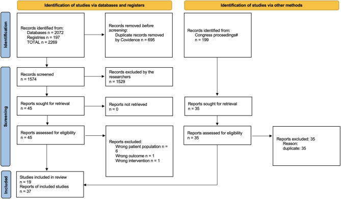
The bibliographic search retrieved 2269 studies from databases and registries. Forty-five full texts were assessed for eligibility, and 19 trials met the eligibility criteria for NMA (Fig. 1).
Figure 1.
PRISMA flow diagram. #: ESSKA 2008–2021, ICRS 2007–2019, OARSI 2008–2021, SFA 2010–2019, AANA 2000–2020.
The mean average trial sample size was 60.5 knees, for a total of 1149 knees in all assessed trials. The MF had the largest size (421 knees, 36.6%), followed by ACI3 (208 knees, 18.1%) and ACI1 (204 knees, 17.7%). Most patients were male (59%), with a mean age of 33.5, and ten trials included patients with OCD. Six trials allowed for the inclusion of concomitant surgery, and 11 allowed for prior MF or cartilage debridement (Supplementary Appendix 2, section A). Only one trial had three arms (29), and all others had two arms.
Primary outcome
The primary outcome was represented by all available Patient Reported Outcome Measures (PROMs) over short, mid-, and long term. The most used scale was IKDC (9/19 trials, 47.3%), followed by KOOS and Lysholm (7/19 trials, 36.8%).
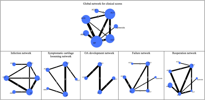
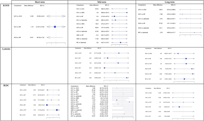
The global network for the primary outcome is shown in Fig. 2. The main comparator was MF, whereas the main comparison was between ACI3 and MF (four comparisons). The results of the pairwise comparison and NMA forest plots of pooled data did not indicate any differences among the functional outcomes (Supplementary Appendix 2, section B). The rankograms of multiple functional scores rated MF as the first treatment at short-, mid-, and long-term (shown in Supplementary Appendix 2, section B). Nevertheless, the forest plots of pooled data did not show any difference among PROMs at any timepoint (Fig. 3).
Figure 2.
Network diagrams for the clinical scores and adverse events. The network diagrams show the direct comparisons (lines) among techniques (nodes). The size of the nodes is proportional to the sample size of each treatment. The thickness of the lines is proportional to the number of available studies, indicated by the white number. ACI1, autologous chondrocyte implantation with periosteal membrane; ACI2, autologous chondrocyte implantation with collagen membrane; ACI3, autologous chondrocyte implantation with scaffold; AMIC, autologous matrix-induced chondrogenesis; MF, microfracture; OA, osteoarthritis; OCT, osteochondral transplantation; spheroids refer to chondrospheres.
Figure 3.
Forest plots of the most used clinical scores. There were no available studies using Lysholm over the short term. In forest plots, no difference for the mean differences corresponds to 0. The I2 for KOOS: over short term 25%, mid-term 8%, long term 10%. The I2 for Lysholm: mid-term 16%, long term 15%. The I2 for IKDC: at short term 16%, mid-term 2%, long term 2%. ACI1, autologous chondrocyte implantation with periosteal membrane; ACI2, autologous chondrocyte implantation with collagen membrane; ACI3, autologous chondrocyte implantation with scaffold; AMIC, autologous matrix-induced chondrogenesis; MF, microfracture; OA, osteoarthritis; OCT, osteochondral transplantation; spheroids refer to chondrospheres.
Secondary outcome
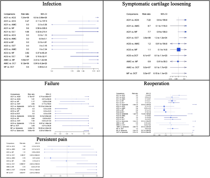
The safety analysis represented our secondary outcome, retrieved at a mean of 57.8 postoperative months in the overall NMA (Supplementary Appendix 2, section A). The forest plots of pooled data on the postoperative infection rate allowed the analysis of 15 paired comparisons, whose two showed a difference advantageous for ACI2 in the comparison versus ACI1 and AMIC (risk ratio (RR): 7.24e+08 (95% CI: 6.5–3.06e+26) and RR: 0 (95% CI: 0–0.1), respectively), and one for MF (AMIC vs MF – RR: 1.93e+07 (95% CI: 2.43–1.2e+26)), with I2 of 23% (Fig. 4). The rank probabilities for the infection classified ACI2 as first treatment followed by MF (Supplementary Appendix 2, section B). A description of failure was reported in 8/19 trials, where 6/8 defined it as a need for cartilage revision surgery (Supplementary Appendix 2, section A). Of the 15 available comparisons of the failure forest plot, 4/5 comparisons involving chondrospheres showed fewer failures with chondrospheres (ACI1 vs chondrospheres – RR: 1.6e+13 (95% CI: 37.5–7.5e+32); ACI3 vs chondrospheres – RR: 6.0e+12 (95% CI: 12.7 to 3.3e+32); MF vs chondrospheres – RR: 1.2e+13 (95% CI: 28.3–4.7e+32); OCT vs chondrospheres – RR: 2e+12 (95% CI: 5.7–9.8e+31); I2: 0%). The rank probabilities classified chondrospheres as the first treatment, followed by ACI2. Of the 21 paired comparisons of the forest plot for reoperations, only six showed a difference, in all cases favoring chondrospheres (vs ACI1 – RR: 4.0e+10, (95% CI: 35.9–4.6e+25); vs ACI2 – RR: 1.3e+10, (95% CI: 10.5–1.5e+25); vs ACI3 – RR: 3.3e+10, (95% CI: 32.3–3.5e+25); vs AMIC – RR: 2.8e+10, (95% CI: 26.3–3.6e+25); vs MF – RR: 3.3e+10, (95% CI: 25.7–3.8e+25); vs OCT – RR: 2.7e+10, (95% CI: 22.5–2.9e+25); I2: 0%). The rank probabilities classified the chondrospheres as the best treatment, followed by ACI2 and OCT.
Figure 4.
Forest plots of adverse events. In forest plots, no difference for the relative risk corresponds to one. The I 2 for infection was 23%, for symptomatic cartilage loosening was 29%, and for failure, reoperation, and persistent pain was 0%. ACI1, autologous chondrocyte implantation with periosteal membrane; ACI2, autologous chondrocyte implantation with collagen membrane; ACI3, autologous chondrocyte implantation with scaffold; AMIC, autologous matrix-induced chondrogenesis; MF, microfracture; OA, osteoarthritis; OCT, osteochondral transplantation; spheroids refer to chondrospheres.
Among ten comparisons of the forest plot for persistent pain, four showed a difference, favoring OCT (vs ACI1 – RR: 4.3e+07 (95% CI: 4.2–2.6e+24); vs ACI3 – RR: 3.55e+07 (95% CI: 2.62–2.15e+23); vs AMIC – RR: 2.13e+08 (95% CI: 7.41–1.3e+24); vs MF – RR: 3.6e+07 (95% CI: 2.24–2.15e+23), I2: 0%). The forest plot on symptomatic cartilage loosening shows mainly wide CI without any significant difference among treatments (Fig. 4). The complete report, including the raw data and the detailed reporting of adverse events is shown in sections A and B of Supplementary Appendix 2, respectively.
Subgroup analyses
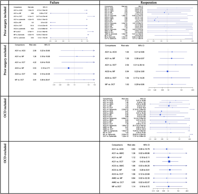
For the subgroup analyses (Fig. 5), we chose to assess the most used PROM, IKDC, as well as failures and reoperations. The subgroups for failure with the inclusion/exclusion of OCD were not feasible. Chondrospheres were the only treatment favored for fewer reoperations when OCD and prior cartilage surgery were included, in 6/21 comparisons each, but no difference was shown when OCD and prior cartilage surgery were excluded. The same result was confirmed in the subgroup analysis for failures. No difference was shown for IKDC in any subgroup (Supplementary Appendix 2, section B).
Figure 5.
Forest plots of the subgroup analysis showing difference among groups. In forest plots, no difference for the relative risk corresponds to one and for mean differences to zero. ACI1, autologous chondrocyte implantation with periosteal membrane; ACI2, autologous chondrocyte implantation with collagen membrane; ACI3, autologous chondrocyte implantation with scaffold; AMIC, autologous matrix-induced chondrogenesis; MF, microfracture; OA, osteoarthritis; OCT, osteochondral transplantation; spheroids refer to chondrospheres.
Risk-of-bias assessment
We assessed the risk of bias for the primary outcome and ITT. An adequate random sequence generation was specified in 89.5% (17/19) of trials. Outcome assessors and patients were blinded only in one study (29), resulting in a high risk of bias in 94.7% (18/19) of trials. In 52.6% (10/19) of trials, the analysis was intention-to-treat. Missing outcome data were responsible for a high risk of bias in 26.3% (5/19) of trials. A low risk of bias was shown for the selection of reported results in 94.7% (18/19) of trials. The overall risk of bias was high in all evaluated trials. Consequently, the GRADE analysis for the primary outcome showed low confidence in the results of the NMA (Supplementary Appendix 2, section C). Funnel plots for IKDC, failure, and reoperation are shown in Supplementary Appendix 2, section C.
Discussion
The current systematic review with NMA showed no differences in PROMs among the assessed treatments. The secondary (safety) outcome analysis, showing lower failure and reoperation rates with chondrospheres, must be interpreted with caution since adverse event data were incomplete for multiple techniques.
To date, the results of available NMA have shown different results for the safety analyses. Riboh et al. (21) showed lower reoperation rates with OCT at 5 years and with ACI2 at 10 years, while Migliorini et al. (22) showed reoperation rates with AMIC at a median follow-up of 36 months. Zamborsky et al. (23) showed fewer failures with ACI at 10 years and Migliorini et al. (22) showed failures with AMIC at 3 years.
The absence of a difference in PROMs at any time point is concordant with the NMA of Riboh et al. (21), and a previous meta-analysis comparing MF and three generations of ACI at a 5-year follow-up (30). The NMA of Migliorini et al. (22) showed better Lysholm and Tegner scores for AMIC at a median follow-up of 36 months, and that of Zamborsky et al. (23) for OCT at > 3 years.
The discordant results of the efficacy and safety analyses of the available NMAs can be explained by their different methodological features. The eligibility criteria of three previous NMAs included pediatric patients (21, 22, 23) and non-randomized trials (22), which we excluded in the present study. The nodes also differed among NMAs: ACI1 and ACI2 were pooled together in the study by Zamborsky et al. (23), whereas Migliorini et al. (22) grouped ACI3 and chondrospheres. We decided to distinguish not only each ACI generation (1, 2, 3) but also the latest generation of commercially available scaffold-free ACI3 (chondrospheres) from scaffold-based ACI3 like MACI, due to the different technologies necessary for their production, techniques of implantation, and economic impact. Indeed, the production, cell costs, and transportation were estimated by NICE to be £10 000 per patient for chondrospheres and a maximum of £16 000 per patient for ACI1, ACI2, and MACI (31, 32). Moreover, the type of surgical implantation, being under a periosteum or a collagen membrane (ACI1 vs ACI2), and sutured or not sutured to the surrounding cartilage (ACI1–3) can have an impact on the healing process, and therefore may affect the clinical outcome. This is the case with ACI1, where graft hypertrophy and bone spur development have been described together with cell dedifferentiation leading to the development of osteoarthritis (33, 34, 35). Another difference among NMAs is the analysis of clinical scores. Riboh et al. (21) considered only Lysholm and Tegner, Zamborsky et al. (23) used all available PROMs distinguishing among excellent or good versus poor results, and Migliorini et al. (22) used the standardized mean differences (SMD). We decided to use all available scores first, but once shown no difference among treatments, we decided not to proceed with SMD.
Differently from the other NMAs, we carried out subgroup analyses addressing two effect modifiers debated in the literature: OCD and prior cartilage surgery (microfracture, drilling, debridement, or abrasion).
For unstable OCD with surgical indication, it is debated if debridement and microfracture (the prevalent comparison in our NMA) can adequately restore the subchondral bone and joint congruity and relieve symptoms (36). Literature on bone-stimulating treatment prior to ACI showed discordant results, associated with both good and negative ACI outcomes (37, 38). In our study, the subgroup analysis assessing IKDC did not show any difference with or without prior cartilage treatment or OCD, which was coherent with the main analysis. On the other hand, the subgroup analysis evaluating failures and reoperations showed a difference with fewer failures and reoperations for chondrospheres in most of the comparisons when OCD or prior cartilage surgery were considered and showed no difference excluding them, confirming the results of the main safety analysis. From the results of our main and subgroup analyses, we can question whether PROMs are good indicators of outcomes to establish the superiority of treatments in trials. Moreover, there is no evidence proving which PROM is best for the assessment of knee cartilage surgery, nor do the guidelines for trial development in the knee cartilage field restrict the choice of PROMs (39). The heterogeneity in the use of PROMs added methodological differences among NMAs, possibly responsible for their discordant results. Similarly, the reporting of adverse events was very heterogeneous or incomplete in some cases. It must be noted that only one trial for chondrospheres and for ACI2 was available, which could add a bias related to their limited amount of data compared to other treatments used in multiple trials. For this reason and due to the low confidence in the NMA results provided by GRADE, further studies should demonstrate if chondrospheres do provide a benefit in terms of safety compared to other ACI generations. To improve the quality of trials and the summary of evidence (systematic reviews), we suggest that future evidence or guidelines for the design and conduction of trials on knee cartilage surgery standardize the PROMs and the reporting of adverse events. This is relevant because the technologies for knee cartilage defects have a remarkable economic impact. Future systematic reviews and cost-effectiveness studies should guide practitioners towards the use of the best and most cost-effective treatment.
Our study has the following limitations: the assessment of the risk of bias and GRADE was based on PROMs, in a batch of 18/19 open-label, non-blinded trials, resulting in overall high risk of bias and low confidence in the NMA results; the differences highlighted for the safety analysis in the main and subgroup analyses must be interpreted carefully, as explained before; a subgroup analysis on patellofemoral defects was not possible due to the absence of related raw data. Moreover, matching treatments with defect size could have been interesting, but we considered the mean average of the defect size provided by the trial authors not to be the best indicator of defect size. In fact, even for average medium-size defects (< 4 cm2) the size range could be > 10 cm2.
Conclusion
This systematic review with NMA compared MF, AMIC, OCT, ACI1, ACI2, ACI3 scaffold-based, and chondrospheres in adult patients with focal knee cartilage defects. There were no differences among treatments in terms of PROMs, and the results of our secondary (safety) analysis, showing lower failures and reoperations with chondrospheres are limited by the incomplete and heterogeneous reporting of adverse events in the assessed trials. Recommendations and guidelines aimed to define the best outcome measure in trials on cartilage repair and regeneration, as well as the standardization of PROMs use and adverse event reporting are needed to draw solid conclusions on the efficacy and safety of available techniques for knee cartilage repair and regeneration.
Supplementary Materials
ICMJE Conflict of Interest Statement
Two authors have declared a potential conflict of interest as specified in the ICMJE disclosure form: research funding from the Fondation privée des HUG (DH) and reimbursements for educational activities as a consultant from Arthrex and Amplitude (EC). The institution of three authors (SV, PT, DH) receives funding from the International Olympic Committee, because it belongs to the Réseau Francophone Olympique pour la Recherche en Médecine du Sport (ReFORM, IOC Research Medical Center).
Funding Statement
This study did not receive any specific grant from funding agencies in the public, commercial, or not-for-profit sectors.
Author contribution statement
Bibliographic search: SV, BA, PT, EC; data collection and extraction: SV, BA; data analysis: PB; statistical expertise: PB; conception of the design of the study, article review, final approval: all authors.
Acknowledgements
We thank Mrs Ann Kelly, Mr Jack Young and Mrs Rachel Couban, librarians of the Health Sciences Library at McMaster University, for revising the bibliographic search strategy. We want to thank the following authors for providing clarifications and unpublished data on trials’ study population: PD Dr med. Basad Erhan, Dr Clavé Arnaud (PhD), Dr Dozin Beatrice, Prof. Dr med. Moradi Babak, Prof. Saris Daniel B F, Mr Shive Matthew (PhD), and Dr Volz Martin. We express our gratitude to the support center of The Journal of Bone and Joint Surgery and Clinical Orthopaedics and Related Research, in the person–– of Prof. Leopold Seth, for helping us retrieve COI data, and the SAGE Publishing Support Centre for providing us with publications’ appendix. We are grateful to PD Combescure Christophe for his statistical support (Methodology Unit, University of Geneva, Switzerland) and to Dr Haqeeqat Singh Gurm, MD, for his writing assistance (Guys and St. Thomas’ Trust, UK).
References
- 1.Curl WWKJ Gordon S Rushing J Smith BP & Poehling GG. Cartilage injuries: a review of 31,516 knee arthroscopies. Journal of Arthroscopic and Related Surgery 199713456–460. [DOI] [PubMed] [Google Scholar]
- 2.Widuchowski W Widuchowski J & Trzaska T. Articular cartilage defects: study of 25,124 knee arthroscopies. Knee 200714177–182. ( 10.1016/j.knee.2007.02.001) [DOI] [PubMed] [Google Scholar]
- 3.Hjelle K Solheim E Strand T Muri R & Brittberg M. Articular cartilage defects in 1,000 knee arthroscopies. Arthroscopy 200218730–734. ( 10.1053/jars.2002.32839) [DOI] [PubMed] [Google Scholar]
- 4.Gomoll AHL Lattermann C & Farr J. A “Unifying Theory” treatment algorithm for cartilage defects. Cartilage Restoration: Practical Clinical Applications. Springer; 2018. [Google Scholar]
- 5.Flanigan DC Harris JD Trinh TQ Siston RA & Brophy RH. Prevalence of chondral defects in athletes' knees: a systematic review. Medicine and Science in Sports and Exercise 2010421795–1801. ( 10.1249/MSS.0b013e3181d9eea0) [DOI] [PubMed] [Google Scholar]
- 6.Steadman JR Rodkey WG Singleton SB & Briggs KK. Microfracture technique forfull-thickness chondral defects: technique and clinical results. Operative Techniques in Orthopaedics 19977300–304. ( 10.1016/S1048-6666(9780033-X) [DOI] [Google Scholar]
- 7.Anders S Volz M Frick H & Gellissen J. A randomized, controlled trial comparing autologous matrix-induced chondrogenesis (AMIC(R)) to microfracture: analysis of 1- and 2-year follow-up data of 2 centers. Open Orthopaedics Journal 20137133–143. ( 10.2174/1874325001307010133) [DOI] [PMC free article] [PubMed] [Google Scholar]
- 8.Stanish WD McCormack R Forriol F Mohtadi N Pelet S Desnoyers J Restrepo A & Shive MS. Novel scaffold-based BST-CarGel treatment results in superior cartilage repair compared with microfracture in a randomized controlled trial. Journal of Bone and Joint Surgery 2013951640–1650. ( 10.2106/JBJS.L.01345) [DOI] [PubMed] [Google Scholar]
- 9.Hangody L Kish G Karpati Z Udvarhelyi I Szigeti I & Bely M. Mosaicplasty for the treatment of articular cartilage defects: application in clinical practice. Orthopedics 199821751–756. ( 10.3928/0147-7447-19980701-04) [DOI] [PubMed] [Google Scholar]
- 10.McCormick F Yanke A Provencher MT & Cole BJ. Minced articular cartilage–basic science, surgical technique, and clinical application. Sports Medicine and Arthroscopy Review 200816217–220. ( 10.1097/JSA.0b013e31818e0e4a) [DOI] [PubMed] [Google Scholar]
- 11.Christensen BB Lind M & Foldager CB. Particulated cartilage auto- and allograft. In Cartilage Restoration. Farr J & Gomoll A Eds. Cham: Springer; 2018. ( 10.1007/978-3-319-77152-6_23) [DOI] [Google Scholar]
- 12.Brittberg M Faxen E & Peterson L. Carbon fiber scaffolds in the treatment of early knee osteoarthritis. A prospective 4-year followup of 37 patients. Clinical Orthopaedics and Related Research 1994. (307) 155–164. [PubMed] [Google Scholar]
- 13.Bentley G Biant LC Carrington RW Akmal M Goldberg A Williams AM Skinner JA & Pringle J. A prospective, randomised comparison of autologous chondrocyte implantation versus mosaicplasty for osteochondral defects in the knee. Journal of Bone and Joint Surgery 200385223–230. ( 10.1302/0301-620x.85b2.13543) [DOI] [PubMed] [Google Scholar]
- 14.Behrens P Ehlers EM Kochermann KU Rohwedel J Russlies M & Plotz W. New therapy procedure for localized cartilage defects. Encouraging results with autologous chondrocyte implantation. MMW Fortschritte Der Medizin 199914149–51. [PubMed] [Google Scholar]
- 15.Zeifang F Oberle D Nierhoff C Richter W Moradi B & Schmitt H. Autologous chondrocyte implantation using the original periosteum-cover technique versus matrix-associated autologous chondrocyte implantation: a randomized clinical trial. American Journal of Sports Medicine 201038924–933. ( 10.1177/0363546509351499) [DOI] [PubMed] [Google Scholar]
- 16.Kessler MW Ackerman G Dines JS & Grande D. Emerging technologies and fourth generation issues in cartilage repair. Sports Medicine and Arthroscopy Review 200816246–254. ( 10.1097/JSA.0b013e31818d56b3) [DOI] [PubMed] [Google Scholar]
- 17.Shah SS Lee S & Mithoefer K. Next-generation marrow stimulation technology for cartilage repair: basic science to clinical application. JBJS Reviews 20219e20.00090. ( 10.2106/JBJS.RVW.20.00090) [DOI] [PubMed] [Google Scholar]
- 18.Mithoefer K Scopp JM & Mandelbaum BR. Articular cartilage repair in athletes. Instructional Course Lectures 200756457–468. [PubMed] [Google Scholar]
- 19.Hinckel BB, Thomas D, Vellios EE, Hancock KJ, Calcei JG, Sherman SL, Eliasberg CD, Fernandes TL, Farr J, Lattermann C, et al. Algorithm for treatment of focal cartilage defects of the knee: classic and new procedures. Cartilage 202113(1_suppl) 473S–95S. ( 10.1177/1947603521993219) [DOI] [PMC free article] [PubMed] [Google Scholar]
- 20.Safran MR & Seiber K. The evidence for surgical repair of articular cartilage in the knee. Journal of the American Academy of Orthopaedic Surgeons 201018259–266. ( 10.5435/00124635-201005000-00002) [DOI] [PubMed] [Google Scholar]
- 21.Riboh JC Cvetanovich GL Cole BJ & Yanke AB. Comparative efficacy of cartilage repair procedures in the knee: a network meta-analysis. Knee Surgery, Sports Traumatology, Arthroscopy 2017253786–3799. ( 10.1007/s00167-016-4300-1) [DOI] [PubMed] [Google Scholar]
- 22.Migliorini F Eschweiler J Schenker H Baroncini A Tingart M & Maffulli N. Surgical management of focal chondral defects of the knee: a Bayesian network meta-analysis. Journal of Orthopaedic Surgery and Research 202116543. ( 10.1186/s13018-021-02684-z) [DOI] [PMC free article] [PubMed] [Google Scholar]
- 23.Zamborsky R & Danisovic L. Surgical techniques for knee cartilage repair: an updated large-scale systematic review and network meta-analysis of randomized controlled trials. Arthroscopy 202036845–858. ( 10.1016/j.arthro.2019.11.096) [DOI] [PubMed] [Google Scholar]
- 24.Hutton B, Salanti G, Caldwell DM, Chaimani A, Schmid CH, Cameron C, Ioannidis JPA, Straus S, Thorlund K, Jansen JP, et al. The PRISMA extension statement for reporting of systematic reviews incorporating network meta-analyses of health care interventions: checklist and explanations. Annals of Internal Medicine 2015162777–784. ( 10.7326/M14-2385) [DOI] [PubMed] [Google Scholar]
- 25.Salanti G Del Giovane C Chaimani A Caldwell DM & Higgins JPT. Evaluating the quality of evidence from a network meta-analysis. PLoS One 20149e99682. ( 10.1371/journal.pone.0099682) [DOI] [PMC free article] [PubMed] [Google Scholar]
- 26.Lu G & Ades AE. Combination of direct and indirect evidence in mixed treatment comparisons. Statistics in Medicine 2004233105–3124. ( 10.1002/sim.1875) [DOI] [PubMed] [Google Scholar]
- 27.Higgins JPT Thomas J Chandler J Cumpston M Li T Page MJ & Welch VA. Cochrane Handbook for Systematic Reviews of Interventions Version 6.3. Cochrane, 2022. [Google Scholar]
- 28.Hozo SP Djulbegovic B & Hozo I. Estimating the mean and variance from the median, range, and the size of a sample. BMC Medical Research Methodology 2005513. ( 10.1186/1471-2288-5-13) [DOI] [PMC free article] [PubMed] [Google Scholar]
- 29.Lim HC Bae JH Song SH Park YE & Kim SJ. Current treatments of isolated articular cartilage lesions of the knee achieve similar outcomes. Clinical Orthopaedics and Related Research 20124702261–2267. ( 10.1007/s11999-012-2304-9) [DOI] [PMC free article] [PubMed] [Google Scholar]
- 30.Negrin LL & Vécsei V. Do meta-analyses reveal time-dependent differences between the clinical outcomes achieved by microfracture and autologous chondrocyte implantation in the treatment of cartilage defects of the knee? Journal of Orthopaedic Science 201318940–948. ( 10.1007/s00776-013-0449-3) [DOI] [PubMed] [Google Scholar]
- 31.Autologous chondrocyte implantation for treating symptomatic articular cartilage defects of the knee (Technology appraisal guidance (TA477)). 2017. Available at: https://www.nice.org.uk/guidance/ta477. [Google Scholar]
- 32.Autologous chondrocyte implantation using chondrosphere for treating symptomatic articular cartilage defects of the knee (Technology appraisal guidance (TA508)). 2018. Available at: https://www.nice.org.uk/guidance/ta508. [Google Scholar]
- 33.Demoor M Maneix L Ollitrault D Legendre F Duval E Claus S Mallein-Gerin F Moslemi S Boumediene K & Galera P. Deciphering chondrocyte behaviour in matrix-induced autologous chondrocyte implantation to undergo accurate cartilage repair with hyaline matrix. Pathologie-Biologie 201260199–207. ( 10.1016/j.patbio.2012.03.003) [DOI] [PubMed] [Google Scholar]
- 34.Hunziker EB & Stahli A. Surgical suturing of articular cartilage induces osteoarthritis-like changes. Osteoarthritis and Cartilage 2008161067–1073. ( 10.1016/j.joca.2008.01.009) [DOI] [PMC free article] [PubMed] [Google Scholar]
- 35.Kreuz PC Steinwachs M Erggelet C Krause SJ Ossendorf C Maier D Ghanem N Uhl M & Haag M. Classification of graft hypertrophy after autologous chondrocyte implantation of full-thickness chondral defects in the knee. Osteoarthritis and Cartilage 2007151339–1347. ( 10.1016/j.joca.2007.04.020) [DOI] [PubMed] [Google Scholar]
- 36.Edmonds EW & Polousky J. A review of knowledge in osteochondritis dissecans: 123 years of minimal evolution from Konig to the ROCK study group. Clinical Orthopaedics and Related Research 20134711118–1126. ( 10.1007/s11999-012-2290-y) [DOI] [PMC free article] [PubMed] [Google Scholar]
- 37.Minas T Gomoll AH Rosenberger R Royce RO & Bryant T. Increased failure rate of autologous chondrocyte implantation after previous treatment with marrow stimulation techniques. American Journal of Sports Medicine 200937902–908. ( 10.1177/0363546508330137) [DOI] [PubMed] [Google Scholar]
- 38.McNickle AG L'Heureux DR Yanke AB & Cole BJ. Outcomes of autologous chondrocyte implantation in a diverse patient population. American Journal of Sports Medicine 2009371344–1350. ( 10.1177/0363546509332258) [DOI] [PubMed] [Google Scholar]
- 39.Mithoefer K, Saris DBF, Farr J, Kon E, Zaslav K, Cole BJ, Ranstam J, Yao J, Shive M, Levine D, et al. Guidelines for the design and conduct of clinical studies in knee articular cartilage repair: international Cartilage Repair Society recommendations based on current scientific evidence and standards of clinical care. Cartilage 20112100–121. ( 10.1177/1947603510392913) [DOI] [PMC free article] [PubMed] [Google Scholar]
Associated Data
This section collects any data citations, data availability statements, or supplementary materials included in this article.


 This work is licensed under a