Abstract
Background
The study assessed the association between COVID-19 and new-onset obstructive airway diseases, including asthma, chronic obstructive pulmonary disease, and bronchiectasis among vaccinated individuals recovering from COVID-19 during the Omicron wave.
Methods
This multicenter retrospective cohort study comprised 549,606 individuals from the U.S. Collaborative Network of TriNetX database, from January 8, 2022, to January 17, 2024. The hazard of new-onset obstructive airway diseases between COVID-19 and no-COVID-19 groups were compared following propensity score matching using the Kaplan–Meier method and Cox proportional hazards model.
Results
After propensity score matching, each group contained 274,803 participants. Patients with COVID-19 exhibited a higher risk of developing new-onset asthma than that of individuals without COVID-19 (adjusted hazard ratio (aHR), 1.27; 95% CI, 1.22–1.33; p < 0.001). Stratified analyses by age, SARS-CoV-2 variant, vaccination status, and infection status consistently supported this association. Non-hospitalized individuals with COVID-19 demonstrated a higher risk of new-onset asthma (aHR, 1.27; 95% CI, 1.22–1.33; p < 0.001); however, no significant differences were observed in hospitalized and critically ill groups. The study also identified an increased risk of subsequent bronchiectasis following COVID-19 (aHR, 1.30; 95% CI, 1.13–1.50; p < 0.001). In contrast, there was no significant difference in the hazard of chronic obstructive pulmonary disease between the groups (aHR, 1.00; 95% CI, 0.95–1.06; p = 0.994).
Conclusion
This study offers convincing evidence of the association between COVID-19 and the subsequent onset of asthma and bronchiectasis. It underscores the need for a multidisciplinary approach to post-COVID-19 care, with a particular focus on respiratory health.
Supplementary Information
The online version contains supplementary material available at 10.1186/s12916-024-03589-4.
Keywords: Asthma, Bronchiectasis, COPD, COVID-19, Obstructive airway diseases
Background
Over the past 3 years, severe acute respiratory syndrome coronavirus 2 (SARS-CoV-2) has infected more than 773 million individuals globally [1]. Despite coronavirus disease (COVID-19) being responsible for over seven million fatalities, the case fatality rate stood at a mere 0.9% [1], suggesting that more than 99% of patients with COVID-19 have survived the acute phase of SARS-CoV-2 infection. Nevertheless, a significant number of COVID-19 survivors develop post-COVID-19 conditions, also termed long COVID, which present a grave threat to global health [2–4]. Reports have documented a broad range of persistent symptoms linked to post-COVID-19 conditions, impacting the cardiovascular and pulmonary systems, gastrointestinal tract, neurocognitive functions, psychological well-being, and musculoskeletal system [3–7].
The respiratory system is frequently involved in post-COVID-19 conditions, as demonstrated by clinical and radiologic findings, along with pulmonary function tests [8, 9]. Predominantly, studies have concentrated on the restrictive ventilatory defect, which is marked by lung fibrosis, a diminished diffusion capacity of the lung for carbon monoxide, and reduced lung volume [8–11]. In addition, residual abnormalities on computed tomography for patients with COVID-19 are not uncommon. One meta-analysis showed that fibrotic-like changes had the highest event rates of 0.44 and 0.38 during both short-term (1–6 months) and long-term (12–24 months) follow-up periods [12]. Moreover, patients with severe COVID-19 exhibited significantly higher rates of various abnormalities, including bronchiectasis, fibrotic-like changes, and reticulation, at long-term follow-ups compared to those in the non-severe subgroup [12]. Conversely, although prior research has identified a link between respiratory virus infections and the subsequent onset of asthma [13–15], the evaluation of obstructive airway disease risk following COVID-19 has been limited [16–18].
Kim et al. using the Korean National Health Insurance claim-based database demonstrated that COVID-19 could be associated with a higher risk of new-onset asthma (adjusted hazard ratio (aHR), 2.14; 95% CI, 1.88–2.45) [16]. However, the study was conducted between 2020 and June 30, 2021, in South Korea and focused specifically on asthma. Their findings were thus limited to the Asian population and the non-Omicron wave. Since then, the virulence, epidemiology, and disease severity of the currently prevalent Omicron variant have changed significantly from those of the early strains [19, 20]. An updated investigation among non-Asian populations is also needed to provide a broader understanding of the association between COVID-19 and obstructive airway diseases. Therefore, we conducted this study using the TriNetX network to assess the association between COVID-19 and new-onset obstructive airway diseases, particularly asthma, following COVID-19 during the Omicron wave.
Results
Characteristics of study subjects
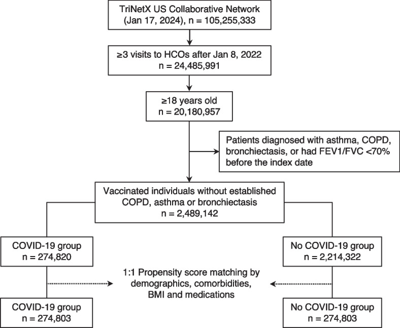
Among the vaccinated individuals without pre-existing asthma, COPD, or bronchiectasis, the study included 274,820 enrollees in the COVID-19 group and 2,214,332 in the no-COVID-19 group (Fig. 1). Prior to PSM, the average age of patients with COVID-19 was 55.5 years, compared to 54.8 years for those without. The COVID-19 group was 57.4% female, while the no-COVID-19 group was 58.9% female; 64% of the COVID-19 group and 67.5% of the no-COVID-19 group were Caucasians. Age, sex, and race did not differ significantly between the groups (all standardized differences < 0.1). However, the COVID-19 group showed a higher prevalence of comorbidities, including DM, dyslipidemia, hypertension, ischemic heart disease, heart failure, cerebrovascular disease, CKD, neoplasm, anemia, and systemic connective tissue disease, as well as allergic rhinitis, compared to their counterparts (Table 1). Additionally, a greater percentage of the COVID-19 group had histories of tobacco use/nicotine dependence, a BMI of ≥ 30 kg/m2, and socioeconomic status (SES)- or psychosocial-related health hazards. Regarding medication, a higher proportion of the COVID-19 group had received systemic glucocorticoids, antihistamines, beta-blockers, aspirin, and NSAIDs within 1 year preceding the index date (Table 1).
Fig. 1.

Flowchart of cohort construction. BMI body mass index, COVID-19 coronavirus disease 2019, FEV1 forced expiratory volume in the first second, FVC forced vital capacity, HCO healthcare organization, COPD chronic obstructive pulmonary disease
Table 1.
Baseline characteristics
| Variables | Before matching | After matching | ||||
|---|---|---|---|---|---|---|
| COVID-1 (n = 274,820) | No COVID-19 (n = 2,214,322) | Std. diff. | COVID-19 (n = 274,803) | No COVID-19 (n = 274,803) | Std. diff. | |
| Demographics | ||||||
| Age (mean ± SD) | 55.5 ± 18.5 | 54.8 ± 18.8 | 0.037 | 55.5 ± 18.5 | 55.8 ± 18.3 | 0.014 |
| Female | 161,968 (58.9) | 1,271,264 (57.4) | 0.031 | 161,954 (58.9) | 161,511 (58.5) | 0.003 |
| White | 185,452 (67.5) | 1,417,970 (64.0) | 0.073 | 185,438 (67.5) | 185,767 (67.6) | 0.003 |
| African American | 31,209 (11.4) | 291,711 (13.2) | 0.055 | 31,208 (11.4) | 31,720 (11.1) | 0.006 |
| Asian | 21,816 (7.9) | 184,713 (8.3) | 0.015 | 21,816 (7.9) | 21,761 (7.7) | 0.001 |
| Comorbidities | ||||||
| Diabetes mellitus | 47,704 (17.4) | 208,628 (9.4) | 0.235 | 47,691 (17.4) | 47,293 (17.1) | 0.004 |
| Dyslipidemia | 105,886 (38.5) | 460,850 (20.8) | 0.395 | 105,870 (38.5) | 110,171 (38.3) | 0.032 |
| Hypertension | 107,739 (39.2) | 494,876 (22.4) | 0.371 | 107,723 (39.2) | 110,598 (39.3) | 0.021 |
| Ischemic heart diseases | 27,825 (10.1) | 109,008 (4.9) | 0.198 | 27,815 (10.1) | 26,617 (10.1) | 0.015 |
| Heart failure | 12,402 (4.5) | 41,265 (1.9) | 0.151 | 12,395 (4.5) | 10,877 (4.4) | 0.027 |
| Peripheral vascular disease | 6,295 (2.3) | 22,709 (1.0) | 0.099 | 6,289 (2.3) | 5,680 (2.2) | 0.015 |
| Cerebrovascular diseases | 14,040 (5.1) | 52,719 (2.4) | 0.144 | 14,032 (5.1) | 12,813 (5.5) | 0.021 |
| Dementia | 3,446 (1.3) | 10,705 (0.5) | 0.083 | 3,443 (1.3) | 3,131 (1.1) | 0.010 |
| Alzheimer's disease | 1,938 (0.7) | 7,280 (0.3) | 0.053 | 1,937 (0.7) | 1,825 (0.0) | 0.005 |
| Chronic kidney disease | 23,891 (8.7) | 86,687 (3.9) | 0.198 | 23,884 (8.7) | 22,449 (8.8) | 0.019 |
| Inflammatory liver diseases | 3,907 (1.4) | 12,246 (0.6) | 0.088 | 3,904 (1.4) | 3,328 (1.1) | 0.018 |
| Liver cirrhosis | 3,057 (1.1) | 11,227 (0.5) | 0.068 | 3,057 (1.1) | 2,608 (1.1) | 0.016 |
| Neoplasms | 61,041 (22.2) | 272,071 (12.3) | 0.265 | 61,026 (22.2) | 61,983 (22.2) | 0.008 |
| Anemia | 25,937 (9.4) | 85,755 (3.9) | 0.225 | 25,928 (9.4) | 23,833 (9.9) | 0.027 |
| Systemic CTDs | 7,369 (2.7) | 27,394 (1.2) | 0.104 | 7,365 (2.7) | 6,643 (2.2) | 0.017 |
| Rheumatoid arthritis | 4,177 (1.5) | 15,896 (0.7) | 0.076 | 4,170 (1.5) | 3,873 (1.1) | 0.009 |
| Atopic dermatitis | 1,509 (0.6) | 6,073 (0.3) | 0.043 | 1,509 (0.6) | 1,346 (0.0) | 0.008 |
| Allergic rhinitis | 19,379 (7.1) | 69,874 (3.2) | 0.178 | 19,371 (7.1) | 19,381 (7.7) | 0.000 |
| Tobacco use | 11,697 (4.3) | 38,852 (1.8) | 0.147 | 11,688 (4.3) | 10,181 (4.4) | 0.028 |
| Nicotine dependence | 12,913 (4.7) | 60,134 (2.7) | 0.105 | 12,908 (4.7) | 12,503 (4.4) | 0.007 |
| Alcohol-related disorders | 5,919 (2.2) | 20,583 (0.9) | 0.099 | 5,913 (2.2) | 5,185 (2.2) | 0.019 |
| SES and psychosocial-related health hazards | 9,816 (3.6) | 28,838 (1.3) | 0.148 | 9,805 (3.6) | 8,539 (3.3) | 0.026 |
| HIV | 2,797 (1.0) | 14,075 (0.6) | 0.042 | 2,797 (1.0) | 2,501 (1.1) | 0.011 |
| BMI ≥30 kg/m2 | 39,113 (14.2) | 168,388 (7.6) | 0.214 | 39,100 (14.2) | 40,247 (14.1) | 0.012 |
| Medications | ||||||
| Glucocorticoids | 88,008 (32.0) | 371,437 (16.8) | 0.361 | 88,992 (32.0) | 88,114 (32.3) | 0.001 |
| Antihistamines | 55,601 (20.2) | 217,363 (9.8) | 0.295 | 55,587 (20.2) | 55,187 (20.2) | 0.004 |
| Beta blockers | 53,298 (19.4) | 258,025 (11.7) | 0.215 | 53,287 (19.4) | 52,790 (19.1) | 0.005 |
| Aspirin | 26,372 (9.6) | 112,237 (5.1) | 0.174 | 26,366 (9.6) | 24,916 (9.9) | 0.018 |
| 25,176 (9.2) | 109,365 (4.9) | 0.165 | 25,166 (9.2) | 24,976 (9.9) | 0.002 | |
BMI Body mass index, COVID-19 Coronavirus disease 2019, CTD Connective tissue disease, HIV Human immunodeficiency virus, NSAID Non-steroidal anti-inflammatory drugs, SD Standard deviation, SES Socioeconomic status, Std. diff Standardized difference
After PSM, each group consisted of 274,803 patients. Characteristics for all the considered covariates, including demographics, comorbidities, and medications, were balanced between the two groups with standardized differences < 0.1 (Table 1). The group with COVID-19 was designated as the study group, whereas the group without COVID-19 was designated as the control group.
Primary outcome
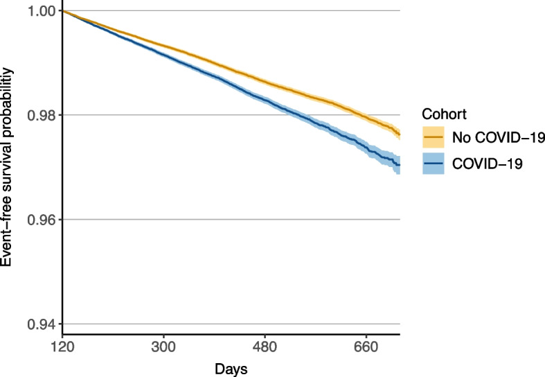
During the follow-up period, 4216 new asthma cases were diagnosed in the study group, compared with 3352 in the control group. The study group had a significantly higher risk of developing new-onset asthma than the control group (aHR, 1.27; 95% CI, 1.22–1.33; p < 0.001; see Table 2). Additionally, Kaplan–Meier analysis showed a lower probability of event-free survival for the study group relative to the control group (log-rank p < 0.001; see Fig. 2).
Table 2.
Adjusted hazard ratios of primary and secondary outcomes
| Outcomes | No. of patients with outcome | Adjusted hazard ratio (95% CI) | p-value | |
|---|---|---|---|---|
| COVID-19 (n = 274,803) | No COVID-19 (n = 274,803) | |||
| Primary outcome | ||||
| Asthma | 4216 | 3352 | 1.27 (1.22–1.33)* | <0.001 |
| Secondary outcomes | ||||
| Bronchiectasis | 438 | 345 | 1.30 (1.13–1.50)* | <0.001 |
| COPD | 2180 | 1964 | 1.00 (0.95–1.06)* | 0.994 |
CI Confidence interval, COPD Chronic obstructive pulmonary disease
*Proportionality (Schoenfeld test p >0.05)
Fig. 2.

Kaplan–Meier curves of event-free survival for new-onset asthma (log-rank test p < 0.001)
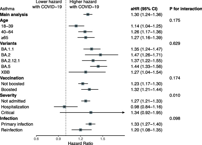
Stratified analyses by age group, SARS-CoV-2 variant, vaccine status, and infection status consistently demonstrated similar trends. Patients with COVID-19 exhibited a higher risk of new-onset asthma, as illustrated in Fig. 3. Notably, there were no significant differences in effect size estimates across the analyses, except for those based on disease severity (p for interaction = 0.011). A significantly higher risk of new-onset asthma was observed only in non-hospitalized patients with COVID-19, whereas no significant differences were observed between hospitalized and critically ill groups (Fig. 3).
Fig. 3.
Subgroup analysis of new-onset asthma between COVID-19 and no-COVID-19 groups
Secondary outcomes
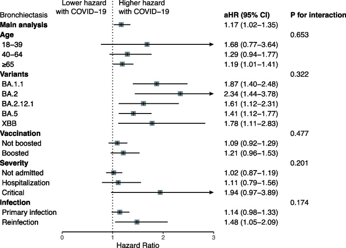
We also identified that patients with COVID-19 had a higher risk of bronchiectasis (aHR, 1.30; 95% CI, 1.13–1.50, p < 0.001, Table 2). There was a trend towards an increased risk of bronchiectasis in patients with critical COVID-19 compared to those not hospitalized, but no statistically significant between-subgroup heterogeneity was observed for bronchiectasis as evidenced by the interaction tests (all p values for interaction > 0.05) and the largely overlapping confidence intervals (Fig. 4). On the other hand, there was no significant difference in the incidence of COPD between the groups (aHR, 1.00; 95% CI, 0.95–1.06, p = 0.994, Table 2).
Fig. 4.
Subgroup analysis of new-onset bronchiectasis between COVID-19 and no-COVID-19 groups
Sensitivity analysis
Our sensitivity analysis using different cutoff landmarks (2, 3, and 6 months after the index date) to define new-onset obstructive airway diseases and the start of follow-up, showed consistent results. These results suggested that COVID-19 is associated with an increased risk of new-onset asthma and bronchiectasis, but not COPD (Additional file 1: Table S4).
Regarding the negative and positive control outcomes, we observed a positive association between COVID-19 and symptoms characteristic of the post-COVID-19 condition, as expected, compared to the no-COVID-19 group. No significant associations were observed with the negative control outcomes, as depicted in Additional file 1: Fig. S1.
Discussion
This large-scale study evaluated the respiratory sequelae of COVID-19, including asthma, COPD, and bronchiectasis. We found a potential link between COVID-19 and an increased risk of new-onset asthma. Specifically, patients with COVID-19 had a 27% higher risk of developing asthma during the follow-up period compared to those without COVID-19. Further analyses, stratified by age, SARS-CoV-2 variant, vaccination status, and infection status, as well as sensitivity analyses using different landmarks, consistently supported these findings. In summary, our study presents compelling evidence regarding the association between COVID-19 and the subsequent development of asthma, highlighting the potential long-term respiratory impacts of SARS-CoV-2 infection and enhancing our knowledge of the disease’s progression beyond the acute phase.
Stratified analysis by disease severity revealed an intriguing nuance. Although the overall risk of new-onset asthma was significantly higher in the COVID-19 group, this association was particularly pronounced in non-hospitalized individuals. The absence of a significant difference in hospitalized and critically ill groups might be partly due to a smaller patient count in these categories. Progression to viral pneumonia could indicate more severe inflammation of the lower airway and could be associated with the risk of subsequent obstructive airway diseases, but its occurrence, the chronological relationship with SARS-CoV-2 infection, and the etiologies could not be ascertained in the current database, thereby precluding further analysis on this variable. However, the interaction test suggested a significant difference, indicating a complex relationship between COVID-19 disease severity and the risk of obstructive airway diseases that warrants further exploration.
Our findings are consistent with those reported by Kim et al., who used the Korean National Health Insurance claim-based database. They found that 1.6% of the COVID-19 cohort and 0.7% of the matched cohort developed new-onset asthma, with an aHR of 2.14 (95% CI, 1.88–2.45) [16]. However, unlike the Korean study [16] conducted during 2020–2021, our analyses were based on data collected after 2022 and feature a more ethnically diverse population from the TriNetX platform. Consequently, our results provided updated insights during the Omicron wave and are likely to be more generalizable and relevant to the current context.
Respiratory tract viral infections, including respiratory syncytial virus and measles, have been identified as potential causes of bronchiectasis [21], but the link between SARS-CoV-2 and post-infectious bronchiectasis is not well established. Although a meta-analysis by Guinto et al. [22] reported a 16.8% prevalence of bronchiectasis (95% CI, 9.10–26.1%) based on imaging studies during post-COVID-19 follow-up, our study is the first to demonstrate an increased risk of subsequent bronchiectasis following COVID-19, with an aHR of 1.30 (95% CI, 1.13–1.50). These findings suggest that bronchiectasis may be a respiratory sequela of COVID-19. Respiratory viruses were found more frequently in bronchiectasis exacerbations and the proposed mechanisms included disturbances in host-defense responses, heightened inflammation, and changes in bacterial virulence [23], which could be involved in the vicious cycle model of development and progression of bronchiectasis, for which the primary insult is often unknown [24]. Our findings accordingly supported that clinicians should be vigilant for this potential complication after COVID-19.
The current study aligns with previous research [16, 22] highlighting the respiratory system’s vulnerability to post-COVID-19 conditions. Notably, the increased incidence of new-onset asthma and bronchiectasis observed in the study group suggests a potential link between SARS-CoV-2 infection and the development of obstructive airway diseases. This represents a contradiction with the results of previous studies [25, 26] which primarily focused on restrictive ventilatory defects associated with lung fibrosis. However, further investigation is crucial to validate these findings and elucidate the potential mechanisms involved.
This study boasts several strengths. First, the analyses utilized the TriNetX platform, a sizable database, which facilitated the inclusion of a substantial patient cohort. Second, we conducted numerous stratification analyses and sensitivity tests, with the majority yielding consistent results. Third, although patients with COVID-19 exhibited a higher prevalence of comorbidities such as diabetes mellitus, cardiovascular diseases, and systemic connective tissue disorders—conditions potentially elevating the risk of obstructive airway diseases—these factors were meticulously adjusted in relation to study outcomes. The application of PSM effectively equalized baseline characteristics between the COVID-19 and control groups, thus bolstering the study’s internal validity.
Despite the study’s strengths, we must acknowledge certain limitations. The reliance on electronic health records and administrative data raises the possibility of misclassification and underreporting. Furthermore, the study does not investigate potential mechanisms behind the observed associations, which leaves a gap for future research to clarify the pathophysiological connections between SARS-CoV-2 infection and obstructive airway diseases. Long-term patient follow-up is also essential to comprehend the persistence and progression of these respiratory conditions over time. Meanwhile, FEV1 and disease staging data for asthma or COPD were not documented for most patients in the TriNetX database, thereby introducing potential information bias and precluding further analysis on these variables. Finally, the possibility of residual confounding could not be fully eliminated though we had accounted for numerous clinically significant confounders in our analyses.
Conclusions
This study provides essential insights into the long-term respiratory consequences of COVID-19, highlighting the need for continued monitoring and care for individuals recovering from SARS-CoV-2 infection. The associations found with new-onset asthma and bronchiectasis underscore the importance of a multidisciplinary approach to post-COVID-19 care, with a particular focus on respiratory issues. Further research is necessary to investigate the underlying mechanisms and to develop therapeutic strategies to mitigate the effects of SARS-CoV-2 on the respiratory system.
Methods
Data source
The present study utilized data from the US Collaborative Network of the TriNetX database, which gathered de-identified patient-level information from electronic health records. A health care organization (HCO) typically referred to an academic healthcare center that compiled data from its associated facilities, including the main and satellite hospitals, and outpatient clinics. The collected data encompassed patient demographics, clinical diagnoses (coded using ICD-10-CM), medical procedures (categorized according to ICD-10-PCS or Current Procedural Terminology), medications (coded based on the Veterans Affairs Drug Classification System and RxNorm medication codes), laboratory tests (organized with Logical Observation Identifiers Names and Codes), and records of healthcare utilization. The US Collaborative Network contained data from over 100 million patients across 61 HCOs in the United States. Analysis of patient-level data was conducted on the TriNetX platform, and the results were provided to researchers in a summarized format. The TriNetX database performs intensive data preprocessing procedures to minimize missing values and maps the data to a consistent framework. Details regarding the database can be found online [21].
The current study’s use of the TriNetX database received ethical approval from the Chi-Mei Hospital’s Institutional Review Board (no: 11202–002). We conducted the study in accordance with the Declaration of Helsinki and reported our findings following the Strengthening the Reporting of Observational Studies in Epidemiology (STROBE) guidelines.
Study population
Participants of this study were ≥ 18-year-olds and visited HCOs ≥ 3 times after January 8, 2022. This date coincides with the emergence of the Omicron variant as the predominant strain in the United States [26], ensuring that the data for our analysis were collected within a comparable timeframe. We limited our inclusion to those vaccinated against COVID-19 to reflect the high vaccination rates among U.S. adults, which had surpassed 90% in U.S. adults [27], and to minimize biases related to health-seeking behavior using vaccination status as a proxy. To avoid misinterpretation, we excluded individuals with pre-existing diagnoses of asthma, chronic obstructive pulmonary disease (COPD), bronchiectasis, or forced expiration in the first second (FEV1)/forced vital capacity (FVC) < 70% before the index date. The index date was defined as the date of COVID-19 diagnosis for the COVID-19 group and the date of the first visit to HCOs during the inclusion period for the no-COVID-19 group.
The enrollees were further divided into those diagnosed with COVID-19 (the COVID-19 group) during the study timeframe and those who never had COVID-19 (the no-COVID-19 group). One-to-one propensity score matching (PSM) was conducted, involving 34 variables, including demographics, comorbidities, and medication usage, to balance the two groups. Both groups were followed for a maximum of 2 years or until the date of data analysis on January 17, 2024. Details regarding the codes used to identify demographics, diagnoses, procedures, and medications are provided in Additional file 1: Table S2.
Covariates
In the current analysis, we considered the following variables to balance baseline characteristics between the COVID-19 and non-COVID-19 groups: age, sex, race, diabetes mellitus (DM), dyslipidemia, cardiovascular diseases (hypertension, ischemic heart diseases, heart failure, peripheral vascular disease, cerebrovascular diseases), dementia, chronic kidney disease (CKD), hepatitis, cirrhosis, autoimmune diseases (systemic connective tissue disorders, rheumatoid arthritis), anemia, neoplasms, human immunodeficiency virus (HIV) disease, atopic dermatitis, allergic rhinitis, tobacco use/nicotine dependence, alcohol-related disorders, potential health hazards related to socioeconomic and psychosocial circumstances, and BMI (≥ 30 kg/m2). We also included medications that could alter allergic or inflammatory responses or are potentially associated with bronchospasm (glucocorticoids, antihistamines, beta-adrenergic blockers, aspirin and other non-steroidal anti-inflammatory drugs (NSAIDs)). The codes used to define the covariates are provided in Additional file 1: Table S3.
Prespecified outcomes
The primary outcome of this study was the hazard of new-onset asthma during the follow-up period, which commenced 4 months after the index date to preclude the confounding effects of post-viral bronchial hyperreactivity syndrome. Secondary outcomes encompassed the hazard of bronchiectasis and COPD within the same follow-up timeframe. To ascertain the specificity of our results, we incorporated negative control outcomes, including traumatic intracranial injury, skin cancer, schizophrenia, and cataract. Conversely, the positive control outcome was the post-COVID-19 condition, characterized by a collection of symptoms such as fatigue, headache, dizziness, myalgia, sleep disturbance, emotional distress, cognitive impairment, palpitation, shortness of breath, and changes in bowel habits [28]. Additional file 1: Table S4 contains the definitions and codes pertinent to these outcomes.
Statistical analysis
Baseline characteristics for the COVID-19 and no-COVID-19 groups are presented as means with standard deviations (SD) or as counts and percentages. We compared categorical variables using the χ2 test and assessed continuous variables with the independent two-sample t-test. We conducted one-to-one PSM using the greedy nearest neighbor algorithm with a caliper of 0.1 pooled standardized differences to balance baseline characteristics. Variables were considered adequately matched post-PSM if the standardized difference between groups was less than 0.1. We computed survival probabilities using the Kaplan–Meier method and calculated adjusted hazard ratios (aHR) for the outcomes using the Cox proportional hazards model, including corresponding 95% confidence intervals (CI) and p-values. We tested the proportional hazards assumption with the generalized Schoenfeld residuals method. Outcome variables were categorized as present or absent; thus, missingness was not applicable. We excluded cases lost to follow-up to reduce potential biases or inaccuracies from incomplete data.
We conducted sensitivity analyses using different cutoff landmarks (2, 3, and 6 months post-index date) to initiate follow-up and define new-onset asthma. To investigate potential differences in effect sizes among clinically relevant subgroups, we performed pre-specified subgroup analyses based on age (≥ 18 to < 40, ≥ 40 to < 65, or ≥ 65 years), variants (BA.1.1, BA.2, BA.2.12.1, BA.5, or XBB, corresponding to periods when a variant predominated in the U.S. [27]), booster vaccination status, COVID-19 severity (not admitted, hospitalized, or critical, the latter defined by the need for endotracheal intubation, mechanical ventilation, extracorporeal membrane oxygenation, or intensive care unit admission), and infection status (primary infection or reinfection). All tests were two-sided with a significance threshold of 0.05. Statistical analyses were performed using TriNetX analytics tools and R (version 4.2.2; R Foundation for Statistical Computing, Vienna, Austria).
Supplementary Information
Additional file 1: Table S1. Sensitivity analysis for primary and secondary outcomes with different cutoff landmarks to define new-onset obstructive airway diseases and the start of follow-up. Table S2. Demographic, diagnostic, procedural, medication, visit, and laboratory codes used in the definition of the cohort. Table S3. Demographic, diagnostic, laboratory, and medication codes used in the definition of covariates. Table S4. Diagnostic codes used in the definition of outcomes. FigS1. Results for negative and positive control outcomes.
Acknowledgements
We express our gratitude to Chung-Han Ho for his consultation on statistical analysis.
Abbreviations
- aHR
Adjusted hazard ratio
- BMI
Body mass index
- CI
Confidence interval
- COPD
Chronic obstructive pulmonary disease
- COVID-19
Coronavirus disease 2019
- CKD
Chronic kidney disease
- DM
Diabetes mellitus
- FEV1
Forced expiration in the first second.
- FVC
Forced vital capacity
- HCO
Health care organization
- HIV
Human immunodeficiency virus
- ICD-10-CM
International Classification of Diseases, Tenth Revision, Clinical Modification
- ICD-10-PCS
International Classification of Diseases, Tenth Revision, Procedure Coding System
- NSAID
Non-steroidal anti-inflammatory drugs
- PSM
Propensity score matching
- SARS-CoV-2
Severe acute respiratory syndrome coronavirus 2
- SD
Standard deviation
Authors’ contributions
MHC conceptualized, designed the study performed the data analysis and drafted the manuscript. WH, YWT, WHH, JYW, THL and PYH assisted data correction and created figures. MHC, and CCL contributed to project design and edited the manuscript. MHC and CCL was responsible for the data interpretation. MHC and CCL finalized the manuscript. All authors approved the final version of the manuscript.
Funding
No specific funding was received from any bodies in the public, commercial, or not-for-profit sectors to carry out the work described in this article.
Availability of data and materials
All data generated or analyzed during this study are included in this published article and will be available upon request to CCL.
Data availability
No datasets were generated or analysed during the current study.
Declarations
Ethics approval and consent to participate
The Institutional Review Board of the Chi Mei Medical Center approved the study protocol (no. 11202–002). Written informed consent was not required because TriNetX contains anonymized data.
Consent for publication
None.
Competing interests
None.
Footnotes
Publisher’s Note
Springer Nature remains neutral with regard to jurisdictional claims in published maps and institutional affiliations.
References
- 1.World Health Organization. Who COVID-19 dashboard. Available at: https://data.who.int/dashboards/covid19/cases?n=c Accessed January 18, 2024.
- 2.Frallonardo L, Segala FV, Chhaganlal KD, Yelshazly M, Novara R, Cotugno S, et al. Incidence and burden of long covid in Africa: a systematic review and meta-analysis. Sci Rep. 2023;13(1):21482. 10.1038/s41598-023-48258-3 [DOI] [PMC free article] [PubMed] [Google Scholar]
- 3.Liu TH, Huang PY, Wu JY, Chuang MH, Hsu WH, Tsai YW, et al. Comparison of post-acute sequelae following hospitalization for COVID-19 and influenza. BMC Med. 2023;21(1):480. 10.1186/s12916-023-03200-2 [DOI] [PMC free article] [PubMed] [Google Scholar]
- 4.Kelly JD, Curteis T, Rawal A, Murton M, Clark LJ, Jafry Z, et al. SARS-CoV-2 post-acute sequelae in previously hospitalised patients: systematic literature review and meta-analysis. Eur Respir Rev. 2023;32(169): 220254. 10.1183/16000617.0254-2022 [DOI] [PMC free article] [PubMed] [Google Scholar]
- 5.Natarajan A, Shetty A, Delanerolle G, Zeng Y, Zhang Y, Raymont V, et al. A systematic review and meta-analysis of long COVID symptoms. Syst Rev. 2023;12(1):88. 10.1186/s13643-023-02250-0 [DOI] [PMC free article] [PubMed] [Google Scholar]
- 6.Liu TH, Huang PY, Wu JY, Chuang MH, Hsu WH, Tsai YW, et al. Post-covid-19 condition risk in patients with intellectual and developmental disabilities: a retrospective cohort study involving 36,308 patients. BMC Med. 2023;21(1):505. 10.1186/s12916-023-03216-8 [DOI] [PMC free article] [PubMed] [Google Scholar]
- 7.Chuang MH, Wu JY, Liu TH, Hsu WH, Tsai YW, Huang PY, et al. Efficacy of nirmatrelvir and ritonavir for post-acute COVID-19 sequelae beyond 3 months of SARS-CoV-2 infection. J Med Virol. 2023;95(4): e28750. 10.1002/jmv.28750 [DOI] [PubMed] [Google Scholar]
- 8.Cha MJ, Solomon JJ, Lee JE, Choi H, Chae KJ, Lee KS, et al. Chronic lung injury after COVID-19 pneumonia: clinical, radiologic, and histopathologic perspectives. Radiology. 2024;310(1): e231643. 10.1148/radiol.231643 [DOI] [PMC free article] [PubMed] [Google Scholar]
- 9.Pietruszka-Wałęka E, Rząd M, Żabicka M, Rożyńska R, Miklusz P, Zieniuk-Lesiak E, et al. Impact of symptomatology, clinical and radiological severity of COVID-19 on pulmonary function test results and functional capacity during follow-up among survivors. J Clin Med. 2023;13(1):45. 10.3390/jcm13010045 [DOI] [PMC free article] [PubMed] [Google Scholar]
- 10.Eizaguirre S, Sabater G, Belda S, Calderón JC, Pineda V, Comas-Cufí M, et al. Long-term respiratory consequences of COVID-19 related pneumonia: a cohort study. BMC Pulm Med. 2023;23(1):439. 10.1186/s12890-023-02627-w [DOI] [PMC free article] [PubMed] [Google Scholar]
- 11.Suppini N, Fira-Mladinescu O, Traila D, Motofelea AC, Marc MS, Manolescu D, et al. Longitudinal analysis of pulmonary function impairment one year post-covid-19: a single-center study. J Pers Med. 2023;13(8):1190. 10.3390/jpm13081190 [DOI] [PMC free article] [PubMed] [Google Scholar]
- 12.Babar M, Jamil H, Mehta N, Moutwakil A, Duong TQ. Short- and long-term chest-CT findings after recovery from COVID-19: a systematic review and meta-analysis. Diagnostics (Basel). 2024;14(6):621. 10.3390/diagnostics14060621 [DOI] [PMC free article] [PubMed] [Google Scholar]
- 13.Rantala A, Jaakkola JJ, Jaakkola MS. Respiratory infections precede adult-onset asthma. PLoS ONE. 2011;6(12): e27912. 10.1371/journal.pone.0027912 [DOI] [PMC free article] [PubMed] [Google Scholar]
- 14.Jackson DJ, Gangnon RE, Evans MD, Roberg KA, Anderson EL, Pappas TE, et al. Wheezing rhinovirus illnesses in early life predict asthma development in high-risk children. Am J Respir Crit Care Med. 2008;178(7):667–72. 10.1164/rccm.200802-309OC [DOI] [PMC free article] [PubMed] [Google Scholar]
- 15.Kusel MM, de Klerk NH, Kebadze T, Vohma V, Holt PG, Johnston SL, et al. Early-life respiratory viral infections, atopic sensitization, and risk of subsequent development of persistent asthma. J Allergy Clin Immunol. 2007;119(5):1105–10. 10.1016/j.jaci.2006.12.669 [DOI] [PMC free article] [PubMed] [Google Scholar]
- 16.Kim BG, Lee H, Yeom SW, Jeong CY, Park DW, Park TS, et al. Increased risk of new-onset asthma after COVID-19: a nationwide population-based cohort study. J Allergy Clin Immunol Pract. 2024;12(1):120–32.e5. 10.1016/j.jaip.2023.09.015 [DOI] [PubMed] [Google Scholar]
- 17.Lee H, Kim BG, Chung SJ, Park DW, Park TS, Moon JY, et al. New-onset asthma following COVID-19 in adults. J Allergy Clin Immunol Pract. 2023;11(7):2228–31. 10.1016/j.jaip.2023.03.050 [DOI] [PMC free article] [PubMed] [Google Scholar]
- 18.Cherrez-Ojeda I, Osorio MF, Robles-Velasco K, Calderón JC, Cortés-Télles A, Zambrano J, et al. Small airway disease in post-acute COVID -19 syndrome, a non-conventional approach in three years follow-up of a patient with long covid: a case report. J Med Case Rep. 2023;17(1):386. 10.1186/s13256-023-04113-7 [DOI] [PMC free article] [PubMed] [Google Scholar]
- 19.Dhama K, Nainu F, Frediansyah A, Yatoo MI, Mohapatra RK, Chakraborty S, et al. Global emerging omicron variant of SARS-CoV-2: Impacts, challenges and strategies. J Infect Public Health. 2023;16(1):4–14. 10.1016/j.jiph.2022.11.024 [DOI] [PMC free article] [PubMed] [Google Scholar]
- 20.Tian D, Sun Y, Xu H, Ye Q. The emergence and epidemic characteristics of the highly mutated SARS-CoV-2 omicron variant. J Med Virol. 2022;94(6):2376–83. 10.1002/jmv.27643 [DOI] [PMC free article] [PubMed] [Google Scholar]
- 21.Chalmers JD, Polverino E, Crichton ML, Ringshausen FC, De Soyza A, Vendrell M, et al. Bronchiectasis in europe: data on disease characteristics from the european bronchiectasis registry (embarc). Lancet Respir Med. 2023;11(7):637–49. 10.1016/S2213-2600(23)00093-0 [DOI] [PubMed] [Google Scholar]
- 22.Guinto E, Gerayeli FV, Eddy RL, Lee H, Milne S, Sin DD. Post-COVID-19 dyspnoea and pulmonary imaging: a systematic review and meta-analysis. Eur Respir Rev. 2023;32(169): 220253. 10.1183/16000617.0253-2022 [DOI] [PMC free article] [PubMed] [Google Scholar]
- 23.Gao YH, Guan WJ, Xu G, Lin ZY, Tang Y, Lin ZM, et al. The role of viral infection in pulmonary exacerbations of bronchiectasis in adults: a prospective study. Chest. 2015;147(6):1635–43. 10.1378/chest.14-1961 [DOI] [PMC free article] [PubMed] [Google Scholar]
- 24.Chalmers JD, Sethi S. Raising awareness of bronchiectasis in primary care: overview of diagnosis and management strategies in adults. NPJ Prim Care Respir Med. 2017;27(1):18. 10.1038/s41533-017-0019-9 [DOI] [PMC free article] [PubMed] [Google Scholar]
- 25.Duong-Quy S, Vo-Pham-Minh T, Tran-Xuan Q, Huynh-Anh T, Vo-Van T, Vu-Tran-Thien Q, et al. Post-COVID-19 pulmonary fibrosis: facts-challenges and futures: A narrative review. Pulm Ther. 2023;9(3):295–307. 10.1007/s41030-023-00226-y [DOI] [PMC free article] [PubMed] [Google Scholar]
- 26.Stewart I, Jacob J, George PM, Molyneaux PL, Porter JC, Allen RJ, et al. Residual lung abnormalities after COVID-19 hospitalization: interim analysis of the UKILD post-COVID-19 study. Am J Respir Crit Care Med. 2023;207(6):693–703. 10.1164/rccm.202203-0564OC [DOI] [PMC free article] [PubMed] [Google Scholar]
- 27.Centers for Disease Control and Prevention. COVID data tracker: COVID-19 vaccinations in the United States. Available at: https://covid.cdc.gov/covid-data-tracker/#vaccinations_vacc-people-booster-percent-pop5. Accessed January 22, 2024.
- 28.Nalbandian A, Desai AD, Wan EY. Post-COVID-19 condition. Annu Rev Med. 2023;74:55–64. 10.1146/annurev-med-043021-030635 [DOI] [PubMed] [Google Scholar]
Associated Data
This section collects any data citations, data availability statements, or supplementary materials included in this article.
Supplementary Materials
Additional file 1: Table S1. Sensitivity analysis for primary and secondary outcomes with different cutoff landmarks to define new-onset obstructive airway diseases and the start of follow-up. Table S2. Demographic, diagnostic, procedural, medication, visit, and laboratory codes used in the definition of the cohort. Table S3. Demographic, diagnostic, laboratory, and medication codes used in the definition of covariates. Table S4. Diagnostic codes used in the definition of outcomes. FigS1. Results for negative and positive control outcomes.
Data Availability Statement
All data generated or analyzed during this study are included in this published article and will be available upon request to CCL.
No datasets were generated or analysed during the current study.


