Abstract
Cancer is highlighted as a major global health challenge in the XXI century. The cyclooxygenase-2 (COX-2) enzyme rises as a widespread tumor progression marker. Celecoxib (CXB) is a selective COX-2 inhibitor used in adjuvant cancer therapy, but high concentrations are required in humans. In this sense, the development of nanocarriers has been proposed once they can improve the biopharmaceutical, pharmacokinetic and pharmacological properties of drugs. In this context, this article reviews the progress in the development of CXB-loaded nanocarriers over the past decade and their prospects. Recent advances in the field of CXB-loaded nanocarriers demonstrate the use of complex formulations and the increasing importance of in vivo studies. The types of CXB-loaded nanocarriers that have been developed are heterogeneous and based on polymers and lipids together or separately. It was found that the work on CXB-loaded nanocarriers is carried out using established techniques and raw materials, such as poly (lactic-co-glicolic acid), cholesterol, phospholipids and poly(ethyleneglycol). The main improvements that have been achieved are the use of cell surface ligands, the simultaneous delivery of different synergistic agents, and the presence of materials that can provide imaging properties and other advanced features. The combination of CXB with other anti-inflammatory drugs and/or apoptosis inducers appears to hold effective pharmacological promise. The greatest advance to date from a clinical perspective is the ability of CXB to enhance the cytotoxic effects of established chemotherapeutic agents.
Graphical abstract
Keywords: Cancer, COX-2, Celecoxib, Nanocarriers, Nanoparticles, Drug delivery systems
Introduction
Cancer is highlighted as a major global health challenge in the XXI century, and it is considered the second most prominent non-communicable disease death toll. The World Health Organization estimated for the year 2020 a total of 10 million deaths occurring for complications related to cancer [1]. The appearance, development, and profile of a malignant tumor is related to etiological factors such as smoking, alcoholism, unbalanced diet, infection-induced cell transformation, and the presence of hereditary oncogenic genes. These factors are capable of leading healthy cells to oncogenic pathways, assisting in disease development and progression [1, 2]. Cancer treatments involve primally surgical resection, radiotherapy, and cytotoxic chemotherapy [3]. However, these procedures are considered invasive to the patients, with a high impact on the quality of life and do not guarantee complete healing [4–6]. Thus, extensive research is made with a focus in substances capable of inhibiting the action of oncogenic molecules, a strategy viewed as a therapeutic perspective less harmful and more efficient for the treatment of patients with cancer.
The cyclooxygenase-2 (COX-2) is an enzyme and an oncogenic molecule related to the maintenance and development of head and neck squamous cell carcinoma (HNSCC) [7, 8]. It also has a pro-tumoral association in breast, gastric, pancreatic, and other varieties of cancer [9, 10]. COX-2 is part of an aberrant way of the arachidonic acid metabolism, catalyzing the production of prostaglandin E2 (PGE2), and its pro-tumoral profile is related to an activation cascade that leads to the formation of the chronic inflammatory component in the tumor microenvironment [9, 11]. The role of the COX-2 seems to induce tolerance and protection of the cancer cells to the immune system mechanisms [8, 12]. As such, the enzyme role in carcinogenic processes gives rise to it be considered a chemotherapeutic target.
COX-2 selective inhibitors or "coxibs" are anti-inflammatory drugs that do not show the adverse effects associated with unspecific inhibition of COX-1, where celecoxib (CXB) was the first of their kind [13]. In the light of the COX-2 association with the tumoral progression, the CXB was quickly incorporated in the adjuvant chemotherapy treatment of cancer [14–16], being introduced in the treatment protocols of breast and colon cancer 2 decades ago [17, 18]. It seems that inhibition of PGE2 biosynthesis through direct inhibition of COX-2 is CXB main mechanism for tumor inhibition, possibly preventing the immunosuppressive role of the prostaglandin in the tumor growth due to suppression of the Akt/ERK signaling pathways [19, 20]. Inhibition of tumor-associated angiogenesis is other mechanism discussed to came from COX-2 inhibition by CXB [21]. Moreover, CXB has shown anti-tumoral activity independent of COX [22, 23]. Induction of apoptosis through antagonistic effects with the antiapoptotic proteins Mcl-1 and survivin arose as a possible key aspect for COX-independent CXB activity [24].
Moreover, CXB has low solubility in aqueous media, low permeability, and short half-life, which requires the use of high doses for an efficient pharmacological effect [25]. In addition, it has been observed that the therapeutic doses of CXB can lead to severe adverse reactions in the cardiovascular system [26–28]. Thus, CXB was withdrawn from the market by the determination of the US Food and Drug Administration in 2004 (FDA, 2004). The use of the drug was re-established only when studies showed that their adverse effects are dependant of a high dose. Currently, the CXB has a controlled use, through the patients’ cardiovascular evaluation, limiting their application in the cancer treatment [27, 29]. In this way, the use of CXB-loaded nanocarriers has been idealized due to its reported ability to control the drug release and the possibility of drug targeting [30].
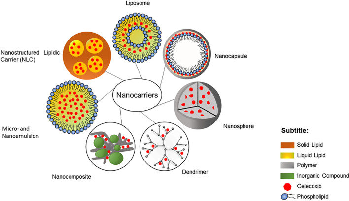
Nanocarriers are colloidal dispersions able to carry different types of drugs by providing a proper media for the molecule to dissolve or adsorb, increasing their apparent bioavailability. Incorporation of drugs into nanocarriers can also protect then of the degradation or early excretion by the organism. In fact, the use of nanocarriers is considered a valid strategy in overcoming the limitation of solubility for many lipophilic drugs, such as CXB [31, 32]. There are many types of nanocarriers (Fig. 1) and each one of them possesses properties that make them suitable for different uses, such as controlled and/or sustained release, improvement of the biodistribution and the ability to cross the blood–brain barrier [33–35].
Fig. 1.
Schematic representation of nanocarriers reported in the literature associated with CXB
Regarding the use of nanotechnology on the treatment of cancer, it has been observed that nanocarriers have an advantage in infiltrating and remain in the tumoral microenvironment exploiting tumor-induced angiogenesis, in an enhanced permeability and retention (EPR) effect [36]. Although the EPR effect does not show the same effectivity on in vivo evaluations and on clinical trials [37], it is relevant in augmenting the site-specific accumulation of the drug compared to other approaches [38].
In this context, it was expected that the use of CXB-loaded nanocarriers could be evaluated for the treatment of different types of cancer. Thus, the goal of this study was to review the formulations of CXB-loaded nanocarriers developed in the last decade and the most recent advances in this perspective (2011–2024), focusing on their morphological and physicochemical properties. Also, it was tried to trace the improvement of the systems developed and the future perspectives for the subject.
Data collection
For this study, only original research manuscripts published since 2011 until 27 April 2024, indexed at PubMed, Web of Science, and Scopus databases, were considered. The search for the manuscript was based on the use of the terms "COX-2; nanotechnology; cancer; COX-2 inhibitor, celecoxib; and nanocarriers" in different combinations. Also, the references of the selected original manuscripts were checked.
Articles that showed significant conflicts of interest or results without statistical data have been disregarded. Common characteristics of nanocarriers formulations such as long-term stability, biocompatibility, and CXB loading capacity, had to be reported for the inclusion of the study in this review. Technical elements of the studies were recorded such as year of publication; targeted tumor type; pharmaceutical target; route of administration; and the stage of development of the nanocarrier. Also, the methods of obtention of the nanocarriers; main components; the presence of co-encapsulation; and the main results of the studies were here noted.
Results and discussion
Data processing
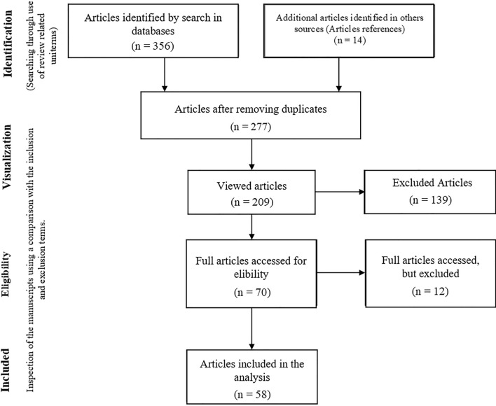
The articles were selected based on inclusion and exclusion criteria using the guidelines of acquisition protocol PRISMA (Preferred Reporting Items for Systematic reviews and meta-analyses) (Fig. 2). The search was able to identify 356 articles for the subsequent analyses. The exclusion of the repeated studies and those which are not under the inclusion criteria generated a list of 58 eligible manuscripts for this review.
Fig. 2.
Scheme of the screening employed to select the manuscripts used in this review
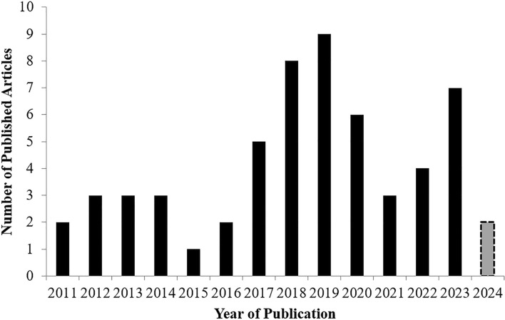
The use of CXB-loaded into nanocarriers in the context of cancer is recent, and growth in the number of studies in the subject starting in 2016 and peaking in 2019 (Fig. 3). The reduced number of publications between 2020 and 2022 can be related in the impact of the COVID-19 pandemic in both scientific activity and publications [39]. However, it seems that pre-pandemic levels are returning with a rise of publications in 2023. At the time of writing this review, two more papers were published in 2024.
Fig. 3.
Published studies (2011–2024) on the subject of CXB-loaded nanocarrier. Review process occurred until April/2024
Among these studies there were no clinical trials, which indicates that no current studies are applying CXB-loaded nanocarriers in the treatment of cancer. Thus, all the studies in this review are in the pre-formulation and pre-clinical phases. Therefore, it is important to notice that there were 253 studies on the use of celecoxib for the treatment of cancer applied in the clincaltrials.gov database, despite not directly employing a nanosized delivery system.
In vivo studies were performed in 54% of the selected manuscripts, becoming more common since 2017 (Table 1). Articles that contain only in vitro assays correspond to 43%, with a remarkable presence in the first 5 years (Table 1). Two articles only showed the pre-formulation studies, without any in vitro or in vivo test. This scenario underlines the fact that research into the use of CXB-loaded nanocarriers is still in its infancy.
Table 1.
Main characteristics of the analyzed studies containing CBX-loaded nanocarriers in the context of cancer regarding trial approaches, targeted tumor type, proposed administration of the nanocarrier and specific therapeutic target
| Citation | Publication date | Trial approach | Type of targeted tumor | Proposed administration | Specific therapeutic target |
|---|---|---|---|---|---|
| Kim et al. [40] | 2011 | In vitro | Glioma | Intravenous | Tumor cells |
| Venkatesan et al. [41] | 2011 | In vitro | Colorectal | Intravenous | Tumor cells |
| Bragagni et al. [42] | 2012 | In vitro | Skin | Transdermal | Tumor cells |
| Kang et al. [43] | 2012 | In vitro | N.S. | N.S. | Tumor cells |
| O’Hanlon et al. [44] | 2012 | In vitro | N.S. | Parenteral | Microenvironment* |
| Erdoğ et al. [45] | 2013 | In vitro | Colorectal | Intravenous | Tumor cells |
| Patel, A. R. et al. [46] | 2013 | In vivo | Lung | Inhalation | Tumor cells |
| Patel, S.K, et al. [47] | 2013 | In vitro | N.S. | Intravenous | Microenvironment |
| Emami et al. [48] | 2014 | In vitro | Lung | Inhalation | Tumor cells |
| Ju et al. [49] | 2014 | In vivo | Breast | Intravenous | Vasculogenesis** |
| Vera et al. [50] | 2014 | In vitro | Glioblastoma | Intravenous | Tumor cells |
| Limasale et al. [51] | 2015 | In vitro | N.S. | Intravenous | Tumor cells |
| Said-Elbahr et al. [52] | 2016 | In vivo | Lung | Inhalation | Tumor cells |
| Yen et al. [53] | 2016 | Pre-formulation | Oral carcinoma | Epidermal | Susceptible non-tumoral cells† |
| Elzoghby et al.a [31] | 2017 | In vivo | Breast | Intravenous | Tumor cells |
| Elzoghby et al.,b [35] | 2017 | In vivo | Breast | Intravenous | Tumor cells |
| Gowda et al. [54] | 2017 | In vivo | Melanoma | Intravenous | Tumor cells |
| Riahi et al. [55] | 2017 | In vivo | N.S. | Intravenous | Tumor cells |
| Wu et al. [56] | 2017 | In vitro | N.S. | N.S. | Tumor cells |
| AbdElhamid et al.a [57] | 2018 | In vivo | Breast | Parenteral, N.S. | Tumor cells |
| AbdElhamid et al.b [58] | 2018 | In vivo | Breast | Intravenous | Tumor cells |
| Abdelmoneen et al. [59] | 2018 | In vivo | Hepatocellular carcinoma | Oral | Tumor cells |
| Hong et al. [60] | 2018 | Pre-formulation | N.S. | Intravenous | Tumor cells |
| Shi et al. [61] | 2018 | In vivo | N.S. | Intravenous | Tumor cells |
| Singh [62] | 2018 | In vitro | Skin | N.S. | Tumor cells |
| Uram et al. [63] | 2018 | In vitro | Glioblastoma multiforme | Topical, N.S. | Tumor cells |
| Yu et al. [64] | 2018 | In vitro | Breast | N.S. | Tumor cells |
| Ahmed et al. [65] | 2019 | In vivo | Melanoma | Epidermal | Tumor cells |
| Huang et al. [66] | 2019 | In vivo | N.S. | Intravenous | Microenvironment and tumor cells |
| Liu et al. [67] | 2019 | In vivo | N.S. | Parenteral, N.S. | Tumor cells |
| Sun et al. [68] | 2019 | In vivo | Breast | Intravenous | Microenvironment |
| Tian et al. [69] | 2019 | In vitro | Prostate | N.S. | Tumor cells |
| Üner et al. [70] | 2019 | In vitro | N.S. | N.S. | Tumor cells |
| Zhang et al. [71] | 2019 | In vivo | Breast | Intravenous | Tumor cells |
| Uram et al. [72] | 2019 | In vitro | Glioblastoma multiforme | Topical | Tumor cells |
| Zhang et al. [73] | 2019 | In vivo | Breast | Intravenous | Microenvironment and tumor cells |
| Liu et al. [74] | 2020 | In vivo | Breast | Parenteral, subcutaneous | Microenvironment and tumor cells |
| Xu et al. [75] | 2020 | In vivo | Melanoma | Intravenous | Microenvironment |
| Xv et al. [76] | 2020 | In vivo | Melanoma | Parenteral, subcutaneous | Tumor cells |
| Sun et al. [77] | 2020 | In vivo | Breast | Intravenous | Tumor cells |
| Lee et al. [78] | 2020 | In vivo | Non-small lung cancer | Intravenous | Microenvironment and tumor cells |
| Uram et al. [79] | 2020 | In vivo | Glioblastoma multiforme | Intravenous | Tumor cells |
| Ahmed et al. [80] | 2021 | In vitro | Breast | Intravenous | Tumor cells |
| Farooq et al. [81] | 2021 | In vitro | N.S. | N.S. | Tumor cells |
| Yan et al. [82] | 2021 | In vivo | N.S. | Intravenous | Microenvironment and tumor cells |
| Said-Elbahr et al. [83] | 2022 | In vivo | Lung | Inhalation | Tumor cells |
| Guo et al. [84] | 2022 | In vivo | Breast | Parenteral, subcutaneous | Microenvironment and tumor cells |
| Alajani et al. [85] | 2022 | In vitro | N.S. | Oral | Tumor cells |
| Wróbel et al. [86] | 2022 | In vitro | Glioblastoma | N.S. | Tumor cells |
| Mabrouk et al. [87] | 2023 | In vivo | Oral carcinoma | Topical | Microenvironment and tumor cells |
| Jahani et al. [88] | 2023 | In vivo | Melanoma | Intravenous | Microenvironment and tumor cells |
| Cao et al. [89] | 2023 | In vivo | N.S. | Intravenous | Microenvironment and tumor cells |
| Abdellatif et al. [90] | 2023 | In vitro | Skin | Topical | Tumor cells |
| Basheer et al. [91] | 2023 | In vitro | Breast | N.S. | Tumor cells |
| Mendes et al. [92] | 2023 | In vitro | Glioblastoma | Parenteral | Tumor cells |
| Kaur et al. [93] | 2023 | In vitro | Bone metastasis | N.S. | Tumor cells |
| Liu et al. [94] | 2024 | In vivo | Melanoma | Intravenous | Microenvironment and tumor cells |
| Cai et al. [95] | 2024 | In vivo | N.S. | Intravenous | Microenvironment and tumor cells |
*Tumor surroundings intensely modulated by tumor cells and/or the immune system
**De fato blood vessels or mimics induced by the tumor
†Cells of patients who are in risk groups for determinate cancer type. N.S. stands for “not specified”
Sixteen studies (28%) showed CXB-loaded nanocarriers targeting different tumor types, thus revealing the potential versatility of the CXB in cancer protocols due to COX-2 participation in many cancer types. On the other hand, it is already possible to observe a trend when looking into the data: (1) there is the remarkable presence of carriers developed for breast cancer (14 studies, 24%); (2) almost half of the papers used an intravenous (i.v.) approach (28 studies), and (3) with a focus on the cytotoxic action of the drug in the tumor cells (41 studies, 70%).
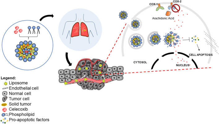
These facts do not hide that the use of CXB-loaded nanocarriers also is observed in alternatives ways of chemotherapy, such as in the overall model of application or in the direct use of the CXB, where the intended role of CXB-loaded systems is summarized in Fig. 4. Wu et al., (2017), Liu et al., (2019), and Zhang et al., (2019) developed formulations in a way that they act in chemo-resistant tumor cells [56, 67, 71]. Yen et al., (2016) produced formulations targeting susceptible normal cells in a preventive perspective to patients in the risk group of oral cancer [53]. At least fifteen studies proposed the modulation or destruction of components of the tumor microenvironment (26%), from which 11 studies acknowledged that the immunomodulation could contribute to the inhibition of the tumoral cells [66, 71, 74, 78, 82, 84, 87–89, 94, 95]. In fact, it seems that there is a growing understanding that the potential of CXB in cancer treatment protocols lies in the sensibilization of the tumor to the cytotoxic approaches, providing an immunosupportive tumor-associated microenvironment through COX-2 inhibition. Studies that focused the CXB component into the inhibition of COX-2 on the microenvironment corresponded to 45% of the papers published since 2020.
Fig. 4.
Schematic illustration of CXB for COX-2 inhibition and selective antitumor action. CXB can be conjugated with phospholipid to generate nano-sized liposomes. After intravenous administration, the system focuses on tumor tissues. Then, the liposome is internalized by tumor cells with subsequent lysosomal scape and CXB release. CXB can control chronic inflammation and prevent the accumulation of immune cells, cytokines and prostaglandins after direct COX-2 inhibition COX-2. In addition, CXB may induce release of proapoptotic factors by a COX-2 independent mechanism resulting in cellular apoptosis
Nanocarriers
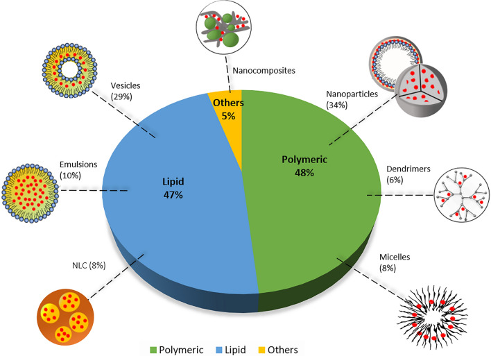
Nanocarriers are one of the main perspectives for the optimization of CXB use in cancer therapeutics. In this review, the formulations observed can be majorly classified as polymeric-based or lipid-based formulations (Fig. 5.). In a lower amount, there are also reports on hybrid nanoparticles and nanocomposites.
Fig. 5.
Profile of the CBX-loaded nanocarriers over their general classification based on the major component and distribution of the types of polymeric particles, lipid-based formulations and nanocomposites particles. *NLC: nanostructured lipidic nanocarrier
From Fig. 5, it can be inferred that the development of CXB-loaded nanocarriers does not deviate from most traditional formulation perspectives [96]. As an example, the poly (lactic-co-glycolic acid) (PLGA), a common polymer for drug delivery systems, had a remarkable presence, being used in 6 of 8 nanosphere nanoparticles.
The PLGA is a biopolymer, biocompatible, composed of lactic acid and glycolic acid monomers, that are degraded by the organism due to its carbohydrate-derived components being metabolized by the Krebs cycle [97]. Use of PLGA is also boosted by the possibility of surface modifications such as PEGylation or immune-targeting. This polymer can also be used to entrap hydrophobic drugs such as CXB through a variety of techniques such as nanoprecipitation, emulsification-solvent evaporation and spray-drying. The drug is easily incorporated through solubilization in a suitable organic solvent, such as acetone, alongside the polymer, which is them mixed with an aqueous phase, with the organic solvent being later removed through evaporation [33, 98]. Usage of surfactant seems to have improved CXB entrapment efficiency into PLGA nanoparticles [40, 48, 50, 52].
From the perspective of the improving biopharmaceutical and pharmacokinetic properties of CXB, Kim et al., (2011) and Vera et al., (2014) produced CXB-loaded PLGA nanospheres targeting gliomas. Kim et al. revealed the capacity of the nanoparticles to promote the cytotoxicity of CXB in a similar manner as the free drug [40], and Vera et al. showed that the cytotoxicity of the nanoencapsulated CXB is concentration-dependent. In addition, the potential use of polysorbate 80 as a permeation enhancer for the blood–brain barrier has also been reported [50]. Liu et al., (2024) used the known properties of PLGA to load CXB while coating the system with a C166 cell line membrane to ensure white blood cell adhesion and preferential delivery to the tumor site [94].
On the range of lipid-based systems, vesicular structures (18 formulations), mainly liposomes (10 formulations), were majorly studied (Fig. 4b). The liposomes used for CXB incorporation were based on the use of phosphatidylcholine and cholesterol. Liposome’s structure, as seem in Fig. 1, is composed of a hydrophilic core and an amphiphilic bilayer membrane containing phospholipids and structural lipids. While hydrophilic molecules can be dispersed in the core, hydrophobic drugs such as CXB are incorporated into the bilayer, alongside the lipophilic tails of the phospholipids and cholesterol. The liposomes can be obtained through methods such as lipid film hydration, the most common in the screened papers, and ethanol injection [99]. The CXB is incorporated by dissolution into organic solvents during the process. Following in the footsteps of the Doxil®, the use of polyethylene glycol (PEG) was reported for 9 out of 10 of the developed liposomes [45, 49, 51, 54, 55, 60, 62, 69, 88]. This factor was expected due to the knowledge of the stealthiness provided by this polymer when onto the surface of the nanocarriers [100–102].
Other type of vesicles applied for CXB loading were ethosomes, transfersomes [42], cubosomes [87], transethosomes [90], and niosomes [91]. For these formulations, there was a remarkable presence of surfactants such as Span® 60, Poloxamer® 407 and Tween® 20, aiming for a improved formulation stability for topical administration [42, 87, 90] in possible subsequent in vivo and clinical studies.
Hybrids between lipid and polymeric particles, assigned as nanocapsules or just nanoparticles, were also relevant among the screened papers. These systems were characterized by multilayers with a oily core such as palmitic acid [93] a phospholipid emulsion [31, 35, 57, 58] or liposome [68]. The core would them present one or more polymeric coatings with multipurpose functions including structural and controlled release purposes, such as usage of protamine [31, 35] and chondroitin sulfate [57, 58], targeting components such as hyaluronic acid for CD44 recognition [31, 68, 93], or theranostics purposes with lactoferrin [57] or gelatin [58] conjugated with quantum dots.
In the main formulations analyzed in this report, no negative interactions between CXB and the common nanocarrier components were detected, indicating that the drug is well suited for transport with standard methods and reagents. On the other hand, most of the techniques used required the use of organic solvents to dissolve CXB, including chloroform and methanol. While this is a necessary step due to the hydrophobic nature of the drug, it can be a bottleneck in the purification processes of the nanocarrier for clinical use. Therefore, the use of safer solvents such as ethanol is recommended.
Approaches to enhance CXB therapeutic efficiency
Targeting
To develop efficient nanocarriers different approaches were used. Most of them were based on the use of specific molecules on their surface. Initially, it was tried to overcome the well-known negative sides of PEGylation in both liposomal and polymeric systems, such as the reduced effectivity after the first dose because of antigenic recognition [103], and the decrease of the absorption capacity and endosomal escape in the target cells [104]. As such, all the systems in this topic intended to confer additional qualities to the traditional designed systems through innovative surface modifications.
Initially, Ju et al. (2014) used the transduction domain peptide of HIV-1 protein (PTDHIV-1) linked to the PEG chains of a liposome. This addition showed the enhancement of the cell recognition and permeation of the formulation and also provided a better entry into the nucleus, which is a intracellular target of epirubicin, co-encapsulated with the CXB in the study [49].
Another protein-based approach was used by Limasale et al., (2015), who branched an antibody against the epidermal growth factor receptor (EGFR) on the surface of liposomes, due to the hypothesis that cancer cells overexpress EGFR on their surface. The immunenanoliposome, as it was called, showed cytotoxicity against colon (HCT-116, SW620 and HT-29) and breast cancer (MDA-MB-468) cell lines. This effect was similar to the one observed by the free CXB and greater than non-immunogenic liposomes [51]. It is interesting to observe that in a previous study published by the same research group, the liposomes used as a control showed cytotoxicity against the same colon cancer cell lines [45], which are not observed in the latter study.
Two nanocapsules developed by Abdelhamid et al. in parallel works also had unique targeting mechanisms In one of the studies, a particle was developed as an emulsion enveloped by a layer of chondroitin sulfate (CS) and a layer of lactoferrin that are carrying CXB and honokiol (Abdelhamid et al. 2018a). The particle developed in the second study has a layer of gelatin type A replacing the lactoferrin and are co-encapsulated with rapamycin (Abdelhamid et al. 2018b). The first study proposed that the recognition of the lactoferrin and the CS by CD44 induced greater uptake of the particle and, inside the endosome, initialized a gradual release of the drugs. In the second study, gelatin acted as a protection of the nanocapsule in the bloodstream, and its degradation by the metalloproteinases, overexpressed in breast cancer cells, allowed the endocytosis of the nanocarrier by the uptake promoted by the CS [57, 58]. Similar perspective promoted the coating of Sun et al. (2019) liposome and Lee et al. chitosan nanoparticle in hyaluronic acid, aiming for its CD44 recognition [68, 78]. Kaur et al. also applied this concept, but instead by developing hyaluronic acid conjugated to alendronate nanoparticles [93].
Theranostics
The previously mentioned studies from of Abdelhamid et al. discern from each other by the presence of quantum dots of cadmium-tellurite in the nanocapsule. Quantum dots are components with high fluorescent capacity and easy detection, having as main application the capacity to generate traceable particles with imaging and diagnostic function [105]. Thus, the authors approached in their studies the theranostic capacity of their CXB-loaded nanocarriers: in the same time that the therapeutic action is provided by the tumor-specific cytotoxicity, induced by the selective and facilitated cell uptake, the quantum dots presence allowed the observation or detection in the tumor site [57, 58].
Theranostic nanocarriers were also idealized by O’hanlon et al. (2012) and S. K Patel et al. (2013), who were in the same research group. In this case, nanoemulsions were developed with a perfluoropolyether core which has the isotope F19 of fluorine. Also, a near-infrared dye (NIR dye) was co-encapsulated with the CXB. The nanocarrier accumulation in the tumor would allow the production of an image through nuclear magnetic resonance with the PFPE or through fluorescence using the NIR. Therefore, it is important to notice that the studies focused on different targets, first, tumor cells [44], and, second tumor-adjacent macrophages [47].
Controlled release mechanisms
In their studies, Abdelhamid et al. also attributed the increase of the carrier uptake and selective drug-release mechanisms by the addition of a layer of lactoferrin—recognized by CD44 in the tumor cell surface—or gelatin—recognized by cell surface metalloproteinases, both overexpressed in the surface of the tumor cells [51, 52]. Huang et al. (2019) also sought to explore the surface metalloproteinases of tumor cells using a peptide susceptible to degradation by these enzymes in a triblock system with PEG and poly(ε-caprolactone). A nanosphere was developed and engineered in a way to have amphiphilic characteristics. Their hypothesis was based on the idea that the degradation of the peptide would lead a CXB release from the nanocarrier to the tumor microenvironment [66]. Similar perspective was employed by Cai et al. (2024) for multi-modal combined therapy, with CXB acting as an anti-inflammatory also towards microenvironment modulation. The drug was incorporated in the outermost layer of a complex nanosphere system, and associated to gelatin for metalloproteinases-associated release [95].
Others authors employed pH-induced release, such as Cao et al. (2023). By complexing CXB with Poly-L-arginine arranged in micelles, it was expected that the acidic environment of the tumor microenvironment would promote the drug release and subsequent inhibition of the COX-2 related immunosuppressive profile [89]. This hypothesis was confirmed with the formulation containing co-loaded CXB presenting higher tumor suppression in vivo, related to sensibilization for the cytotoxic treatment. This highlighting that multiple coatings are not needed to achieve microenvironment pH-mediated CXB release. Wu et al. (2017) and Zhang et al. (2019) also obtained nanoparticles susceptible to pH response for controlled drug release. Wu et al., observed that the release was achieved by the increased solubility of calcium carbonate and calcium phosphate of the nanocomposite in the acidic pH of the tumor cells and the endosomes [56]. While Zhang et al. reported the presence of a tertiary amine group, which enabled the release of the drug in the acidic environment of the endosome. This work also showed a joint release mechanism, based on the susceptibility of the disulfide linkage in the nanocarrier to the control of redox in the cell fulfilled from the glutathione (GSH) [71]. Another study that exploited release through susceptibility to redox using a disulfide linkage was performed by Liu et al. (2019). It is interesting to note that the three above described studies are targeting the reversion of the mechanism of resistance of the tumor cells against the drugs, with a major priority in control and selective release, dependant on the tumor cells' response to the adsorption of the carrier [56, 67, 71].
Excipient choice
Some types of tumor target have an urge for specifics approaches, usually related to their primary location in the body. CXB-loaded nanocarriers targeting lung and various skin cancers are examples of systems designed with special attention to the target location. Lung cancer is the most prevalent cancer type in the world [3] and nanocarriers whose targeted site is in the lung have a preference for administration through inhalation. In the works of Emami et al. (2015) and Said-Elbahr et al. (2016), it is possible to observe the ability of PLGA nanoparticles to deliver CXB through inhalation when they are associated to an adequate content of surfactant.
Emami et al. (2015) used the Taguchi method for the development of an optimized CXB-loaded nanocarrier with PLGA. The polymer was associated with poly(vinyl alcohol) as a surfactant to try to obtain a more uniform particle size distribution. Therefore, it was detected that the particle has instability by spray administration, so the authors inoculated it in a microparticle of lactose, in a system called "nano-in-micro". The lactose particles were able to release the intact nanocarrier to continue its course [48].
The PLGA nanoparticle developed by Said-Elbahr et al. (2016) was produced with poloxamer 188 to increase the stability and enhance absorption. The authors demonstrated in vivo that the obtained nanosphere has good inoculation through the Air Jet nebulization method, with high lung accumulation and relevant presence in metastatic tumor sites of lung cancer, although this indicates reduced site-specific selectivity [52].
Co-loading of CXB with other drugs
Targeting breast cancer
Certain types of cancer received more attention in health concerns because of their prevalence and group risks, as the breast cancer, with high prevalence among women and associated with relevant hereditary characteristics, who justified a robust investment in novel effective and less harmful therapeutics [3]. The work of Ju et al. (2014) revealed that it is possible to have an interesting niche for CXB-loaded nanocarriers in this cancer type through targeting of vasculogenic mimicry channels. These pro-tumor segments were recently discovered, and their production seems to be related with a tumor-induced angiogenesis, since they are observed in processes of breast cancer relapse and metastasis [49, 106]. A PEGylated liposome loaded with CXB and epirubicin was produced and the authors inferred that the exploitation of the EPR effect, greater circulation time, increased permeation of the PTDHIV-1 peptide, and synergic effect between the drugs could explain the better results found in vitro and in vivo when compared to the free drugs. It was observed the reversibility of the epirubicin resistance in the epirubicin-resistant cell lines, high CXB-dependant cytotoxicity against the formed channels cells, as well as a decrease in the invasive cell capacity [49].
The synergy between the dependant and independent of COX-2 CXB anti-cancer effects, and co-loaded drugs is one of the most effective and promissory strategies in the analyzed studies. The CXB mechanisms of action are capable of help or being helped by other drugs who act crosswise in the various metabolic ways affected by the COX-2 inhibitor [35, 54, 65, 68, 71, 78, 80]. It was frequently observed that drugs with anti-inflammatory effects and/or apoptosis inductors have excellent synergic effects when associated with the CXB, mainly when altering the modulation of TNFα, NF-κB, and STAT3.
In the work of Sun et al. (2019) a CXB and curcumin-loaded nanocapsule was developed aiming the suppression of the metastatic process in breast cancer. The carrier has the cell-penetrating peptide (TAT) linked with the NF-κB essential modulator (NEMO)-binding domain peptide (NBD) and demonstrate action in inhibition of pro-tumoral inflammatory components in the tumor microenvironment, responsible for the important process of invasion and metastasis. The inhibition by these combined drugs focused on the action of the NF-κB factor showed good results in vitro and in vivo. The study demonstrates that the proprieties of the curcumin allied with the anti-inflammatory and anti-tumoral effects of the CXB were able to revert the expression of inflammatory factors by the tumor and to induce significant cytotoxicity decreasing the migration capacity by the invasive cells [68].
Another study focused on the inhibition of the metastatic process on breast cancer was realized by Yu et al. (2018). They developed a PEGylated nanosphere loaded with CXB and Brefeldin A. This nanocarrier showed promising results once it inhibited the tumoral growth and invasion capacity by a synergic regulation of the Golgi apparatus in mice [64].
The last two mentioned studies exemplify a trend observed through the analysis of the selected articles: a wide range of drugs are being studied due to similar or crossed pharmacodynamics with the CXB. In addition, a distinction can be made by strategies that (i) exploit the modulation provided by the CXB and (ii) evaluated the use of new molecules for cancer therapeutic protocols. At the moment, only the use of traditional drugs have robust literature support for safety use, novel molecules are showed as alternative perspectives for a late exploration after greater coverage.
Synergy between the CXB and established chemotherapeutic drugs
Among established drug, it was observed the use of epirubicin and the doxorubicin between the different drugs co-loaded with the CXB in the developed nanocarriers. The doxorubicin (DOX) is an anthracycline that acts by inhibition of the replication and transcription of DNA by genome biding, being largely used in cancer therapy. The DOX has liposomal formulation commercially available and several ongoing clinical trials for new products [61, 65, 107]. The epirubicin is also an anthracycline used mostly as an adjuvant in chemotherapy of breast cancer, however, it is observed that invasive cells of this type of cancer acquire resistance to this drug [49].
DOX was the chosen drug for co-loading in four novel polymeric particles developed by [67, 71, 78, 80]. These studies exploited the ability of CXB to apparently revert the DOX resistance by the tumor cells. The combined mechanisms of action of the drugs and the use of selective release mechanisms showed a significant effect in the sensibility of tumor cells to the treatment. It was observed the effects of apoptosis induction, inhibition of recidivist carcinogenesis pathways, tumor expansion, and in vivo greater reduction of tumor volume. DOX was also applied in combination therapy with CXB-containing nanocarriers in different regimens of administration, aiming for optimal use of the modulation of the tumor microenvironment by the coxib [74, 75, 84].
Previously mentioned formulations of Cao et al. [89] and Cai et al. [95] also employed DOX as the cytotoxic chemotherapeutic. In an interesting perspective, Cao et al. incorporated a plasmid for IL-12 into their micelles, producing micelleplexes, aiming for gene modulation to contribute into the immune modulation and potentialize the CXB effects, triggering a tumor-repressive cascade. The potential of genetic therapy is well-know, but clinical results are still in need. Cai et al. on other hand aimed for a multi-modal therapeutic approach, with CXB-induced immunomodulation, and combined effects of photothermal and photodynamic therapies and DOX for intracellular action against induced murine cervical carcinoma (U14 cell line).
Huang et al., (2019) tested the synergy between the CXB and the paclitaxel (PTX). The PTX is one of the most employed drugs in cancer adjuvant therapy—it has a mitosis inhibitor effect. The study observed that CBX, as an anti-inflammatory in the microenvironment and tumor cells, helped in the chemotherapeutic action of PTX, suppressing chemoresistance mechanisms like the activation of anti-apoptosis factors. Interesting results in vitro and in vivo were showed. The in vitro tests displayed a significant tumor cell line inhibition by the reduction of exogenous PGE2; while the in vivo experiments revealed a reduction of the tumor volume with an increase in the survival rate. Additionally, no significant mass reduction in the mice treated with the nanoparticles was observed when compared with the free drugs [66].
Letrozole (LTZ), used by Elzoghby et al. in two studies, is also a drug used in breast cancer treatment protocols, mainly with women in postmenopausal. As an aromatase inhibitor it was expected that LTZ would have a synergic effect with the CXB since the targeted enzyme has shown stimulus hormone-dependant by COX-2 in breast cancer. The authors sought to develop nanoparticles able to exploit this synergy and the resistance reversion induced by the CXB. The promising results in both studies encourage the addition of CXB in cancer therapeutic protocols as an antitumor modulator [31, 35].
Synergy between the CXB and non-established chemotherapeutic drugs
In terms of novel drug alternatives, it is natural that co-loading another substance with the CXB will lead to other hydrophobic drugs. The brefeldin A used by Yu et al. (2018) is a lactone of fungal origin with very poor aqueous medium solubility [64]. Another fungal origin lactone used was the rapamycin (RAP). It's estimated that the inhibition of the mammalian target of rapamycin (mTOR) possess decisive antitumor characteristics, but the RAP high hydrophobicity was a limiting to use. Co-loading in a nanocapsule with the CXB showed notable synergic antitumor characteristics [58].
The use of drugs obtained by a natural perspective, such as the brefeldin A and the rapamycin, are well established and important alternatives for the discovery of new active principles and the development of new drugs. These novel drugs can also benefit or increase the antitumor characteristics of the CXB. Furthermore, these substances also have a poor hydrosolubility and toxicity to healthy cells, thus they can profit from an administration who guarantees selectivity and good bioavailability.
One of these substances, the diosmin are a high hydrophobic drug commonly used as an anticoagulant. The drug was shown capable of modulating the expression of molecules relevant to the hepatocellular carcinoma context in a crossway with the CXB, confirming the synergic interaction by both drugs [59]. Similar results are founded by co-loading of CXB and the hydrophobic compound honokiol, with strong apoptosis induction in vitro and in vivo targeting breast cancer cells [57].
The plumbagin and the genistein are two phytochemicals who also are co-loaded with CXB in studies selected for that review. The plumbagin is a toxin capable of provoking critical alterations in the genome with a high risk tied to its use [54]. In fact, in the study of Gowda et al., although synergic antitumor effect with the CXB has noted, non-specific toxicity dependant of plumbagin was also observed, so that methods to include greater tumor selectivity are necessary for possible use.
Genistein is an isoflavone capable to connect in estrogens receptors and has the capacity of inhibition of GLUT1 channels, an important perspective for prostate cancer. Moreover, in the study realized by Tian et al., the synergic effect with the CXB induced stress in the antioxidant metabolism by blocking the GSH and formation of reactive oxygen species (ROS) in the tumor cells [69].
Nanocarriers which are in early development are also identified in the analyzed studies. Uram et al. (2018, 2019) produced a biotinized 3ª generation PAMAM dendrimer for the carrying of a peroxisome proliferator-activated receptor-gamma (PPARγ) agonist Fmoc-L-Leucine along with CXB. Dendrimers are a particle type with notable pharmacological potential and fast development evolution [34]. The nanoparticle of Uram et al. showed the inhibition of the growth of glioblastoma cells [63, 72]. In the in vitro evaluation against skin cancers, there was more cytotoxicity to the fibroblasts cell line (BJ) than against squamous cell carcinoma line (SCC-15), a factor attributed to the toxicity of the PPARγ agonist and the lack of biotin receptors in the later cancer cell line [63]. In the later study, IC50 of formulation in the BJ cell line was of 1.29 µM against 1.25 µM in the glioblastoma U-118 MG cell line, showing high tumor cytotoxicity but worrying results against a healthy cell line. Lastly, a G3 dendrimer containing 31 subunits of CXB showed high cytotoxicity against U-118 MG cells, but also possessed high cytotoxicity against Caenorhabditis elegans when compared with the CXB alone [79]. Similar results were still found when co-loading with simvastatin and introduction of R-glycidol in the system [86].
Among the nanocomposites, the complex formed by Wu et al., (2017) was capable of an efficient CXB and buthionine sulfoximine delivery with a controlled pH-dependent release. The study showed a reversion of the chemotherapy resistance of the tested cancer cell lines, and a synergetic effect among the co-loaded drugs and an increase of the effect of DOX applied in parallel.
Future perspectives in CXB-loaded nanocarriers
The increase in in vivo tests published in the last 3 years of the analysis shows the possibility of clinical trials using CXB-loaded nanocarriers in the current decade. The developed formulations revealed innovative approaches considering the surface modification of the systems used, the techniques for engineering the carriers, and the presence of other substances within the CXB-loaded carriers.
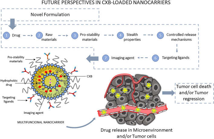
One of the main challenges to overcome the barriers of the clinical use of CXB in cancer therapy is its low water solubility, an aspect cited in all the analyzed studies. In this sense, conventional nanocarriers, which already helped to enhance other drugs properties (e.g.: Ambisome®), may be able to overcome these drawbacks. However, different aspects also have to be considered for the feasibility of an innovative CXB-loaded nanocarrier. Thus, the main points we identified in this review were (i) Quality-by-design approach looking at stability in blood and serum, site-specific targeting and absorption enhancers; and (ii) co-loading CXB with other drugs. Additionally, mechanisms that can induce controlled release and/or provide diagnostic properties to the carriers received considerable attention. In this way, it is possible to infer that the CXB-loaded nanocarrier development process effectively can be guided by:
Careful selection of the raw materials used to produce the nanocarriers;
Choice of a drug with cross-action to the COX-2 dependent and independent mechanisms of the CXB, with preference to another hydrophobic drug, in the way that the same encapsulation mechanisms can be explored;
Selection of materials that can ensure stability of the nanocarrier;
Selection of nanocarriers with stealth properties;
Selection of targeting ligands capable to enhance the nanocarrier entry into the tumor cells;
Selection of materials capable to promote a controlled release mechanism (e.g.: pH-dependent); and
Selection of materials able to provide images from their location site.
The studies have shown that the constant introduction of new functionalities in the decades between 2010 and 2020 has increased the size and complexity of drug delivery systems. While the continued advances in nanocarrier patterning are commendable, there are concerns about the suitability of these systems for the industrial production required for clinical trials. While more complex systems are effective, they can be held back by the limitations of the technology available for scaling up laboratory formulations. The fact that, to our knowledge, there is no clinical trial with a CXB-loaded nanocarrier can be partly attributed to this. Alongside, no complex in-vivo models were employed in the studies reviewed in this paper, with a vast dominance of tumor-bearing mice of murine origin or xenografted from human cell lines. As the relevance of the EPR effect has declined in some part due to the lack between murine models and clinical application, the advance in the models for the in vivo assays are essential to allow the newly fabricated formulations and/or particles to meet the safety guarantees for clinical assays, regardless of the status of the CXB alone.
Finally, it must be acknowledged that the cytotoxic effect of CXB alone is often lower compared to conventional anticancer protocols at feasible concentrations of the drug. However, this does not mean that the use of CXB for cancer chemotherapy is doomed to failure. It has been shown that the immunomodulation resulting from the inhibition of COX-2 can induce a sensitization effect that enhances the effect of cytotoxic agents administered by co-loading or other methods. This has opened in vivo perspectives for both established chemotherapeutic agents such as doxorubin and novel pharmaceuticals and has even recently been cited in relation to postoperative immunomodulation [94]. We believe that the pathway of immunomodulation by CXB, suitable in nanocarriers and in combination with other cytotoxic agents, chemotherapies or not, is the most promising prospect for the clinical application of this drug in cancer protocols.
Conclusion
The reports in the literature dealing with CXB-loaded nanocarriers in cancer therapy assume that these molecules are an important alternative to increase the contribution of CXB against tumor growth. Although the research and development of CXB-loaded nanocarriers is recent, the promising results are not limited to a specific type of nanocarrier or cancer. Among the major technical advances, effective prototypes developed in the last 3 years stand out. The combination of CXB with other anti-inflammatory drugs and/or apoptosis inducers seems pharmacologically promising. The greatest advance to date from a clinical perspective is the ability of CXB to potentiate the cytotoxic effects of established chemotherapeutic agents. The aspects identified and discussed in this review confirm the growth of an emerging field based on the use of synergistic molecules supported by nanotechnology for the use of conventional drugs in adjuvant cancer therapy.
Acknowledgements
The authors thank CNPq (National Council for Scientific and Technological Development), FAPESB (Foundation for Research Support of the State of Bahia), PRPPG (Dean of Research/Pro Rectory of Research and Postgraduate Studies/Federal University of Bahia) and CAPES (Coordination of Superior Level Staff Improvement).
Author contributions
All authors contributed to the study conception and design. Data collection and analysis were performed by [M de JOS], [JTS] [RF de A-C] and [DSV-B]. Design graphs and design tables were performed by [M de JOS]. Figures and images design were performed by [M de JOS], [JTS] and [RF de A-C]. The first draft of the manuscript was written by [M de JOS] and [DSV-B]. All authors commented on previous versions of the manuscript. The manuscript was critically reviewed by [RLC], [HRM] and [DSV-B]. Reformulation/updating of the figures and graphical abstract for the final version following the reviewers' comments were performed by [M de JOS]. Reformulation of the manuscript text according to the reviewers' comments were performed by [M de JOS], [HRM] and [DSV-B]. All authors read and approved the final manuscript.
Funding
Conselho Nacional de Desenvolvimento Científico e Tecnológico (BR) Award Number: Processo: 424854/20163 | Recipient: Deise Souza Vilas-Bôas, Ph.D. (Grants); Fundação de Amparo à Pesquisa do Estado da Bahia (BR) Award Number: Termo Outorga Nº JCB0048/2013 |Recipient: Deise Souza Vilas-Bôas, Ph.D. (Grants); Pró-Reitoria de Pesquisa e Pós-Graduação, Universidade Federal da Bahia (BR) Award Number: Edital PROPCI/PROPG 004/2016 Projeto nº11347 Recipient: Recipient: Deise Souza Vilas-Bôas, Ph.D. (Grants) and Miguel de Jesus Oliveira Santos, MsC (Undergraduate Scholarship).
Data availability
The authors declare that all collected data are available. Please, do not hesitate to contact ourselves if you require any information.
Declarations
Ethics approval and consent to participate
Due to the study design, the need for ethical approval by an ethics committee and consent to participate does not apply.
Ethical responsibilities of authors
The authors warrant that this manuscript is an original work and the submitted version of the manuscript is not under consideration elsewhere. Further, the authors attest it has not yet been published as well as will not be published in another journal.
Consent for publication
All authors consent and approval this publication.
Competing interests
The authors do not declare any conflict of interest.
Footnotes
Publisher's Note
Springer Nature remains neutral with regard to jurisdictional claims in published maps and institutional affiliations.
References
- 1.Bray F, Ferlay J, Soerjomataram I, et al. Global cancer statistics 2018: GLOBOCAN estimates of incidence and mortality worldwide for 36 cancers in 185 countries. CA Cancer J Clin. 2018. 10.3322/caac.21492. 10.3322/caac.21492 [DOI] [PubMed] [Google Scholar]
- 2.Omitola OG, Soyele OO, Sigbeku O, et al. A multi-centre evaluation of oral cancer in southern and Western Nigeria: an African oral pathology research consortium initiative. Pan Afr Med J. 2017. 10.11604/pamj.2017.28.64.13089. 10.11604/pamj.2017.28.64.13089 [DOI] [PMC free article] [PubMed] [Google Scholar]
- 3.Bernard WS, Christopher PW (2020) World cancer report 2020.
- 4.Shylasree TS, Bryant A, Athavale R. Chemotherapy and/or radiotherapy in combination with surgery for ovarian carcinosarcoma. Cochrane Database Syst Rev. 2013. 10.1002/14651858.CD006246.pub2. 10.1002/14651858.CD006246.pub2 [DOI] [PMC free article] [PubMed] [Google Scholar]
- 5.Kann BH, Lester-Coll NH, Park HS, et al. Adjuvant chemotherapy and overall survival in adult medulloblastoma. Neuro Oncol. 2016. 10.1093/neuonc/now150. 10.1093/neuonc/now150 [DOI] [PMC free article] [PubMed] [Google Scholar]
- 6.Lin Y, Zhou J, Cheng Y, et al. Comparison of survival benefits of combined chemotherapy and radiotherapy versus chemotherapy alone for uterine serous carcinoma: a meta-analysis. Int J Gynecol Cancer. 2017. 10.1097/IGC.0000000000000856. 10.1097/IGC.0000000000000856 [DOI] [PMC free article] [PubMed] [Google Scholar]
- 7.Abrahao AC, Castilho RM, Squarize CH, et al. A role for COX2-derived PGE2 and PGE2-receptor subtypes in head and neck squamous carcinoma cell proliferation. Oral Oncol. 2010;46:880–7. 10.1016/j.oraloncology.2010.09.005. 10.1016/j.oraloncology.2010.09.005 [DOI] [PMC free article] [PubMed] [Google Scholar]
- 8.Li X, Yang B, Han G, Li W. The EP4 antagonist, L-161,982, induces apoptosis, cell cycle arrest, and inhibits prostaglandin E2-induced proliferation in oral squamous carcinoma Tca8113 cells. J Oral Pathol Med. 2017. 10.1111/jop.12572. 10.1111/jop.12572 [DOI] [PubMed] [Google Scholar]
- 9.Cui Y, Shu XO, Li HL, et al. Prospective study of urinary prostaglandin E2 metabolite and pancreatic cancer risk. Int J Cancer. 2017. 10.1002/ijc.31007. 10.1002/ijc.31007 [DOI] [PMC free article] [PubMed] [Google Scholar]
- 10.Hosseini F, Mahdian-Shakib A, Jadidi-Niaragh F, et al. Anti-inflammatory and anti-tumor effects of α-L-guluronic acid (G2013) on cancer-related inflammation in a murine breast cancer model. Biomed Pharmacother. 2018. 10.1016/j.biopha.2017.12.111. 10.1016/j.biopha.2017.12.111 [DOI] [PubMed] [Google Scholar]
- 11.Todoric J, Antonucci L, Karin M. Targeting inflammation in cancer prevention and therapy. Cancer Prev Res. 2016;9:895–905. 10.1158/1940-6207.CAPR-16-0209 [DOI] [PMC free article] [PubMed] [Google Scholar]
- 12.Chang LY, Wan HC, Lai YL, et al. Areca nut extracts increased the expression of cyclooxygenase-2, prostaglandin E2and interleukin-1α in human immune cells via oxidative stress. Arch Oral Biol. 2013. 10.1016/j.archoralbio.2013.05.006. 10.1016/j.archoralbio.2013.05.006 [DOI] [PubMed] [Google Scholar]
- 13.van Ryn J, Pairet M. Selective cyclooxygenase-2 inhibitors: pharmacology, clinical effects and therapeutic potential. Expert Opin Investig Drugs. 1997;6:609–14. 10.1517/13543784.6.5.609. 10.1517/13543784.6.5.609 [DOI] [PubMed] [Google Scholar]
- 14.Kawamori T, Rao CV, Seibert K, Reddy BS. Chemopreventive activity of celecoxib, a specific cyclooxygenase-2 inhibitor, against colon carcinogenesis. Cancer Res. 1998. 10.1007/s00262-006-0268-x. 10.1007/s00262-006-0268-x [DOI] [PubMed] [Google Scholar]
- 15.Fischer SM, Lo HH, Gordon GB, et al. Chemopreventive activity of celecoxib, a specific cyclooxygenase-2 inhibitor, and indomethacin against ultraviolet light-induced skin carcinogenesis. Mol Carcinog. 1999. 10.1002/(SICI)1098-2744(199908)25:4%3c231::AID-MC1%3e3.0.CO;2-F. [DOI] [PubMed] [Google Scholar]
- 16.Hsu AL, Ching TT, Wang DS, et al. The cyclooxygenase-2 inhibitor celecoxib induces apoptosis by blocking Akt activation in human prostate cancer cells independently of Bcl-2. J Biol Chem. 2000. 10.1074/jbc.275.15.11397. 10.1074/jbc.275.15.11397 [DOI] [PubMed] [Google Scholar]
- 17.North GLT. Celecoxib as adjunctive therapy for treatment of colorectal cancer. Ann Pharmacother. 2001. 10.1345/aph.10133. 10.1345/aph.10133 [DOI] [PubMed] [Google Scholar]
- 18.Howe LR, Subbaramaiah K, Brown AMC, Dannenberg AJ. Cyclooxygenase-2: a target for the prevention and treatment of breast cancer. Endocr Relat Cancer. 2001;150:97–114. 10.1677/erc.0.0080097 [DOI] [PubMed] [Google Scholar]
- 19.Zelenay S, van der Veen AG, Böttcher JP, et al. Cyclooxygenase-dependent tumor growth through evasion of immunity. Cell. 2015;162:1257–70. 10.1016/j.cell.2015.08.015. 10.1016/j.cell.2015.08.015 [DOI] [PMC free article] [PubMed] [Google Scholar]
- 20.Tai Y, Zhang L-H, Gao J-H, et al. Suppressing growth and invasion of human hepatocellular carcinoma cells by celecoxib through inhibition of cyclooxygenase-2. Cancer Manag Res. 2019;11:2831–48. 10.2147/CMAR.S183376. 10.2147/CMAR.S183376 [DOI] [PMC free article] [PubMed] [Google Scholar]
- 21.Rosas C, Sinning M, Ferreira A, et al. Celecoxib decreases growth and angiogenesis and promotes apoptosis in a tumor cell line resistant to chemotherapy. Biol Res. 2014;47:27. 10.1186/0717-6287-47-27. 10.1186/0717-6287-47-27 [DOI] [PMC free article] [PubMed] [Google Scholar]
- 22.Grösch S, Tegeder I, Niederberger E, et al. COX-2 independent induction of cell cycle arrest and apoptosis in colon cancer cells by the selective COX-2 inhibitor celecoxib. FASEB J. 2001;15:1–22. 10.1096/fj.01-0299fje [DOI] [PubMed] [Google Scholar]
- 23.Kuipers GK, Slotman BJ, Wedekind LE, et al. Radiosensitization of human glioma cells by cyclooxygenase-2 (COX-2) inhibition: independent on COX-2 expression and dependent on the COX-2 inhibitor and sequence of administration. Int J Radiat Biol. 2007. 10.1080/09553000701558985. 10.1080/09553000701558985 [DOI] [PubMed] [Google Scholar]
- 24.Jendrossek V. Targeting apoptosis pathways by Celecoxib in cancer. Cancer Lett. 2013;332:313–24. 10.1016/j.canlet.2011.01.012. 10.1016/j.canlet.2011.01.012 [DOI] [PubMed] [Google Scholar]
- 25.Seedher N, Bhatia S. Solubility enhancement of cox-2 inhibitors using various solvent systems. AAPS PharmSciTech. 2004. 10.1208/pt040333. 10.1208/pt040333 [DOI] [PMC free article] [PubMed] [Google Scholar]
- 26.FitzGerald GA. COX-2 and beyond: approaches to prostaglandin inhibition in human disease. Nat Rev Drug Discov. 2003;2:879–90. 10.1038/nrd1225 [DOI] [PubMed] [Google Scholar]
- 27.Solomon SD, McMurray JJV, Pfeffer MA, et al. Cardiovascular risk associated with celecoxib in a clinical trial for colorectal adenoma prevention. N Engl J Med. 2005. 10.1056/NEJMoa050405. 10.1056/NEJMoa050405 [DOI] [PubMed] [Google Scholar]
- 28.Mosaad SM, Zaitone SA, Ibrahim A, et al. Celecoxib aggravates cardiac apoptosis in L-NAME-induced pressure overload model in rats: immunohistochemical determination of cardiac caspase-3, Mcl-1, Bax and Bcl-2. Chem Biol Interact. 2017. 10.1016/j.cbi.2017.05.012. 10.1016/j.cbi.2017.05.012 [DOI] [PubMed] [Google Scholar]
- 29.Nissen SE, Yeomans ND, Solomon DH, et al. Cardiovascular safety of celecoxib, naproxen, or ibuprofen for arthritis. N Engl J Med. 2016. 10.1056/NEJMoa1611593. 10.1056/NEJMoa1611593 [DOI] [PubMed] [Google Scholar]
- 30.Moghimi SM, Hunter AC, Murray JC. Nanomedicine: current status and future prospects. FASEB J. 2005. 10.1096/fj.04-2747rev. 10.1096/fj.04-2747rev [DOI] [PubMed] [Google Scholar]
- 31.Elzoghby AO, Mostafa SK, Helmy MW, et al. Superiority of aromatase inhibitor and cyclooxygenase-2 inhibitor combined delivery: hyaluronate-targeted versus PEGylated protamine nanocapsules for breast cancer therapy. Int J Pharm. 2017. 10.1016/j.ijpharm.2017.06.077. 10.1016/j.ijpharm.2017.06.077 [DOI] [PubMed] [Google Scholar]
- 32.Son GH, Lee BJ, Cho CW. Mechanisms of drug release from advanced drug formulations such as polymeric-based drug-delivery systems and lipid nanoparticles. J Pharm Investig. 2017;47:287–96. 10.1007/s40005-017-0320-1 [DOI] [Google Scholar]
- 33.Acharya S, Sahoo SK. PLGA nanoparticles containing various anticancer agents and tumour delivery by EPR effect. Adv Drug Deliv Rev. 2011;63:170–83. 10.1016/j.addr.2010.10.008 [DOI] [PubMed] [Google Scholar]
- 34.Akbarzadeh A, Khalilov R, Mostafavi E, et al. Role of dendrimers in advanced drug delivery and biomedical applications: a review. Exp Oncol. 2018;40:178–83. 10.31768/2312-8852.2018.40(3):178-183 [DOI] [PubMed] [Google Scholar]
- 35.Elzoghby AO, Mostafa SK, Helmy MW, et al. Multi-reservoir phospholipid shell encapsulating protamine nanocapsules for co-delivery of letrozole and celecoxib in breast cancer therapy. Pharm Res. 2017. 10.1007/s11095-017-2207-2. 10.1007/s11095-017-2207-2 [DOI] [PubMed] [Google Scholar]
- 36.Matsumura Y, Maeda H. A new concept for macromolecular therapeutics in cancer chemotherapy: mechanism of tumoritropic accumulation of proteins and the antitumor agent smancs. Cancer Res. 1986;46:6387–92. [PubMed] [Google Scholar]
- 37.Danhier F. To exploit the tumor microenvironment: since the EPR effect fails in the clinic, what is the future of nanomedicine? J Control Release. 2016;244:108–21. 10.1016/j.jconrel.2016.11.015 [DOI] [PubMed] [Google Scholar]
- 38.Greish K. Enhanced permeability and retention of macromolecular drugs in solid tumors: a royal gate for targeted anticancer nanomedicines. J Drug Target. 2007;15:457–64. 10.1080/10611860701539584 [DOI] [PubMed] [Google Scholar]
- 39.Riccaboni M, Verginer L. The impact of the COVID-19 pandemic on scientific research in the life sciences. PLoS ONE. 2022;17:e0263001. 10.1371/journal.pone.0263001. 10.1371/journal.pone.0263001 [DOI] [PMC free article] [PubMed] [Google Scholar]
- 40.Kim TH, Jeong YI, Jin SG, et al. Preparation of polylactide-co-glycolide nanoparticles incorporating celecoxib and their antitumor activity against brain tumor cells. Int J Nanomed. 2011;6:2621–31. 10.2147/IJN.S19497 [DOI] [PMC free article] [PubMed] [Google Scholar]
- 41.Venkatesan P, Puvvada N, Dash R, et al. The potential of celecoxib-loaded hydroxyapatite-chitosan nanocomposite for the treatment of colon cancer. Biomaterials. 2011. 10.1016/j.biomaterials.2011.01.027. 10.1016/j.biomaterials.2011.01.027 [DOI] [PubMed] [Google Scholar]
- 42.Bragagni M, Mennini N, Maestrelli F, et al. Comparative study of liposomes, transfersomes and ethosomes as carriers for improving topical delivery of celecoxib. Drug Deliv. 2012. 10.3109/10717544.2012.724472. 10.3109/10717544.2012.724472 [DOI] [PubMed] [Google Scholar]
- 43.Kang SN, Hong SS, Lee MK, Lim SJ. Dual function of tributyrin emulsion: solubilization and enhancement of anticancer effect of celecoxib. Int J Pharm. 2012. 10.1016/j.ijpharm.2012.02.037. 10.1016/j.ijpharm.2012.02.037 [DOI] [PubMed] [Google Scholar]
- 44.O’hanlon CE, Amede KG, O’hear MR, Janjic JM. NIR-labeled perfluoropolyether nanoemulsions for drug delivery and imaging. J Fluor Chem. 2012. 10.1016/j.jfluchem.2012.02.004. 10.1016/j.jfluchem.2012.02.004 [DOI] [PMC free article] [PubMed] [Google Scholar]
- 45.Erdoǧ A, Putra Limasale YD, Keskin D, et al. In vitro characterization of a liposomal formulation of celecoxib containing 1,2-distearoyl-sn-glycero-3-phosphocholine, cholesterol, and polyethylene glycol and its functional effects against colorectal cancer cell lines. J Pharm Sci. 2013. 10.1002/jps.23674. 10.1002/jps.23674 [DOI] [PubMed] [Google Scholar]
- 46.Patel AR, Chougule MB, Townley I, et al. Efficacy of aerosolized celecoxib encapsulated nanostructured lipid carrier in non-small cell lung cancer in combination with docetaxel. Pharm Res. 2013. 10.1007/s11095-013-0984-9. 10.1007/s11095-013-0984-9 [DOI] [PMC free article] [PubMed] [Google Scholar]
- 47.Patel SK, Zhang Y, Pollock JA, Janjic JM. Cyclooxgenase-2 inhibiting perfluoropoly (ethylene glycol) ether theranostic nanoemulsions-in vitro study. PLoS ONE. 2013. 10.1371/journal.pone.0055802. 10.1371/journal.pone.0055802 [DOI] [PMC free article] [PubMed] [Google Scholar]
- 48.Emami J, Pourmashhadi A, Sadeghi H, et al. Formulation and optimization of celecoxib-loaded PLGA nanoparticles by the Taguchi design and their in vitro cytotoxicity for lung cancer therapy. Pharm Dev Technol. 2015. 10.3109/10837450.2014.920360. 10.3109/10837450.2014.920360 [DOI] [PubMed] [Google Scholar]
- 49.Ju RJ, Li XT, Shi JF, et al. Liposomes, modified with PTDHIV-1 peptide, containing epirubicin and celecoxib, to target vasculogenic mimicry channels in invasive breast cancer. Biomaterials. 2014. 10.1016/j.biomaterials.2014.05.040. 10.1016/j.biomaterials.2014.05.040 [DOI] [PubMed] [Google Scholar]
- 50.Vera M, Barcia E, Negro S, et al. New celecoxib multiparticulate systems to improve glioblastoma treatment. Int J Pharm. 2014. 10.1016/j.ijpharm.2014.07.028. 10.1016/j.ijpharm.2014.07.028 [DOI] [PubMed] [Google Scholar]
- 51.Limasale YDP, Tezcaner A, Özen C, et al. Epidermal growth factor receptor-targeted immunoliposomes for delivery of celecoxib to cancer cells. Int J Pharm. 2015. 10.1016/j.ijpharm.2015.01.016. 10.1016/j.ijpharm.2015.01.016 [DOI] [PubMed] [Google Scholar]
- 52.Said-Elbahr R, Nasr M, Alhnan MA, et al. Nebulizable colloidal nanoparticles co-encapsulating a COX-2 inhibitor and a herbal compound for treatment of lung cancer. Eur J Pharm Biopharm. 2016. 10.1016/j.ejpb.2016.03.025. 10.1016/j.ejpb.2016.03.025 [DOI] [PubMed] [Google Scholar]
- 53.Yen A, Zhang K, Daneshgaran G, et al. A chemopreventive nanodiamond platform for oral cancer treatment. J Calif Dent Assoc. 2016;44:121–7. [PubMed] [Google Scholar]
- 54.Gowda R, Kardos G, Sharma A, et al. Nanoparticle-based celecoxib and plumbagin for the synergistic treatment of melanoma. Mol Cancer Ther. 2017. 10.1158/1535-7163.mct-16-0285. 10.1158/1535-7163.mct-16-0285 [DOI] [PMC free article] [PubMed] [Google Scholar]
- 55.Matbou Riahi M, Sahebkar A, Sadri K, et al. Stable and sustained release liposomal formulations of celecoxib: In vitro and in vivo anti-tumor evaluation. Int J Pharm. 2018. 10.1016/j.ijpharm.2018.01.039. 10.1016/j.ijpharm.2018.01.039 [DOI] [PubMed] [Google Scholar]
- 56.Wu C, Gong MQ, Liu BY, et al. Co-delivery of multiple drug resistance inhibitors by polymer/inorganic hybrid nanoparticles to effectively reverse cancer drug resistance. Colloids Surf B Biointerfaces. 2017. 10.1016/j.colsurfb.2016.10.029. 10.1016/j.colsurfb.2016.10.029 [DOI] [PubMed] [Google Scholar]
- 57.Abdelhamid AS, Zayed DG, Helmy MW, et al. Lactoferrin-tagged quantum dots-based theranostic nanocapsules for combined COX-2 inhibitor/herbal therapy of breast cancer. Nanomedicine. 2018. 10.2217/nnm-2018-0196. 10.2217/nnm-2018-0196 [DOI] [PubMed] [Google Scholar]
- 58.Abdelhamid AS, Helmy MW, Ebrahim SM, et al. Layer-by-layer gelatin/chondroitin quantum dots-based nanotheranostics: combined rapamycin/celecoxib delivery and cancer imaging. Nanomedicine. 2018. 10.2217/nnm-2018-0028. 10.2217/nnm-2018-0028 [DOI] [PubMed] [Google Scholar]
- 59.Abdelmoneem MA, Mahmoud M, Zaky A, et al. Decorating protein nanospheres with lactoferrin enhances oral COX-2 inhibitor/herbal therapy of hepatocellular carcinoma. Nanomedicine. 2018;13:2377–95. 10.2217/nnm-2018-0134. 10.2217/nnm-2018-0134 [DOI] [PubMed] [Google Scholar]
- 60.Hong SS, Thapa RK, Kim JH, et al. Role of zein incorporation on hydrophobic drug-loading capacity and colloidal stability of phospholipid nanoparticles. Colloids Surf B Biointerfaces. 2018. 10.1016/j.colsurfb.2018.07.068. 10.1016/j.colsurfb.2018.07.068 [DOI] [PubMed] [Google Scholar]
- 61.Shi L, Xu L, Wu C, et al. Celecoxib-induced self-assembly of smart albumin-doxorubicin conjugate for enhanced cancer therapy. ACS Appl Mater Interfaces. 2018. 10.1021/acsami.8b00875. 10.1021/acsami.8b00875 [DOI] [PubMed] [Google Scholar]
- 62.Singh S. Liposome encapsulation of doxorubicin and celecoxib in combination inhibits progression of human skin cancer cells. Int J Nanomed. 2018. 10.2147/IJN.S124701. 10.2147/IJN.S124701 [DOI] [PMC free article] [PubMed] [Google Scholar]
- 63.Uram Ł, Filipowicz A, Misiorek M, et al. Biotinylated PAMAM G3 dendrimer conjugated with celecoxib and/or Fmoc-L-Leucine and its cytotoxicity for normal and cancer human cell lines. Eur J Pharm Sci. 2018. 10.1016/j.ejps.2018.08.019. 10.1016/j.ejps.2018.08.019 [DOI] [PubMed] [Google Scholar]
- 64.Yu RY, Xing L, Cui PF, et al. Regulating the Golgi apparatus by co-delivery of a COX-2 inhibitor and Brefeldin A for suppression of tumor metastasis. Biomater Sci. 2018. 10.1039/c8bm00381e. 10.1039/c8bm00381e [DOI] [PubMed] [Google Scholar]
- 65.Ahmed KS, Shan X, Mao J, et al. Derma roller® microneedles-mediated transdermal delivery of doxorubicin and celecoxib co-loaded liposomes for enhancing the anticancer effect. Mater Sci Eng C. 2019. 10.1016/j.msec.2019.02.095. 10.1016/j.msec.2019.02.095 [DOI] [PubMed] [Google Scholar]
- 66.Huang J, Xu Y, Xiao H, et al. Core-shell distinct nanodrug showing on-demand sequential drug release to act on multiple cell types for synergistic anticancer therapy. ACS Nano. 2019;13:7036–49. 10.1021/acsnano.9b02149. 10.1021/acsnano.9b02149 [DOI] [PubMed] [Google Scholar]
- 67.Liu J, Chang B, Li Q, et al. Redox-responsive dual drug delivery nanosystem suppresses cancer repopulation by abrogating doxorubicin-promoted cancer stemness, metastasis, and drug resistance. Adv Sci. 2019. 10.1002/advs.201801987. 10.1002/advs.201801987 [DOI] [PMC free article] [PubMed] [Google Scholar]
- 68.Sun Y, Li X, Zhang L, et al. Cell permeable NBD peptide-modified liposomes by hyaluronic acid coating for the synergistic targeted therapy of metastatic inflammatory breast cancer. Mol Pharm. 2019. 10.1021/acs.molpharmaceut.8b01123. 10.1021/acs.molpharmaceut.8b01123 [DOI] [PubMed] [Google Scholar]
- 69.Tian J, Guo F, Chen Y, et al. Nanoliposomal formulation encapsulating celecoxib and genistein inhibiting COX-2 pathway and Glut-1 receptors to prevent prostate cancer cell proliferation. Cancer Lett. 2019. 10.1016/j.canlet.2019.01.002. 10.1016/j.canlet.2019.01.002 [DOI] [PubMed] [Google Scholar]
- 70.Üner M, Yener G, Ergüven M. Design of colloidal drug carriers of celecoxib for use in treatment of breast cancer and leukemia. Mater Sci Eng C. 2019;103:109874. 10.1016/j.msec.2019.109874. 10.1016/j.msec.2019.109874 [DOI] [PubMed] [Google Scholar]
- 71.Zhang S, Guo N, Wan G, et al. pH and redox dual-responsive nanoparticles based on disulfide-containing poly(β-amino ester) for combining chemotherapy and COX-2 inhibitor to overcome drug resistance in breast cancer. J Nanobiotechnol. 2019;17:109. 10.1186/s12951-019-0540-9. 10.1186/s12951-019-0540-9 [DOI] [PMC free article] [PubMed] [Google Scholar]
- 72.Uram Ł, Misiorek M, Pichla M, et al. The effect of biotinylated PAMAM G3 dendrimers conjugated with COX-2 inhibitor (celecoxib) and PPARγ agonist (Fmoc-L-Leucine) on human normal fibroblasts, immortalized keratinocytes and glioma cells in vitro. Molecules. 2019;24:3801. 10.3390/molecules24203801. 10.3390/molecules24203801 [DOI] [PMC free article] [PubMed] [Google Scholar]
- 73.Zhang T, Liu H, Li Y, et al. A pH-sensitive nanotherapeutic system based on a marine sulfated polysaccharide for the treatment of metastatic breast cancer through combining chemotherapy and COX-2 inhibition. Acta Biomater. 2019;99:412–25. 10.1016/j.actbio.2019.09.001. 10.1016/j.actbio.2019.09.001 [DOI] [PubMed] [Google Scholar]
- 74.Liu J, Sun Y, Liu X, et al. Efficiency of different treatment regimens combining anti-tumor and anti-inflammatory liposomes for metastatic breast cancer. AAPS PharmSciTech. 2020;21:259. 10.1208/s12249-020-01792-z. 10.1208/s12249-020-01792-z [DOI] [PubMed] [Google Scholar]
- 75.Xu C, Yang S, Jiang Z, et al. Self-propelled gemini-like LMWH-Scaffold nanodrugs for overall tumor microenvironment manipulation via macrophage reprogramming and vessel normalization. Nano Lett. 2020;20:372–83. 10.1021/acs.nanolett.9b04024. 10.1021/acs.nanolett.9b04024 [DOI] [PubMed] [Google Scholar]
- 76.Xv L, Qian X, Wang Y, et al. Structural modification of nanomicelles through phosphatidylcholine: the enhanced drug-loading capacity and anticancer activity of celecoxib-casein nanoparticles for the intravenous delivery of celecoxib. Nanomaterials. 2020;10:451. 10.3390/nano10030451. 10.3390/nano10030451 [DOI] [PMC free article] [PubMed] [Google Scholar]
- 77.Sun J, Li J, Liu Q, et al. Tuning mPEG-PLA/vitamin E-TPGS-based mixed micelles for combined celecoxib/honokiol therapy for breast cancer. Eur J Pharm Sci. 2020;146:105277. 10.1016/j.ejps.2020.105277. 10.1016/j.ejps.2020.105277 [DOI] [PubMed] [Google Scholar]
- 78.Lee R, Choi YJ, Jeong MS, et al. Hyaluronic acid-decorated glycol chitosan nanoparticles for pH-sensitive controlled release of doxorubicin and celecoxib in nonsmall cell lung cancer. Bioconjug Chem. 2020;31:923–32. 10.1021/acs.bioconjchem.0c00048. 10.1021/acs.bioconjchem.0c00048 [DOI] [PubMed] [Google Scholar]
- 79.Uram Ł, Markowicz J, Misiorek M, et al. Celecoxib substituted biotinylated poly(amidoamine) G3 dendrimer as potential treatment for temozolomide resistant glioma therapy and anti-nematode agent. Eur J Pharm Sci. 2020;152:105439. 10.1016/j.ejps.2020.105439. 10.1016/j.ejps.2020.105439 [DOI] [PubMed] [Google Scholar]
- 80.Ahmed KS, Liu S, Mao J, et al. Dual-functional peptide driven liposome codelivery system for efficient treatment of doxorubicin-resistant breast cancer. Drug Des Dev Ther. 2021;15:3223–39. 10.2147/DDDT.S317454. 10.2147/DDDT.S317454 [DOI] [PMC free article] [PubMed] [Google Scholar]
- 81.Farooq MA, Jabeen A, Wang B. Formulation, optimization, and characterization of whey protein isolate nanocrystals for celecoxib delivery. J Microencapsul. 2021;38:314–23. 10.1080/02652048.2021.1915398. 10.1080/02652048.2021.1915398 [DOI] [PubMed] [Google Scholar]
- 82.Yan P, Shu X, Zhong H, et al. A versatile nanoagent for multimodal imaging-guided photothermal and anti-inflammatory combination cancer therapy. Biomater Sci. 2021;9:5025–34. 10.1039/D1BM00576F. 10.1039/D1BM00576F [DOI] [PubMed] [Google Scholar]
- 83.Said-Elbahr R, Nasr M, Alhnan MA, et al. Simultaneous pulmonary administration of celecoxib and naringin using a nebulization-friendly nanoemulsion: a device-targeted delivery for treatment of lung cancer. Expert Opin Drug Deliv. 2022;19:611–22. 10.1080/17425247.2022.2076833. 10.1080/17425247.2022.2076833 [DOI] [PubMed] [Google Scholar]
- 84.Guo Z, Sui J, Li Y, et al. GE11 peptide-decorated acidity-responsive micelles for improved drug delivery and enhanced combination therapy of metastatic breast cancer. J Mater Chem B. 2022;10:9266–79. 10.1039/D2TB01816K. 10.1039/D2TB01816K [DOI] [PubMed] [Google Scholar]
- 85.Alajami HN, Fouad EA, Ashour AE, et al. Celecoxib-loaded solid lipid nanoparticles for colon delivery: formulation optimization and in vitro assessment of anti-cancer activity. Pharmaceutics. 2022;14:131. 10.3390/pharmaceutics14010131. 10.3390/pharmaceutics14010131 [DOI] [PMC free article] [PubMed] [Google Scholar]
- 86.Wróbel K, Wołowiec S, Markowicz J, et al. Synthesis of biotinylated PAMAM G3 dendrimers substituted with R-glycidol and celecoxib/simvastatin as repurposed drugs and evaluation of their increased additive cytotoxicity for cancer cell lines. Cancers (Basel). 2022;14:714. 10.3390/cancers14030714. 10.3390/cancers14030714 [DOI] [PMC free article] [PubMed] [Google Scholar]
- 87.Mabrouk AA, El-Mezayen NS, Tadros MI, et al. Novel mucoadhesive celecoxib-loaded cubosomal sponges: anticancer potential and regulation of myeloid-derived suppressor cells in oral squamous cell carcinoma. Eur J Pharm Biopharm. 2023;182:62–80. 10.1016/j.ejpb.2022.12.003. 10.1016/j.ejpb.2022.12.003 [DOI] [PubMed] [Google Scholar]
- 88.Jahani V, Yazdani M, Badiee A, et al. Liposomal celecoxib combined with dendritic cell therapy enhances antitumor efficacy in melanoma. J Control Release. 2023;354:453–64. 10.1016/j.jconrel.2023.01.034. 10.1016/j.jconrel.2023.01.034 [DOI] [PubMed] [Google Scholar]
- 89.Cao Y, Li J, Liang Q, et al. Tumor microenvironment sequential drug/gene delivery nanosystem for realizing multistage boosting of cancer-immunity cycle on cancer immunotherapy. ACS Appl Mater Interfaces. 2023;15:54898–914. 10.1021/acsami.3c11394. 10.1021/acsami.3c11394 [DOI] [PubMed] [Google Scholar]
- 90.Abdellatif AAH, Aldosari BN, Al-Subaiyel A, et al. Transethosomal gel for the topical delivery of celecoxib: formulation and estimation of skin cancer progression. Pharmaceutics. 2023. 10.3390/pharmaceutics15010022. 10.3390/pharmaceutics15010022 [DOI] [PMC free article] [PubMed] [Google Scholar]
- 91.Basheer HA, Alhusban MA, Zaid Alkilani A, et al. Niosomal delivery of celecoxib and metformin for targeted breast cancer treatment. Cancers (Basel). 2023;15:5004. 10.3390/cancers15205004. 10.3390/cancers15205004 [DOI] [PMC free article] [PubMed] [Google Scholar]
- 92.Mendes M, Branco F, Vitorino R, et al. A two-pronged approach against glioblastoma: drug repurposing and nanoformulation design for in situ-controlled release. Drug Deliv Transl Res. 2023;13:3169–91. 10.1007/s13346-023-01379-8. 10.1007/s13346-023-01379-8 [DOI] [PMC free article] [PubMed] [Google Scholar]
- 93.Kaur S, Balakrishnan B, Mallia MB, et al. Technetium-99m labeled core shell hyaluronate nanoparticles as tumor responsive, metastatic skeletal lesion targeted combinatorial theranostics. Carbohydr Polym. 2023;312:120840. 10.1016/j.carbpol.2023.120840. 10.1016/j.carbpol.2023.120840 [DOI] [PubMed] [Google Scholar]
- 94.Liu Y, He J, Li M, et al. Inflammation-driven nanohitchhiker enhances postoperative immunotherapy by alleviating prostaglandin E2-mediated immunosuppression. ACS Appl Mater Interfaces. 2024;16:6879–93. 10.1021/acsami.3c17357. 10.1021/acsami.3c17357 [DOI] [PubMed] [Google Scholar]
- 95.Cai J, Yang Y, Zhang J, et al. Multilayer nanodrug delivery system with spatiotemporal drug release improves tumor microenvironment for synergistic anticancer therapy. Biofabrication. 2024;16:025012. 10.1088/1758-5090/ad22ef. 10.1088/1758-5090/ad22ef [DOI] [PubMed] [Google Scholar]
- 96.Siepmann J, Faham A, Clas SD, et al. Lipids and polymers in pharmaceutical technology: lifelong companions. Int J Pharm. 2019;558:128–42. 10.1016/j.ijpharm.2018.12.080 [DOI] [PubMed] [Google Scholar]
- 97.Dinarvand R, Sepehri N, Manoochehri S, et al. Polylactide-co-glycolide nanoparticles for controlled delivery of anticancer agents. Int J Nanomed. 2011. 10.2147/IJN.S18905. 10.2147/IJN.S18905 [DOI] [PMC free article] [PubMed] [Google Scholar]
- 98.Danhier F, Ansorena E, Silva JM, et al. PLGA-based nanoparticles: an overview of biomedical applications. J Control Release. 2012;161:505–22. 10.1016/j.jconrel.2012.01.043. 10.1016/j.jconrel.2012.01.043 [DOI] [PubMed] [Google Scholar]
- 99.Šturm L, Poklar Ulrih N. Basic methods for preparation of liposomes and studying their interactions with different compounds, with the emphasis on polyphenols. Int J Mol Sci. 2021;22:6547. 10.3390/ijms22126547. 10.3390/ijms22126547 [DOI] [PMC free article] [PubMed] [Google Scholar]
- 100.Martin F, Huang A, Uziely B, et al. Prolonged circulation time and enhanced accumulation in malignant exudates of doxorubicin encapsulated in polyethylene-glycol coated liposomes. Cancer Res. 1994;54:987–92. [PubMed] [Google Scholar]
- 101.Yang Y, Yang Y, Xie X, et al. PEGylated liposomes with NGR ligand and heat-activable cell-penetrating peptide-doxorubicin conjugate for tumor-specific therapy. Biomaterials. 2014. 10.1016/j.biomaterials.2014.01.076. 10.1016/j.biomaterials.2014.01.076 [DOI] [PubMed] [Google Scholar]
- 102.Barenholz Y. Doxil®—the first FDA-approved nano-drug: lessons learned. J Control Release. 2012;160:117–34. 10.1016/j.jconrel.2012.03.020 [DOI] [PubMed] [Google Scholar]
- 103.Ishida T, Ichihara M, Wang XY, et al. Injection of PEGylated liposomes in rats elicits PEG-specific IgM, which is responsible for rapid elimination of a second dose of PEGylated liposomes. J Control Release. 2006. 10.1016/j.jconrel.2006.01.005. 10.1016/j.jconrel.2006.01.005 [DOI] [PubMed] [Google Scholar]
- 104.Fang Y, Xue J, Gao S, et al. Cleavable PEGylation: a strategy for overcoming the “PEG dilemma” in efficient drug delivery. Drug Deliv. 2017;24:22–32. 10.1080/10717544.2017.1388451 [DOI] [PMC free article] [PubMed] [Google Scholar]
- 105.Matea CT, Mocan T, Tabaran F, et al. Quantum dots in imaging, drug delivery and sensor applications. Int J Nanomed. 2017. 10.2147/IJN.S138624. 10.2147/IJN.S138624 [DOI] [PMC free article] [PubMed] [Google Scholar]
- 106.Liu T, Sun B, Zhao X, et al. HER2/neu expression correlates with vasculogenic mimicry in invasive breast carcinoma. J Cell Mol Med. 2013. 10.1111/j.1582-4934.2012.01653.x. 10.1111/j.1582-4934.2012.01653.x [DOI] [PMC free article] [PubMed] [Google Scholar]
- 107.Singh Y, Meher JG, Raval K, et al. Nanoemulsion: concepts, development and applications in drug delivery. J Control Release. 2017;252:28–49. 10.1016/j.jconrel.2017.03.008. 10.1016/j.jconrel.2017.03.008 [DOI] [PubMed] [Google Scholar]
Associated Data
This section collects any data citations, data availability statements, or supplementary materials included in this article.
Data Availability Statement
The authors declare that all collected data are available. Please, do not hesitate to contact ourselves if you require any information.


