Abstract
With the increasing use of primary hip arthroplasty for management of intertrochanteric fractures, firm fixation and union of the greater trochanteric (GT) fragment are required during hip arthroplasty for management of intertrochanteric fractures. Various methods have been suggested to address this issue. However, displacement of the GT is a frequent occurrence. We have introduced a cement-filling technique for performance of hip arthroplasty of the proximal femur for achievement of immediate firm fixation of the GT. Cement filling during performance of hip arthroplasty for management of femoral intertrochanteric fractures is a valuable technique for preventing displacement of the GT and to encourage early mobilization.
Keywords: Femur, Trochanter, Arthroplasty, Bone cement, Greater trochanter
INTRODUCTION
The incidence of femoral intertrochanteric femoral fractures in an aging society is increasing1). Outcomes of surgical treatment in patients with these types of fractures can be influenced by several factors. Factors including the choice of implant, as well as the pattern of fixation and fracture can influence outcomes. Appropriate reduction followed by fixation using implants such as a dynamic hip screw or cephalomedullary nail is the accepted treatment of choice2). Loss of posteromedial support has conventionally been regarded as an unstable pattern3). Use of anteromedial support has also been emphasized in many recent studies reported in the literature4). Relatively high failure rates have been reported for treatment of fractures involving the basal neck component or those without anteromedial support, despite efforts to achieve improved outcomes after surgical fixation of intertrochanteric fractures4,5).
Performance of primary hip arthroplasty for management of intertrochanteric fractures has shown an increasing trend, and many studies have reported favorable outcomes with use of arthroplasty compared to fixation. These studies highlight the benefits of hip arthroplasty, including early full weight-bearing and mobilization, lower risk of implant failure, and higher functional scores3,6,7). Conversion to hip arthroplasty is usually required in cases of fixation failure of an intertrochanteric fracture, which can be challenging in elderly patients due to poor bone quality, proximal bone loss5), and a high risk of complications after undergoing several surgeries8).
In cases of primary arthroplasty for treatment of intertrochanteric fractures or conversion arthroplasty for treatment of failed fixation, the proximal femur, including the greater trochanter (GT), is highly osteoporotic with weak cancellous bone8). Firm fixation and union of the GT fragment is required in performance of primary hip arthroplasty. Various methods to address this issue have been proposed. Femoral revision stems have frequently been favored for diaphyseal fixation8). Locking plates, GT reattachment devices, nonabsorbable sutures, and tension band wiring can be used for fixation of a proximal femoral fragment3,6,9,10). Fixation failure and displacement of the GT were observed during the follow-up period even after application of these techniques. This can eventually result in functional impairment and poor outcomes after hip arthroplasty for management of intertrochanteric fractures.
In this technical note, to augment the weak GT and prevent displacement of the GT fragment, we introduced a technique involving cement filling of the proximal femur bone followed by additional fixation. Good results were achieved using this technique in performance of hip arthroplasty for management of intertrochanteric fractures without displacement of the GT fragment.
This report is a technical note and does not require Institutional Review Board approval. We obtained informed consent from the patients.
TECHNIQUE AND CASE PRESENTATION
1. Indication for Hip Arthroplasty to Treat Femoral Intertrochanteric Fracture
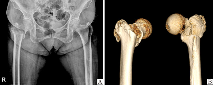
Intertrochanteric fractures with patterns that could not be supported by the anteromedial cortex were considered high risk for fixation failure. The risk of complications is also higher for intertrochanteric fractures involving the basal neck component including post-traumatic avascular necrosis and fixation failure (Fig. 1). Hip arthroplasty was performed for treatment of these types of intertrochanteric fractures.
Fig. 1.
Indication for hip arthroplasty for treatment of a femoral intertrochanteric fracture. (A) Simple radiograph showing an intertrochanteric fracture involving the basal neck component. (B) Computed tomography scan showing a fracture line on the medial side of the intertrochanteric ridge and fracture of the greater trochanter.
2. Surgical Procedure
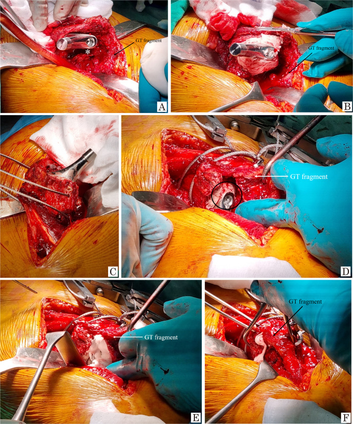
Patients were placed in the lateral decubitus position for performance of hip arthroplasty using the modified Hardinge approach. Routine surgical dislocation of the hip, acetabular preparation, and cup prosthesis insertion were performed sequentially (Fig. 2A). Patterns of intertrochanteric fractures were also identified. Posterior retraction of the GT and posterior fragments was performed using a femoral neck retractor, and the femoral canal was prepared using a stem rasp until achievement of adequate press-fit fixation of the stem (Fig. 2B). The hematoma in the GT was removed during performance of this procedure. The hip joint was reduced for assessment of soft tissue tension and stability using a trial prosthesis. A real stem was inserted for achievement of adequate soft-tissue tension and stability with a temporary trial prosthesis (Fig. 2C). With achievement of press fit fixation of the stem, the space between the stem and the proximal femoral canal is filled with cement in the dough phase (Fig. 3A, B). Two circumferential cables are temporarily placed toward the proximal femur overriding the GT area (Fig. 3C). Following reduction of the hip joint, the void space in the GT area was identified and filled with cement (Fig. 3D, E). Reduction of the GT and the posterior fragment is performed using reduction forceps, followed by removal of excessive protruding cement (Fig. 3F).
Fig. 2.
Surgical procedures. (A) Routine surgical dislocation of the hip, acetabular preparation, and insertion of the cup prosthesis are performed sequentially. (B) The greater trochanteric and posterior fragment are retracted posteriorly using a femoral neck retractor. The femoral canal is prepared using a stem rasp until adequate press fit fixation of the stem is achieved. (C) The real stem is inserted.
Fig. 3.
Surgical procedures – cement filling. (A, B) With achievement of press fit fixation of the stem, the space between the stem and proximal femoral canal (asterisk) is filled with cement in the dough phase. (C) Two circumferential cables are temporarily placed toward the proximal femur overriding the greater trochanteric (GT) area. (D, E) Following reduction of the hip joint, the void space of the GT area (black circle) is identified and additionally filled with cement. (F) Reduction of the GT and the posterior fragment is performed using reduction forceps, with removal of excessive protruding cement.
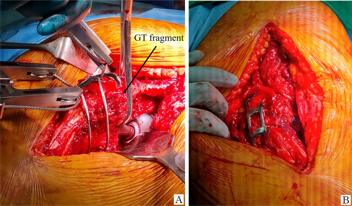
Maintenance of reduction, fixation of a GT reattachment plate to the GT and posterior fragment and tightening of the cable are the final procedures in performance of hip arthroplasty (Fig. 4A). Firm fixation status of the GT fragments was eventually confirmed (Fig. 4B). Because immediate fixation with cement is an important technique, fixation of the GT reattachment plate can be replaced with circumferential wiring after cement filling.
Fig. 4.
Surgical procedures – greater trochanteric (GT) fragment fixation. (A) Maintaining reduction, fixation of a GT reattachment plate to the GT and posterior fragment and tightening of the cable is performed as the final procedure in performance of hip arthroplasty. (B) Firm fixation status of the GT fragment is eventually confirmed.
3. Postoperative Management
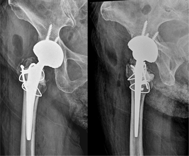
A simple postoperative radiograph is shown in Fig. 5. None of the patients was limited in position from the immediate postoperative period, and progressive weight-bearing was allowed as tolerated three days after surgery.
Fig. 5.
Postoperative simple radiograph. Total hip arthroplasty with cement filling and greater trochanteric reattachment plate fixation was performed.
4. Case Presentation 1
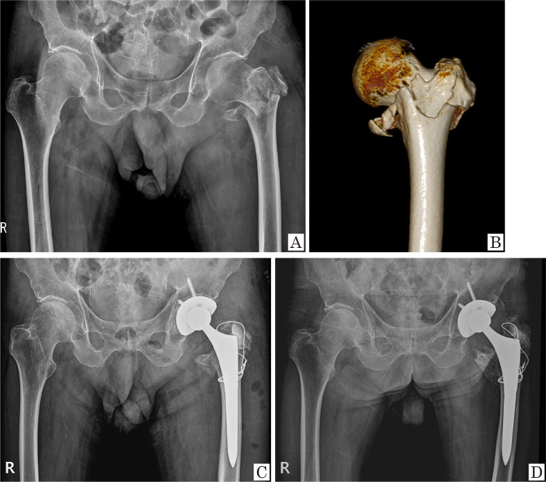
A 55-year-old male presented to our hospital after a slipping injury. Simple radiographs showed a 4-part intertrochanteric fracture, and a computed tomography (CT) scan showed loss of anteromedial support. Primary total hip arthroplasty (THA) was performed with cement filling of the GT and circumwiring areas. Four years after the THA, a well-maintained GT fragment was observed without displacement or visible cement debonding. The patient ambulated without complications (Fig. 6).
Fig. 6.
Case presentation 1. (A) Preoperative simple radiograph. (B) Preoperative computed tomography scan. (C) Immediate postoperative simple radiograph. (D) Four years after follow-up period simple radiograph.
5. Case Presentation 2
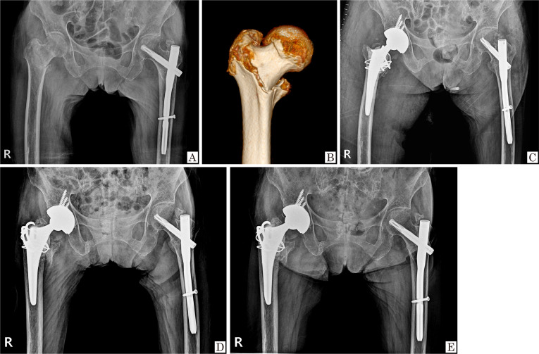
A 94-year-old female presented to our hospital after a slipping injury. The patient was a household walker with a history of a spine problem. Simple radiography and CT tomography showed a basicervical component involving the femoral intertrochanteric fracture. Primary THA was performed with cement filling of the GT area and fixation of the GT reattachment plate. A well-maintained GT fragment was observed at six months and one year after THA. The patient ambulated using a walker without complications (Fig. 7).
Fig. 7.
Case presentation 2. (A) Preoperative simple radiograph. (B) Preoperative computed tomography scan. (C) Immediate postoperative simple radiograph. (D) Six-month follow-up period simple radiograph. (E) One-year follow-up period simple radiograph.
DISCUSSION
Hip arthroplasty is not typically considered as the first treatment of choice for management of intertrochanteric fractures. Although internal fixation of a cephalomedullary nail or dynamic hip screw can be performed in the treatment of several intertrochanteric fractures, many studies have reported a high risk of fixation failure for certain fracture patterns3,4). Arthroplasty has recently been highlighted as an alternative to internal fixation.
Successful fixation and union of the GT are critical factors in performance of hip arthroplasty for management of intertrochanteric fractures. Circumferential wiring, which has been described in several studies, is a popular and effective technique for use in performance of circumferential wiring3,6). Kim et al.3), who classified fracture patterns of the GT, applied different wiring techniques according to the fracture criteria. Figure 8-tension band wiring and a coronal split augmentation technique were introduced by Nho et al.6). Although wire breakage was observed in 11.5% of the patients, there were no complaints of bursitis, and none of the patients required revision surgery6). Despite the simplicity and broad acceptance of this method, some studies have reported its limitations in relation to wiring. Representative complications include nonunion, GT displacement, and wire breakage. Nonabsorbable polyester and ultra-high-molecular-weight polyethylene fiber cable methods for fixation of the osteotomized GT during performance of hip arthroplasty were introduced by Oe et al.9). Use of a locking plate for GT fixation has also been suggested based on a theoretical background that proposes that a unicortical locking screw can provide rigid fixation. However, significant trochanteric pain, nonunion, and the risk of fixation failure after use of a locking plate have been reported10) (Table 1).
Table 1.
Other Techniques for GT Fixation in Hip Arthroplasty
| Study | Implant for GT fixation | Methods | Complications of implant |
|---|---|---|---|
| Kim et al.3) (2019) | Wire Non-absorbable suture |
Figure of 8 wiring using 16-gauge wires and/or cerclage wiring Non-absorbable sutures to further reinforce fixation |
Displacement of GT (20.5%) Wire loosening (2.3%) Wire breakage (9.1%) Fixation failure (6.8%) |
| Nho et al.6) (2023) | Wire | Figure of 8 wiring using 18-gauge wires into double rows GT fixation with double wires by coronal split augmentation |
Wire breakage (11.5%) Stem subsidence (3.2%) |
| Oe et al.9) (2018) | Non-absorbable polyester suture UHMWPE fiber cable |
Osteotomized GT were suture with No. 5 non-absorbable polyester suture UHMWPE fiber cable suture augmentation |
Trochanteric pain (0.4%-5.8%) Trendelenburg’s sign (0.9%-1.3%) |
| Tetreault and McGrory10) (2016) | Locking plate | Plate fixation with multiple proximal locking 3.5 screw in the trochanter Auto- or allobone graft augmentation in the trochanter |
Trochanteric pain (18.8%) Breakage of plate or screw (12.5%) Cable breakage (3.1%) Non-union of trochanter (15.6%) |
GT: greater trochanter, UHMWPE: ultra-high molecular weight polyethylene.
During the follow-up period, fixation failure of the GT was observed after application of circumferential wiring. Finally, we have determined that immediate and rigid fixation may be required in cases involving superior displacement of a GT by the abductor muscle. We eventually designed this cement-filling technique with the expectation that interdigitation between the GT fragment and cement would be effective for immediate prevention of GT displacement. In addition, use of a cement filling technique can have potential advantages. First, fixation of biologic bone ingrowth between the femoral canal and stem could also be achieved in the metaphyseal-diaphyseal junction area when cementless fixation is used. We emphasize that use of this technique offers the advantage of both biologic and cement fixation. In addition, we believe that use of cement filling can prevent expansion of the effective joint space and that a positive effect on osteolysis can be achieved during the long-term follow up period. Increased rates of dislocation can be a concern in performance of hip arthroplasty for treatment of an intertrochanteric fracture, which might be a result of insufficiency of the abductor muscle. We attempted to minimize abductor insufficiency by preventing displacement of the GT. Conduct of studies examining the incidence of complications related to use of this technique is warranted.
There are some limitations to the use of this technique. First, cement filling without GT displacement did not result in immediate union of the GT fragment. Despite this drawback, displacement of the GT can be minimized, and good functional outcomes can be achieved using this technique. To the best of our knowledge, achieving union of the GT fragment in performance of hip arthroplasty for treatment of an intertrochanteric fracture can be difficult even after auto- or allografting to the GT area. Ultimately, fixation failure of the GT is a frequent occurrence, resulting in poor outcomes after hip arthroplasty, meaning that use of this technique is optimal in performance of THA for management of intertrochanteric fracture in old age with osteoporotic bone. Second, because cement is neither a bone substitute nor a promoter of bone union, cement debonding can occur during the follow-up period, or cement between bone fragments can interfere with union of the GT. However, in cases where the GT fragment was maintained without displacement, union of the GT fragment should be expected at the time of cement debonding. Fixation failure of the GT fragment may be rare even after development of cement debonding.
Cement filling of the GT during performance of hip arthroplasty for treatment of femoral intertrochanteric fractures is a useful technique for preventing displacement of the GT and supporting early mobilization. We believe that use of this technique may be useful for surgeons for preventing displacement of the GT and for achievement of more favorable outcomes after hip arthroplasty for treatment of femoral intertrochanteric fractures.
Acknowledgements
We give special thanks to Mi-Kyung Kang for supporting this report.
Funding Statement
Funding No funding to declare.
Footnotes
Conflict of Interest
Kyung-Jae Lee has been a Deputy Editor since January 2023, but had no role in the decision to publish this article. No other potential conflict of interest relevant to this article was reported.
References
- 1.Jegathesan T, Kwek EBK. Are intertrochanteric fractures evolving? Trends in the elderly population over a 10-year period. Clin Orthop Surg. 2022;14:13–20. doi: 10.4055/cios20204. https://doi.org/10.4055/cios20204. [DOI] [PMC free article] [PubMed] [Google Scholar]
- 2.Shin WC, Lee SM, Moon NH, Jang JH, Choi MJ. Comparison of cephalomedullary nails with sliding hip screws in surgical treatment of intertrochanteric fractures: a cumulative meta-analysis of randomized controlled trials. Clin Orthop Surg. 2023;15:192–202. doi: 10.4055/cios22103. https://doi.org/10.4055/cios22103. [DOI] [PMC free article] [PubMed] [Google Scholar]
- 3.Kim MW, Chung YY, Lim SA, Shim SW. Selecting arthroplasty fixation approach based on greater trochanter fracture type in unstable intertrochanteric fractures. Hip Pelvis. 2019;31:144–9. doi: 10.5371/hp.2019.31.3.144. https://doi.org/10.5371/hp.2019.31.3.144. [DOI] [PMC free article] [PubMed] [Google Scholar]
- 4.Chang SM, Zhang YQ, Du SC, et al. Anteromedial cortical support reduction in unstable pertrochanteric fractures: a comparison of intra-operative fluoroscopy and post-operative three dimensional computerised tomography reconstruction. Int Orthop. 2018;42:183–9. doi: 10.1007/s00264-017-3623-y. https://doi.org/10.1007/s00264-017-3623-y. [DOI] [PubMed] [Google Scholar]
- 5.Liu L, Sun Y, Wang L, et al. Total hip arthroplasty for intertrochanteric fracture fixation failure. Eur J Med Res. 2019;24:39. doi: 10.1186/s40001-019-0398-1. https://doi.org/10.1186/s40001-019-0398-1. [DOI] [PMC free article] [PubMed] [Google Scholar]
- 6.Nho JH, Seo GW, Kang TW, Jang BW, Park JS, Suh YS. Bipolar hemiarthroplasty in unstable intertrochanteric fractures with an effective wiring technique. Hip Pelvis. 2023;35:99–107. doi: 10.5371/hp.2023.35.2.99. https://doi.org/10.5371/hp.2023.35.2.99. [DOI] [PMC free article] [PubMed] [Google Scholar]
- 7.Sidhu AS, Singh AP, Singh AP, Singh S. Total hip replacement as primary treatment of unstable intertrochanteric fractures in elderly patients. Int Orthop. 2010;34:789–92. doi: 10.1007/s00264-009-0826-x. https://doi.org/10.1007/s00264-009-0826-x. [DOI] [PMC free article] [PubMed] [Google Scholar]
- 8.Lee YK, Kim JT, Alkitaini AA, Kim KC, Ha YC, Koo KH. Conversion hip arthroplasty in failed fixation of intertrochanteric fracture: a propensity score matching study. J Arthroplasty. 2017;32:1593–8. doi: 10.1016/j.arth.2016.12.018. https://doi.org/10.1016/j.arth.2016.12.018. [DOI] [PubMed] [Google Scholar]
- 9.Oe K, Iida H, Kobayashi F, et al. Reattachment of an osteotomized greater trochanter in total hip arthroplasty using an ultra-high molecular weight polyethylene fiber cable. J Orthop Sci. 2018;23:992–9. doi: 10.1016/j.jos.2018.07.020. https://doi.org/10.1016/j.jos.2018.07.020. [DOI] [PubMed] [Google Scholar]
- 10.Tetreault AK, McGrory BJ. Use of locking plates for fixation of the greater trochanter in patients with hip replacement. Arthroplast Today. 2016;2:187–92. doi: 10.1016/j.artd.2016.09.006. https://doi.org/10.1016/j.artd.2016.09.006. [DOI] [PMC free article] [PubMed] [Google Scholar]


