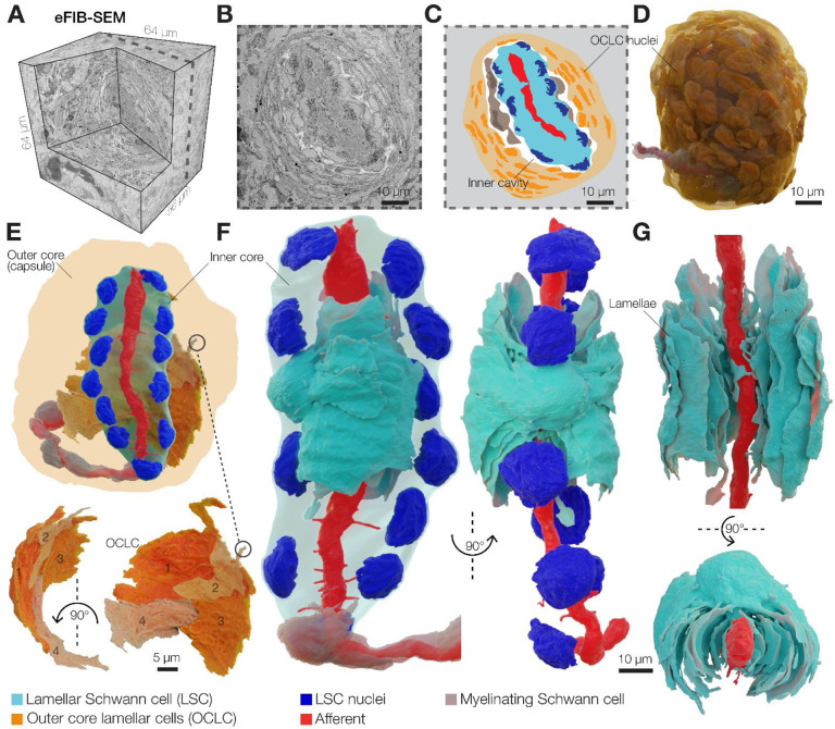
Figure 1. 3D architecture of the Pacinian corpuscle.
(A) A 3D volume of duck bill skin dermis obtained by eFIB-SEM with 8 nm3 resolution.
(B, C) A single eFIB-SEM image (B) and an illustration (C) of a section of an avian Pacinian corpuscle.
(D) 3D reconstruction of the avian Pacinian corpuscle.
(E) 3D reconstruction of the Pacinian corpuscle showing the location of the inner core inside the outer core (top), and reconstruction of four outer core lamellar cells (bottom).
(F, G) 3D reconstruction of the inner core showing the architecture of the afferent terminal and one of 12 lamellar Schwann cells (LSC). Different shades of cyan denote lamellae from the same LSC.

