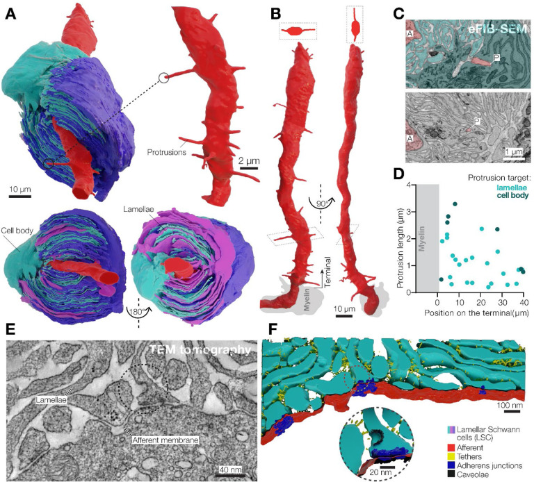
Figure 2. 3D architecture of LSCs and the afferent terminal in the Pacinian corpuscle.
(A) 3D reconstruction of a pair of opposing LSCs from the inner core (upper panel). An additional LSC is located on top of the cyan LSC (bottom panel).
(B) 3D reconstruction of the afferent terminal with 29 protrusions. Two cross section views are shown at the top of the terminal.
(C) A pseudo-colored eFIB-SEM image showing protrusion tips targeting the LSC body (upper panel) and lamellae. A, afferent terminal; P, protrusion tip.
(D) Localization, length and target of afferent protrusions.
(E, F) Transmission electron microscopy image (E) and its 3D reconstruction of the lamellae-afferent contact area.

