Abstract
Background
Congenital tufting enteropathy (CTE) is a rare cause of intractable congenital diarrhea in children, always resulting in parenteral nutrition (PN) dependency. We aimed to report novel mutations in Chinese patients and to illustrate the clinical, histopathological, and molecular features of CTE in China.
Case Description
We report three cases of CTE diagnosed with whole-exome sequencing (WES) and MOC31 [a monoclonal antibody of epithelial cell adhesion molecule (EPCAM)] immunohistochemistry. The main manifestations in the three patients were watery diarrhea and growth retardation. Upper endoscopy in three patients revealed villous atrophy of the duodenal mucosa. Histological examination revealed villus abnormalities and two patients with focal tufting. All of the three patients revealed a complete absence of EPCAM expression through MOC31 immunohistochemistry. Five novel mutations, including c.319delG, c.505_507delGAG, c.491+1G>C, c.60del (p.F20Lfs*17), and c.353G>A, in EPCAM were identified through molecular analysis. In our review, there were 18 different mutations in 11 patients from nine studies, with 12 mutations reported only once. In China, 73% of the patients were compound heterozygotes, and most of the pathogenic variants were in exon 3. All patients presented with congenital diarrhea and needed PN because of growth retardation, even when diarrhea was improved. Of the 11 patients, 3 (27%) died.
Conclusions
CTE is rare and fatal, and lacks characteristic changes during endoscopy. Patients with CTE require early diagnosis via histological examination and genetic detection to improve survival.
Keywords: Congenital tufting enteropathy (CTE), EPCAM mutations, Chinese patients, children, case report
Introduction
Congenital tufting enteropathy (CTE) is a rare digestive disease presenting with inherited intractable diarrhea and is characterized by villus atrophy and the absence of inflammation (1). It was first reported in 1994 by Reifen et al. (2). The prevalence of CTE is estimated to be approximately 1/50,000–100,000 live births in Western Europe (3,4). CTE often presents with chronic watery diarrhea and growth retardation from the first few months after birth, persists throughout life, and is associated with high morbidity and mortality. Most affected individuals depend on parenteral nutrition (PN) to maintain normal growth and development (5).
Considerable progress has been made in molecular diagnoses, particularly with the widespread application of exome sequencing (ES) in clinical practice over the past decade. The first genetic characterization of CTE was performed by Sivagnanam et al., who discovered the epithelial cell adhesion molecule (EPCAM) as the gene contributing to CTE (6). Moreover, mutations in serine peptidase inhibitor Kunitz type 2 (SPINT2) were also found to be associated with CTE (7,8). Up to now, more than 100 EPCAM variants have been identified (4). The exon regions in which the most frequent pathogenic variants are found vary in different parts of the world. Variants in the Middle East are often in exon 5, whereas in East Asia, exon 3 pathogenic variants are more frequent (9,10). We therefore aimed to summarize the clinical, histopathological, and molecular features of our three patients with CTE and review the genetic variants of EPCAM that have been reported in China. Collectively, our cases highlight the application of ES accompanied by MOC31 (a monoclonal antibody of EPCAM) immunohistochemistry to the diagnosis of CTE. We present this article in accordance with the CARE reporting checklist (available at https://tp.amegroups.com/article/view/10.21037/tp-24-97/rc).
Case presentation
Clinical description
All procedures performed in this study were in accordance with the ethical standards of the institutional and/or national research committee(s) and with the Helsinki Declaration (as revised in 2013). Written informed consent was obtained from the patients’ parents or their legal guardians for the publication of this case report and accompanying images. A copy of the written consent is available for review by the editorial office of this journal. The clinical and histological characteristics of patients 1, 2, and 3 are presented in Table 1.
Table 1. Clinical characteristics of the three patients with congenital tufting enteropathy.
| Item | Patient 1 | Patient 2 | Patient 3 |
|---|---|---|---|
| Gender | Male | Male | Female |
| Birth weight (g) | 3,850 | 3,870 | 3,250 |
| Gestation age (weeks) | 40 | 40 | 37 |
| Family history | Negative | Negative | Negative |
| Age at onset of diarrhea | 3 months | 1 week | 1 week |
| Age at admission to our hospital | 17 months | 56 days | Nearly 5 months |
| Weight at admission (g) | 4,000 | 4,000 | 4,100 |
| Age at diagnosis | 19 months | 3 months | 7 months |
| Weight at diagnosis (g) | 4,500 | 4,400 | 4,200 |
| Laboratory examination (blood) at admission to our hospital | |||
| Hemoglobin (g/L) | 83 | 88 | 115 |
| RBC (×1012/L) | 2.8 | 2.8 | 3.74 |
| Albumin (g/L) | 30.0 | 33.7 | 40.3 |
| pH | 7.43 | 7.32 | 7.40 |
| Chloride (mmol/L) | 103 | 105 | 101 |
| Potassium (mmol/L) | 3.2 | 3.4 | 3.4 |
| Sodium (mmol/L) | 133 | 133 | 134 |
| Base excess (mmol/L) | −5.5 | −5 | 0.8 |
| PT (s) | 18.6 | 16.6 | 16.9 |
| APTT (s) | 50.0 | 54.1 | 57.2 |
| PTA†† (%) | 55 | 63 | 61 |
RBC, red blood cell; pH, pondus hydrogenii; PT, prothrombin time; APTT, activated partial thromboplastin time; PTA, prothrombin activity.
Patient 1 came to our hospital (Children’s Hospital of Fudan University) at the age of seventeen months and had intractable diarrhea when he was started on cow’s milk-based formula after three months of breastfeeding after birth. He was hospitalized numerous times because of recurrent electrolyte disturbances. Treatments included probiotics, smectite, feeding with extensively hydrolyzed formula (eHF), PN therapy, and albumin infusion; however, there was no improvement in the patient’s condition. When he was admitted to our center, he showed dehydration and severe growth retardation, with a weight of 4.0 kg (Z score, −5.9) and a height of 62 cm (Z score, −6.5).
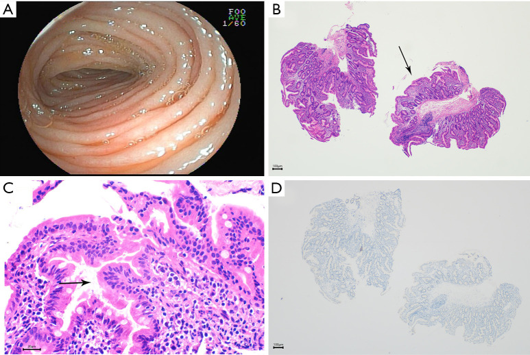
Laboratory tests upon admission revealed electrolyte disturbances (hyponatremia, hypokalemia, hypomagnesemia, and hypocalcemia), anemia, hypoproteinemia, hypolipemia, and coagulant function abnormalities. Other tests, including liver function, renal function, and immunologic function, were normal. Stool culture was negative. An upper endoscopy with biopsies was performed and revealed gross villous atrophy in the duodenum (Figure 1A). Hematoxylin-eosin (H&E) staining of the biopsied tissue showed atrophy of the small intestinal villi, epithelial hyperplasia accompanied by focal clusters of epithelial cells, and increased interstitial chronic inflammatory cells (Figure 1B,1C). Follow-up immunohistochemistry revealed a complete absence of EPCAM expression (Figure 1D) compared with a normal child.
Figure 1.
Images of Patient 1. Upper endoscopy revealed villous atrophy in the duodenal mucosa (A). Staining with hematoxylin and eosin revealed villus atrophy (arrow) (B, magnification, ×40), increased interstitial chronic inflammatory cells (C, magnification, ×400) and epithelial hyperplasia accompanied by focal clusters of epithelial cells (arrow). Immunohistochemistry staining with no brown staining showed absence of EPCAM expression (D, magnification, ×40). EPCAM, epithelial cell adhesion molecule.
When the patient improved, the EN (peptide formula) was reintroduced. However, with an increase in EN, stool output increased. The patient was fed with rice porridge, and stool frequency was maintained at three times per day. However, the patient required PN due to dehydration and malnutrition. During hospitalization, the patient experienced one episode of bloodstream infection associated sepsis, and the sepsis was controlled with antibiotics.
After fifteen days of hospitalization, the parents requested discharge from our hospital and did not receive partial PN after discharge. His mother fed him with rice gruel, gradually with other foods, such as pork. ES revealed that the patient was diagnosed with CTE. He still had mild diarrhea, and grew very slowly.
At seven years of age, he had severe growth retardation with a weight of 11 kg (Z score, −3.6) and a height of 92 cm (Z score, −6.3). Recently, his mother reported that his diet was normal, but he could not drink milk as it led to diarrhea. The frequency of defecation was once a day, and the stool was formed and occasionally unformed. The intelligence development of the boy was normal for children of the same age.
Patient 2 was a boy who was transferred to our hospital at the age of eight weeks because of intractable diarrhea and a failure to thrive. The frequency of watery stools accompanied by mucus was five to eight times per day. Allergic enteritis was initially considered; however, no significant improvement was observed after the patient’s mother made dietary adjustments. After treatment with probiotics, smectite, lactase supplementation, and feeding with an amino acid-based formula (AAF), the diarrhea did not improve. When admitted to our center, his stool frequency was approximately eight to ten times per day and watery; he had severe growth retardation, with a weight of 4.0 kg (Z score, −2.5) and a height of 53 cm (Z score, −2.3).
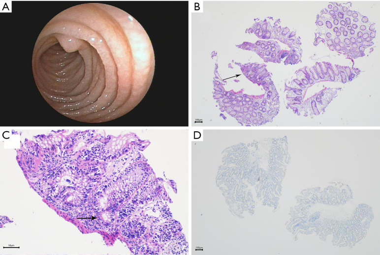
Apart from mild metabolic acidosis, electrolyte disturbances (hyponatremia and hypokalemia), anemia, hypoproteinemia, and coagulant function abnormalities, no other abnormal findings were detected when the child was admitted; the stool culture was negative. This patient had an EGD and colonoscopy performed, which revealed villous atrophy of the duodenal mucosa (Figure 2A). H&E staining of the biopsy specimens revealed eosinophilia of the mucosa of the large intestine and partially distorted crypts (Figure 2B,2C). Follow-up immunohistochemistry revealed the complete absence of EPCAM expression (Figure 2D).
Figure 2.
Images of Patient 2. Upper endoscopy revealed villous atrophy in the duodenal mucosa (A). Staining with hematoxylin and eosin revealed partially distorted crypts (arrow) (B, magnification, ×40) and eosinophilia of the mucosa of the large intestine (arrow) (C, magnification, ×200). Immunohistochemistry staining with no brown staining showed absence of EPCAM expression (D, magnification, ×40). EPCAM, epithelial cell adhesion molecule.
Fasting resulted in the transient relief of diarrhea symptoms, but the stool frequency increased to more than ten times/day after AAF intake. He was administered partial PN. After one month, he was diagnosed with CTE based on ES. The patient underwent partial home PN treatment after discharge, plus a digestive diet. At 27 months of age, he was still on partial PN, with a weight of 12 kg (Z score, −0.39) and a height of 85.5 cm (Z score, −0.66). The fluid volume and energy were reaching 62 mL/kg and 55 kcal/kg, respectively.
Patient 3, an almost five-month-old female, was admitted to our division because of failure to thrive and intractable diarrhea. She experienced recurrent watery diarrhea seven days after birth, with a frequency of eight times per day. When she was administered AAF, the stool frequency was still three to six times per day, with failure to thrive. Treatment with probiotics or smectite had no effect. Her body weight was just 4.1 kg (Z score, −4.0) and her height was 61 cm (Z score, −1.6) when she was transferred to our division.
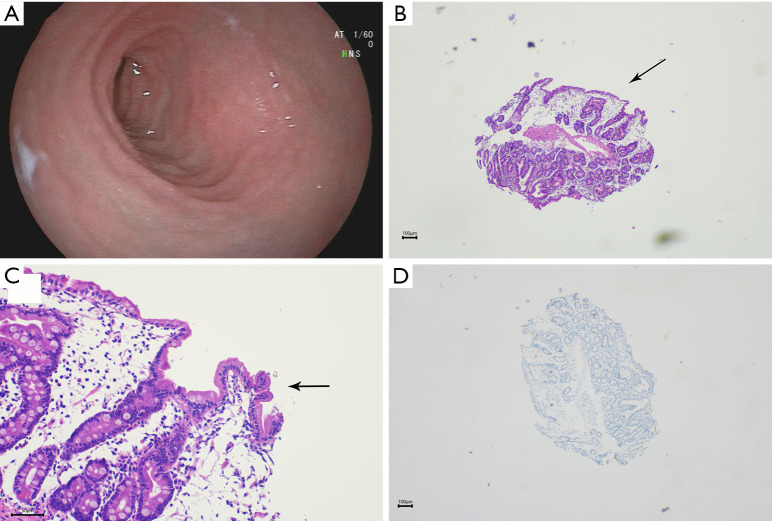
Laboratory tests on admission revealed electrolyte disturbances (hyponatremia and hypokalemia), thyroid dysfunction, and coagulation abnormalities. The results of other tests were normal. Stool culture was negative. Colonoscopy did not reveal any obvious abnormalities, but upper endoscopy gross findings revealed villous atrophy in the duodenal mucosa (Figure 3A). H&E staining of the biopsied tissue showed atrophy of the small intestinal villi and epithelial crowding (Figure 3B,3C). Follow-up immunohistochemistry revealed the complete absence of EPCAM expression (Figure 3D).
Figure 3.
Images of Patient 3. Upper endoscopy revealed villous atrophy in the duodenal mucosa (A). Staining with hematoxylin and eosin revealed atrophy of the small intestinal villi (arrow) (B, magnification, ×40) and epithelial crowding (arrow) (C, magnification, ×200). Immunohistochemistry staining with no brown staining showed absence of EPCAM expression (D, magnification, ×40). EPCAM, epithelial cell adhesion molecule.
The infant was administered extensively hydrolyzed formula feeding orally. Because the diarrhea was not serious, the family removed the child from the hospital. Two months later, ES confirmed the diagnosis of CTE. Due to her slow rate of gaining weight, she underwent partial PN treatment at the age of fourteen months. After two months of intravenous nutrition, the weight and height of the patient increased by 3 kg and 7 cm, respectively. Her body weight increased to 9.75 kg at 16 months of age. When she was two years old, she was still with partial home PN, with volume and energy reaching the level of 56 mL/kg and 58 kcal/kg, respectively. Her height increased to 82 cm (Z score, −1.3), and weight increased to 11 kg (Z score, −0.7).
Genetic analysis
Five novel mutations, including c.319delG, c.505_507delGAG, c.491+1G>C, c.60del (p.F20Lfs*17), and c.353G>A, in the EPCAM gene (NM_002354.3) were identified in this study, as shown in Table 2.
Table 2. Reported mutations in epithelial cell adhesion molecule gene in patients with congenital tufting enteropathy in China.
| Case | Study | Patients (n) | Year | Coding DNA | Mutation type | Zygosity | Symptoms | PN | Outcome |
|---|---|---|---|---|---|---|---|---|---|
| 1 | Tang et al. | P1 | 2016 | c.307G>A (exon 3)/deletion (exon 2–5) | Missense mutation/deletion | Compound heterozygous | Diarrhea, abdominal distension, and impaired growth | PN can be weaned, with 4 times of hospitalization for TPN in the first 2 years of life | Alive with a weight of 27.8 kg and a height of 122.7 cm at 13 years old |
| 2 | Yuan et al. | P1 | 2018 | c.412C>T (exon 3) | Missense mutation | Homozygous | Diarrhea, impaired growth | With no definite history of PN | Alive with a weight of 6.8 kg and a height of 65 cm at age of 46 months |
| 3 | Fang et al. | P1 | 2020 | c.491+1G>A (intron 4) | Splicing mutation | Homozygous | Diarrhea, vomiting, abdominal distension, and impaired growth | Partial PN for 13 months from age of 2 months and weaned at age of 15 months | Deceased at age of 18 months, due to sepsis and multiple organ failure |
| 4 | Zhou et al. | P1 | 2020 | c.657+1G>A (intron 6) | Splicing mutation | Homozygous | Diarrhea and impaired growth | TPN without weaned | Alive at age of 21 months, with a weight of 8.5 kg |
| 5 | Yan et al. | P1 | 2021 | c.184 + 6T>G (intron 2)/large deletion (exon 1–9) | Splicing mutation/deletion | Compound heterozygous | Diarrhea, abdominal distension, and impaired growth | Partial PN from age of 23 months to 3.5 years, without weaned | Deceased at 3.5 years old, due to blood transfusion reaction |
| 6 | Yan et al. | P2 | c.96C>A (exon 2)/c.823delG (exon 7) | Nonsense mutation/frameshift mutation | Compound heterozygous | Diarrhea and impaired growth | Partial PN from the age of 1 month for more than a year without weaned | Alive at 1 year old, with a weight of 3.3 kg | |
| 7 | Yang et al. | P1 | 2022 | c.491+1G>A (intron 4)/c.352_353ins CACC (exon 3) | Splicing mutation/frameshift mutation | Compound heterozygous | Diarrhea and impaired growth | TPN or partial PN from birth, for 3 months without weaned | Deceased due to discontinuing of treatment |
| 8 | Zhao et al. | P1 | 2022 | c.227C>G (exon3)/c.296G>A (exon 3) | Nonsense/missense | Compound heterozygous | Intractable diarrhea from 7 months old, vomiting, abdominal distension, and impaired growth | PN can be weaned. Multiple hospitalizations after birth because of severe malnutrition and growth failure | Alive with a weight of 13 kg and a height of 98 cm at 7 years old |
| 9 | This study | P1 | 2023 | c.319delG (exon3)/c.505_507delGAG (exon 5) | Frameshift mutation/in-frame mutation | Compound heterozygous | Diarrhea and impaired growth | Transient PN, weaned after discharge | Alive with a weight of 11 kg and a height of 92 cm at 7 years old |
| 10 | This study | P2 | 2023 | c.491+1G>C (intron 4)/c.60del (p.F20Lfs*17) (exon 1) | Splicing variant/Frameshift mutation | Compound heterozygous | Diarrhea and impaired growth | Partial PN from age of 2 months without weaned | Alive with a weight of 12 kg and a height of 85.5 cm at age of 27 months |
| 11 | This study | P3 | 2023 | c.353G>A (exon 3)/deletion (exon 1–7) | Missense mutation/deletion | Compound heterozygous | Diarrhea, abdominal distension, and impaired growth | Partial PN from 14 months old without weaned | Alive with a weight of 11 kg and a height of 82 cm at 2 years old |
PN, parenteral nutrition; TPN, total parenteral nutrition.
For patient 1, the results of whole-exome sequencing (WES) identified two novel complex heterozygous mutations in the EPCAM gene. He inherited the mutation c.319delG (frameshift variant) in exon 3 from their mother and c.505_507delGAG (in-frame variant) in exon 5 from their father. Both variants are classified as “likely pathogenic” according to the American College of Medical Genetics and Genomics/Association for Molecular Pathology (ACMG) guidelines.
Patient 2 inherited the mutation c.491+1G>C (splicing variant) in intron 4 from his mother and c.60del (p.F20Lfs*17) (frameshift variant) in exon 1 from his father. Both variants are classified as “pathogenic” according to the ACMG guidelines.
In patient 3, the results of WES confirmed the presence of novel homozygous mutations in exon 3 (c.353G>A) (missense mutation) of EPCAM. The variant is classified as “pathogenic” based on the ACMG guidelines. Then, high-throughput sequencing data suggested that she also had a heterozygous exon 1–7 deletion, which was from her mother. Her father did not undergo genetic testing because he was abroad.
Review of mutations in China
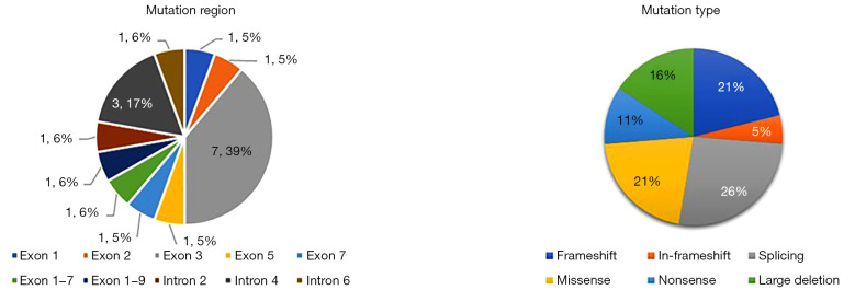
The mutations in EPCAM reported in English and Chinese languages in China were reviewed (Table 2). After a comprehensive review of previous reports on CTE in Chinese patients, we identified eighteen distinct EPCAM mutations in eleven patients from nine studies, including missense, nonsense, frameshift, in-frameshift, splicing mutations, and large deletions, with frameshift and splicing mutations ranked high. Among them, the pathogenic variants c.491+1G>A (intron 4), c.307G>A (exon 3), c.412 C>T (exon 3), c.227 C>G (exon 3), and deletion (exon 1–7, exon 1–9) have been reported in previous studies, with the remaining twelve mutations being reported only once. In China, most of the patients were compound heterozygotes (eight of eleven patients, 73%). Exon 3 pathogenic variants are the most common (Figure 4). Clinically, all patients with EPCAM variants presented with isolated congenital diarrhea and required PN. Only 1 patient had no history of PN, but with growth restoration (Case 2, see Table 2). Of the eleven patients, three (27%) died.
Figure 4.
Summary of the mutations in the epithelial cell adhesion molecule gene in patients with congenital tufting enteropathy in China.
Discussion
We identified five novel EPCAM mutations in our three patients. The main manifestations in our three patients were watery diarrhea and growth retardation. Upper endoscopy revealed villous atrophy of the duodenal mucosa. Histological examination revealed villous abnormalities, including epithelial crowding, disorganization without epithelial lymphocytosis, and focal tufting in two patients. In our review, there were eighteen distinct EPCAM mutations in eleven patients, with twelve mutations reported only once. Exon 3 pathogenic variants are the most common in China. All patients are dependent on PN to maintain survival or normal growth.
Although many CTE patients have been reported worldwide, a total of only eleven cases have been reported in China, with the first being reported in 2018 (9). Patients with CTE manifest with intractable diarrhea during the neonatal period, often combined with other epithelial cell-related diseases or malformations, such as punctate keratitis, nostril atresia, esophageal atresia, no anus (3), or multiple arthritis (11) and cardiomyopathy (12). Simultaneously, growth is impaired. The main manifestations of our three patients were watery diarrhea and growth retardation, along with metabolic acidosis, electrolyte disturbances (hyponatremia, hypokalemia), anemia, and coagulant function abnormalities. Coagulant function abnormalities might be due to hepatic synthetic dysfunction, which couldn’t be corrected by transfusions of vitamin K but by transfusions of frozen plasma. Our patients had one another thing in common—switching to a different type of formula did not improve their diarrhea, but feeding with millet porridge and rice paste might have been more tolerable. Through a review of Chinese patients, we found that, except for intractable diarrhea, one patient experienced pain in multiple joints with swelling of the hands and feet (9), as reported elsewhere (11).
The diagnosis of neonatal-onset diarrhea is particularly difficult. Other congenital diarrheal disorders (CDDs) often present with similar clinical presentations to CTE, such as microvillus inclusion disease (MVID), chloride diarrhea, and sodium diarrhea (13). MVID shows an onset and blunted villi resemblant to CTE, have the same pathogenesis due to the differentiation and polarization of enterocytes as CTE. The presence of surface apical tufts, as opposed to apical inclusion bodies, in enterocytes distinguishes CTE from MVID. Besides, CTE and MVID strains contain different virulence genes. Patients with CTE have changes in EPCAM and SPINT2 and mutations in MYO5B, STX3, and STXBP2 with MVID (14). The diagnoses of these three cases depended on genetic testing and were supplemented by immunohistochemical verification. From this perspective, if there are cases of diarrhea caused by suspected small intestinal epithelial lesions, it is necessary to conduct genetic testing and a multi-point biopsy of the small intestine to improve the early diagnosis rate of the disease. Next generation sequencing (NGS) has contributed to great advances in this field (15). Esposito et al. designed an NGS panel, which can analyze 92 CDD-related genes (16). WES conducted in these 3 patients was now widely applied, which revealed pathogenic variants of EPCAM but other CODE genes in them. In addition, MOC31 immunohistochemistry—a diagnostic stain for CTE with a specificity of 100% for loss of staining—which was applied in our three patients should also be included in the pathological examinations in patients with chronic diarrhea to exclude this possibility (17).
The EPCAM gene maps to chromosome 2p21 (including nine exons and eight introns) and encodes the EPCAM protein. Mutated EPCAM causes the destruction of cell-to-cell connections and defects in cell barrier function (1). By 2022, 140 EPCAM mutations had been found, including missense, nonsense, frameshift, noncoding/splicing mutations, and chromosomal deletions. Homozygous mutations account for 73% of the patients, and most of the remaining patients were compound heterozygotes. Mutations in exon 5 ranked first, followed by exon 3 and intron 5 (18). But mutations in exon 3 are much more common in East Asia (9). In this study, the identification of five novel EPCAM variants provides further evidence that EPCAM is involved in the pathogenesis of CTE. Besides, we found exon 3 pathogenic variants were most common in China, and most of the patients were compound heterozygotes.
PN is the most effective treatment for this condition, and intestinal transplantation (ITx) is required in severe cases (5). Currently, the minimum age for small intestine transplantation in infants is 2.5 years (19). A study by Güvenoğlu et al. indicated that 11.8% of the patients required ITx (18). Patients with frameshift mutations are more severe and more likely to require total PN. In contrast, patients with splice-site mutations more often require partial PN (20). In our review, we did not find this, but we found that although diarrhea in some patients was not serious, they could not achieve normal growth and needed PN. In addition, for patients with mild CTE like Patient 1 and one other patient in our review, feeding with easily digestible foods such as liquid food (porridge) and digestible food (steamed buns) could be attempted and could keep the patients alive. So, a favorable outcome is possible in patients with CTE (21).
Conclusions
We identified five novel EPCAM mutations in three patients. Our clinical report demonstrates details related to CTE. A definite diagnosis of this rare disease may take months or years, and we hypothesize that the prevalence of CTE may have been underestimated in China. We also confirmed through previous observations that patients with CTE have a greater chance of long-term survival, and some can be weaned off PN as they grow up (21,22). However, our study has certain limitations, such as its retrospective nature and small sample size. Further discourse must be undertaken when more cases are reported in the future.
Supplementary
The article’s supplementary files as
Acknowledgments
The authors thank the children’s families for their collaboration. We appreciate the assistance by the Department of Pathology, Zhejiang University School of Medicine Sir Run Run Shaw Hospital (Shao Yifu Hospital), for providing access to the tissue slices. We would like to thank Editage (www.editage.com) for English language editing.
Funding: This work was sponsored by Natural Science Foundation of Shanghai Municipality (Grant Number: 21ZR1410300), Shanghai Rising-Star Program (Grant Number: 22QA1401400), National Key Research and Development Program of China (Grant Number: 2023YFC2706501).
Ethical Statement: The authors are accountable for all aspects of the work in ensuring that questions related to the accuracy or integrity of any part of the work are appropriately investigated and resolved. All procedures performed in this study were in accordance with the ethical standards of the institutional and/or national research committee(s) and with the Helsinki Declaration (as revised in 2013). Written informed consent was obtained from the patients’ parents or their legal guardians for the publication of this case report and accompanying images. A copy of the written consent is available for review by the editorial office of this journal.
Footnotes
Reporting Checklist: The authors have completed the CARE reporting checklist. Available at https://tp.amegroups.com/article/view/10.21037/tp-24-97/rc
Conflicts of Interest: All authors have completed the ICMJE uniform disclosure form (available at https://tp.amegroups.com/article/view/10.21037/tp-24-97/coif). The authors have no conflicts of interest to declare.
References
- 1.Das B, Sivagnanam M. Congenital Tufting Enteropathy: Biology, Pathogenesis and Mechanisms. J Clin Med 2020;10:19. 10.3390/jcm10010019 [DOI] [PMC free article] [PubMed] [Google Scholar]
- 2.Reifen RM, Cutz E, Griffiths AM, et al. Tufting enteropathy: a newly recognized clinicopathological entity associated with refractory diarrhea in infants. J Pediatr Gastroenterol Nutr 1994;18:379-85. [PubMed] [Google Scholar]
- 3.Goulet O, Salomon J, Ruemmele F, et al. Intestinal epithelial dysplasia (tufting enteropathy). Orphanet J Rare Dis 2007;2:20. 10.1186/1750-1172-2-20 [DOI] [PMC free article] [PubMed] [Google Scholar]
- 4.Cai C, Chen Y, Chen X, et al. Tufting Enteropathy: A Review of Clinical and Histological Presentation, Etiology, Management, and Outcome. Gastroenterol Res Pract 2020;2020:5608069. 10.1155/2020/5608069 [DOI] [PMC free article] [PubMed] [Google Scholar]
- 5.Kijmassuwan T, Balouch F. Approach to Congenital Diarrhea and Enteropathies (CODEs). Indian J Pediatr 2024;91:598-605. 10.1007/s12098-023-04929-7 [DOI] [PubMed] [Google Scholar]
- 6.Sivagnanam M, Mueller JL, Lee H, et al. Identification of EpCAM as the gene for congenital tufting enteropathy. Gastroenterology 2008;135:429-37. 10.1053/j.gastro.2008.05.036 [DOI] [PMC free article] [PubMed] [Google Scholar]
- 7.Al Rawahi Y, Al Sunaidi O, Al-Masqari M, et al. Biallelic variants of the first Kunitz domain of SPINT2 cause a non-syndromic form of congenital diarrhea and tufting enteropathy. Am J Med Genet A 2024;194:e63474. 10.1002/ajmg.a.63474 [DOI] [PubMed] [Google Scholar]
- 8.Salomon J, Goulet O, Canioni D, et al. Genetic characterization of congenital tufting enteropathy: epcam associated phenotype and involvement of SPINT2 in the syndromic form. Hum Genet 2014;133:299-310. 10.1007/s00439-013-1380-6 [DOI] [PubMed] [Google Scholar]
- 9.Tang W, Huang T, Xu Z, et al. Novel Mutations in EPCAM Cause Congenital Tufting Enteropathy. J Clin Gastroenterol 2018;52:e1-6. 10.1097/MCG.0000000000000739 [DOI] [PubMed] [Google Scholar]
- 10.Salomon J, Espinosa-Parrilla Y, Goulet O, et al. A founder effect at the EPCAM locus in Congenital Tufting Enteropathy in the Arabic Gulf. Eur J Med Genet 2011;54:319-22. 10.1016/j.ejmg.2011.01.009 [DOI] [PubMed] [Google Scholar]
- 11.Al-Mayouf SM, Alswaied N, Alkuraya FS, et al. Tufting enteropathy and chronic arthritis: a newly recognized association with a novel EpCAM gene mutation. J Pediatr Gastroenterol Nutr 2009;49:642-4. 10.1097/MPG.0b013e3181acaeae [DOI] [PubMed] [Google Scholar]
- 12.Bodian DL, Vilboux T, Hourigan SK, et al. Genomic analysis of an infant with intractable diarrhea and dilated cardiomyopathy. Cold Spring Harb Mol Case Stud 2017;3:a002055. 10.1101/mcs.a002055 [DOI] [PMC free article] [PubMed] [Google Scholar]
- 13.Berni Canani R, Terrin G, Cardillo G, et al. Congenital diarrheal disorders: improved understanding of gene defects is leading to advances in intestinal physiology and clinical management. J Pediatr Gastroenterol Nutr 2010;50:360-6. 10.1097/MPG.0b013e3181d135ef [DOI] [PubMed] [Google Scholar]
- 14.Jayawardena D, Alrefai WA, Dudeja PK, et al. Recent advances in understanding and managing malabsorption: focus on microvillus inclusion disease. F1000Res 2019;8:F1000 Faculty Rev-2061. [DOI] [PMC free article] [PubMed]
- 15.Yan W, Xiao Y, Zhang Y, et al. Monogenic mutations in four cases of neonatal-onset watery diarrhea and a mutation review in East Asia. Orphanet J Rare Dis 2021;16:383. 10.1186/s13023-021-01995-y [DOI] [PMC free article] [PubMed] [Google Scholar]
- 16.Esposito MV, Comegna M, Cernera G, et al. NGS Gene Panel Analysis Revealed Novel Mutations in Patients with Rare Congenital Diarrheal Disorders. Diagnostics (Basel) 2021;11:262. 10.3390/diagnostics11020262 [DOI] [PMC free article] [PubMed] [Google Scholar]
- 17.Ranganathan S, Schmitt LA, Sindhi R. Tufting enteropathy revisited: the utility of MOC31 (EpCAM) immunohistochemistry in diagnosis. Am J Surg Pathol 2014;38:265-72. 10.1097/PAS.0000000000000106 [DOI] [PubMed] [Google Scholar]
- 18.Güvenoğlu M, Şimşek-Kiper PÖ, Koşukcu C, et al. Homozygous Missense Epithelial Cell Adhesion Molecule Variant in a Patient with Congenital Tufting Enteropathy and Literature Review. Pediatr Gastroenterol Hepatol Nutr 2022;25:441-52. 10.5223/pghn.2022.25.6.441 [DOI] [PMC free article] [PubMed] [Google Scholar]
- 19.AlMahamed S, Hammo A. New mutations of EpCAM gene for tufting enteropathy in Saudi Arabia. Saudi J Gastroenterol 2017;23:123-6. 10.4103/1319-3767.203359 [DOI] [PMC free article] [PubMed] [Google Scholar]
- 20.Pathak SJ, Mueller JL, Okamoto K, et al. EPCAM mutation update: Variants associated with congenital tufting enteropathy and Lynch syndrome. Hum Mutat 2019;40:142-61. 10.1002/humu.23688 [DOI] [PMC free article] [PubMed] [Google Scholar]
- 21.Lemale J, Coulomb A, Dubern B, et al. Intractable diarrhea with tufting enteropathy: a favorable outcome is possible. J Pediatr Gastroenterol Nutr 2011;52:734-9. 10.1097/MPG.0b013e31820731db [DOI] [PubMed] [Google Scholar]
- 22.Ashworth I, Wilson A, Aquilina S, et al. Reversal of Intestinal Failure in Children With Tufting Enteropathy Supported With Parenteral Nutrition at Home. J Pediatr Gastroenterol Nutr 2018;66:967-71. 10.1097/MPG.0000000000001894 [DOI] [PubMed] [Google Scholar]


